 فوتون ميديكس












الحواجز البصرية التي تعوق وصول الضوء إلى الحبل الشوكي في حالات الاضطراب الانزلاقي في الأقراص الفقرية لدى الكلاب
Targeted hemoglobin and water resonance bands restore cord microperfusion while microsecond duty cycle pulsing prevents vertebral arch overheating.
A six-year-old Dachshund is rushed into the neurology triage bay on a padded stretcher, dragging its hindquarters in rigid extension with completely flaccid pelvic limbs. Pinching the digits of both hind paws produces no withdrawal or behavioral response, confirming the loss of superficial and deep nociception following an acute T12-T13 thoracolumbar disc extrusion. The owner breaks down at reception: a hemilaminectomy quote exceeds six thousand dollars with no guarantees of walking again, while emergency high-dose dexamethasone protocols risk gastrointestinal perforation and septic peritonitis. Many clinics still attempt conservative care using an underpowered cold laser machine for dogs, only to watch motor neurons die as weak milliwatt beams scatter in the epaxial fascia long before reaching the spinal canal. The clinical team faces an urgent dilemma: deliver enough high-intensity photon fluence past dense vertebral lamina to relieve severe spinal ischemia, or watch irreversible axonal necrosis set in within forty-eight hours.
Overcoming spinal cord compression requires analyzing the photon attenuation curve across dense musculoskeletal and osseous layers. Light passing toward the canine spinal canal must cross skin, subcutaneous fat, dense thoracolumbar fascia, thick longissimus and multifidus muscles, and the cortical bone of the dorsal vertebral arch. Cortical bone and dense fibrous sheaths cause severe Mie and Rayleigh scattering, rapidly stripping photons of their therapeutic energy. To halt secondary spinal ischemia and trigger neurogenesis, photon flux arriving at the compressed dura must exceed the minimum biological threshold. Deploying advanced class 4 ivdd laser therapy bridges this depth deficit, giving clinicians the power and spectral control needed to stimulate cellular repair in hypoxic spinal cords without burning surface tissue.
Optical Decay Across The Thoracolumbar Spine
Navigating photon transmission into the canine vertebral canal involves steep tissue extinction challenges. Unlike peripheral joints surrounded by thin retinacula, the thoracolumbar junction is shielded by heavy epaxial muscle columns and dense mineralized bone. Hydroxyapatite crystals in cortical bone scatter incident light, while muscle beds strip energy through hemoglobin absorption.
[Incident High-Intensity Dual Photon Beam]
                     │
                     ▼ (Melanin Absorption & Fur Coat Scatter)
        [Dense Thoracolumbar Fascial Envelope]
                     │
                     ▼ (Hemoglobin Stripping in Multifidus Muscle Beds)
        [Heavy Paraspinal Epaxial Musculature]
                     │
                     ▼ (Rayleigh & Mie Scattering in Cortical Bone)
        [Dorsal Vertebral Lamina & Articular Facets]
                     │
                     ▼ (Delivered Fluence > Neuroregenerative Threshold)
        [Compressed Ventral Spinal Cord & Ischemic Nerve Roots]
Low-level modalities lose their coherence within the superficial muscle layers, failing to deliver therapeutic energy to compressed motor tracts. To stimulate axonal repair and reduce intramedullary pressure, photon density must remain biologically active inside the epidural space. Delivering effective laser therapy for dogs with ivdd requires matching tissue-specific chromophores to deep spinal targets rather than relying on shallow surface illumination.
Bypassing these stratified barriers demands simultaneous delivery across complementary spectral peaks. Synchronizing these wavelengths allows clinicians to address vascular constriction, soft tissue spasm, and epidural edema within the same treatment session.
Target Tissue Stratum          Optimal Spectrum Band      Primary Physiological Dynamic
-----------------------------------------------------------------------------------------
Spastic Epaxial Musculature    980 nm                     Microvascular dilation, NO induction
Intramedullary Cord Edema      1470 nm                    Selective water drainage, decompression
Ischemic Spinal Neurons        980 nm / 1470 nm Ratio     Mitochondrial ATP synthesis, pain block
The 980nm Hemoglobin Profile And Cord Reperfusion
The 980nm wavelength matches the peak absorption profiles of oxygenated and deoxygenated hemoglobin within paraspinal capillary beds. In acute disc herniation, mechanical compression chokes spinal microvessels, starving motor neurons of oxygen and halting adenosine triphosphate production.
Absorbing 980nm photons triggers localized photothermal dilation within deep capillary networks around the spinal canal. Endothelial cells release nitric oxide, relaxing smooth muscle sphincters and restoring microvascular perfusion. This influx of oxygenated blood flushes acidic metabolites and inflammatory mediators away from compressed nerve roots, stimulating cytochrome c oxidase within neuronal mitochondria. Restoring adenosine triphosphate production provides the metabolic energy needed to maintain membrane potential and preserve surviving axons.
The 1470nm Water Absorption Pathway In Spinal Cord Edema
Mechanical disc rupture produces acute inflammatory fluid pooling within the narrow spinal canal, generating hydrostatic pressure that accelerates secondary spinal cord necrosis. The 1470nm spectrum specifically targets free and bound water molecules in this inflammatory fluid, providing an absorption coefficient significantly higher than standard near-infrared bands.
When 1470nm photons strike edematous paraspinal tissues, targeted energy absorption alters interstitial fluid pressure gradients. Trapped fluid drains into collateral lymphatic networks, decompressing the spinal cord. Combining 1470nm fluid mobilization with 980nm microvascular stimulation delivers rapid neurovascular decompression. This coordinated action makes ivdd laser therapy a dependable non-surgical option, stabilizing acute spinal trauma before irreversible nerve damage occurs.
Duty Cycle Engineering For Vertebral Bone Safety
Delivering high-power photons through thin soft tissue over the spine carries a real risk of thermal buildup. Cortical bone on the dorsal spinous processes and vertebral arches absorbs continuous wave energy rapidly. If an unmodulated beam stays focused over these bony landmarks, thermal accumulation triggers periosteal pain and potential tissue irritation, forcing clinicians to stop treatment before delivering an effective dose to the spinal canal.
تمنع دورات التشغيل الديناميكية المحددة حدوث هذه الارتفاعات الحادة في درجة الحرارة. ويؤدي تعديل الحزمة إلى توزيع نبضات مدتها ميكروثانية مع فترات استرخاء حراري مُعَدَّلة.
Continuous Wave (Thermal Runaway Over Vertebral Arches):
Power ────────────────────────────────────────── (Periosteal Thermal Risk & Pain)

Gated Dynamic Duty Cycle (Structured Thermal Dissipation):
Power ┌───┐    ┌───┐    ┌───┐    ┌───┐    ┌───┐
      │   │    │   │    │   │    │   │    │   │   (Deep Canal Penetration / Zero Burn)
Base  ┘   └────┘   └────┘   └────┘   └────┘   └────
      |Ton|    Toff     |
Because dense paraspinal muscle and bone dissipate heat faster than they absorb peak energy pulses, dynamic duty cycles let clinicians deploy high peak irradiance through the epaxial layers while maintaining safe surface temperatures. Operating between 30 percent and 45 percent duty cycles enables safe, comfortable treatment directly over the dorsal spine.
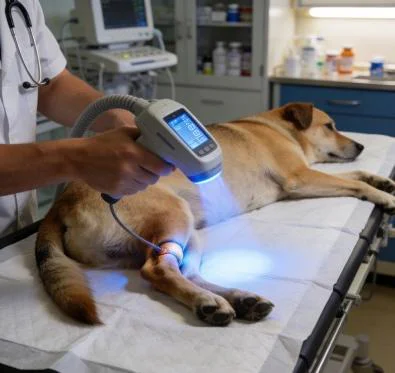
Adjusting pulse frequencies between 10 Hertz and 500 Hertz tailors the biological effect: low frequencies suppress substance P transmission to relieve spinal pain, while higher frequencies stimulate Schwann cell proliferation and axonal remodeling during later rehabilitation phases.
تقرير حالة سريرية ميدانية
The data below comes from Veterinary Neurology and Neurosurgical Rehabilitation unit records, documenting non-surgical recovery metrics in a chondrodystrophic canine with severe acute thoracolumbar disc extrusion.
الملف الشخصي للمريض وتصنيف المرحلة التشخيصية
Case File Number: VNN-2026-1185
Subject: Canine, Miniature Dachshund
Age: 6 Years 2 Months
الجنس: ذكر (مخصي)
Weight: 5.4 Kilograms
Presenting Complaint: Acute inability to bear weight on pelvic limbs following a minor jump from a sofa, severe vocalization, arched thoracolumbar kyphosis, complete paraplegia.
Neurological Examination: Grade 4 non-ambulatory paraparesis progressing to Grade 5 with loss of deep pain perception in both pelvic limbs; absent cutaneous trunci reflex caudal to L1; severe focal hyperpathia from T11 to T13; bilateral patellar hyperreflexia.
Imaging Diagnosis: Spinal CT and high-resolution MRI show acute Hansen Type I disc extrusion at T12-T13 with 55 percent spinal canal cross-sectional compromise, pronounced localized intramedullary hyperintensity on T2-weighted sequences indicating severe focal cord edema. Pre-treatment Modified Frankel Score: 0 (Paraplegic with loss of deep nociception).
| معرّف الجلسة | اليوم | موقع العلاج | توزيع الطول الموجي | ذروة الطاقة (واط) | دورة التشغيل (%) | تردد النبض (هرتز) | إجمالي الطاقة (J) | الملاحظات السريرية |
| S-01 | D-01 | T10-L2 Paraspinal Field | 80% 980 نانومتر / 20% 1470 نانومتر | 6.0 | 30% | 15 | 1,400 | Severe spinal guarding; non-contact linear sweeps applied over dorsal laminae. |
| S-02 | D-02 | T10-L2 & Epaxials | 75% 980 نانومتر / 25% 1470 نانومتر | 7.0 | 35% | 20 | 1,800 | Epaxial muscle spasms soften; deep pain perception returns to lateral right claw. |
| S-03 | D-04 | T11-T13 Spinal Canal Zone | 70% 980 نانومتر / 30% 1470 نانومتر | 8.0 | 35% | 50 | 2,200 | Bilateral deep pain perception verified; cutaneous trunci reflex moves caudally to L3. |
| S-04 | D-06 | T10-L3 & Sciatic Pathway | 65% 980 نانومتر / 35% 1470 نانومتر | 9.0 | 40% | 100 | 2,600 | Superficial pain response returns; voluntary tail movement observed on greeting. |
| S-05 | D-09 | Paraspinal & Hind Limbs | 60% 980 نانومتر / 40% 1470 نانومتر | 10.0 | 40% | 200 | 3,000 | Voluntary motor contractions in quadriceps; assisted standing holds for 8 seconds. |
| S-06 | D-13 | T10-L3 Paraspinal Chain | 50% 980 نانومتر / 50% 1470 نانومتر | 12.0 | 45% | 400 | 3,600 | Unassisted rising on traction mats; dog ambulates 4 meters with mild ataxia. |
| S-07 | D-18 | Regional Neurovascular Axis | 50% 980 نانومتر / 50% 1470 نانومتر | 10.0 | 40% | ديناميكي | 3,200 | Proprioceptive placement corrects within 1.5 seconds; urinary continence restored. |
| S-08 | D-25 | Biomechanical Maintenance | 50% 980 نانومتر / 50% 1470 نانومتر | 8.0 | 35% | ديناميكي | 2,500 | Coordinated voluntary gait; Modified Frankel Score reaches Grade 5. |
مقاييس موضوعية لقياس التعافي الوظيفي
Day 1 (Intake Baseline)
[Frankel Score: 0/5]         ░░░░░░░░░░░░░░░░░░░░
[Deep Pain Sensation]        Absent Bilaterally

Day 6 (Midway Status)
[Frankel Score: 2/5]         ████████░░░░░░░░░░░░
[Deep Pain Sensation]        Intact Bilaterally

Day 25 (Protocol Completion)
[Frankel Score: 5/5]         ████████████████████
[Deep Pain Sensation]        Full Neurological Recovery
By session eight on Day 25, the patient achieved complete functional recovery. The Modified Frankel Score rose from 0 to 5, showing independent ambulation and normal postural reactions. Bilateral conscious proprioception returned to normal, with paw-replacement latency dropping to under one second.
Manual palpation showed complete resolution of thoracolumbar hyperpathia from T11 to T13. Voluntary urinary and fecal continence returned fully, preventing secondary cystitis and allowing the dog to resume daily exercise under a conservative home care protocol.
المزايا العملية مقارنة بالأساليب التقليدية
Managing acute canine IVDD presents difficult clinical decisions in veterinary practice. Emergency decompressive hemilaminectomy involves substantial financial costs, requires specialized neurosurgical facilities, and carries risks of post-operative hematoma, surgical scarring, and prolonged recovery times. Conservative medical care relying solely on high-dose corticosteroids or NSAIDs brings serious risks of gastrointestinal ulceration, pancreatitis, and systemic immunosuppression, without directly resolving the microvascular compression strangling the spinal cord.
Underpowered low-level phototherapy units worsen these clinical delays. Low-wattage hardware requires prolonged session times of thirty to forty minutes to deliver minimal photon quantities through thick epaxial muscles and dense bone. This hands-on demand ties up technicians and examination suites, driving up clinic labor overhead while delivering slow, inconsistent clinical progress.
High-power Class 4 multi-wavelength protocols eliminate these clinical bottlenecks. Delivering up to twelve watts of peak power through balanced 980nm and 1470nm wavelengths cuts hands-on treatment times down to under eight minutes per spinal segment. This delivery speed allows veterinary practices to treat deep neurological structures efficiently, tripling daily room capacity while keeping spinal patients calm on the treatment mat.
Dynamic duty cycle pulsing protects sensitive tissues over the dorsal vertebral arches, preventing thermal spikes on dense, dark-coated limbs. Pet owners see noticeable reductions in paralysis and spinal pain within the first three visits, validating their commitment and keeping compliance high across the complete rehabilitation plan. For veterinary practices, combining deep spinal canal penetration with rapid treatment times turns complex canine neurological cases into a dependable, non-invasive clinical service.