 فوتون ميديكس












التغلب على التوهين الشديد للغضروف الليفي في حالة التهاب المفاصل الجذري للمفصل القاعدي للإبهام
Multi-spectral coherent photons penetrate the dense trapezial fibrocartilage matrix, downregulate localized synovial cytokine synthesis, and suppress mechanoreceptor nociceptive firing via microsecond duty cycle modulation without cutaneous thermal stacking.
A 61-year-old dental hygienist enters the hand and upper extremity clinic resting her dominant right forearm on the examination cushion, unable to pinch a key or open a tight screw cap without wincing. Every attempt to oppose the thumb to the small finger triggers an acute, grinding ache deep within the base of the first metacarpal. Palpation over the carpometacarpal (CMC) junction reveals localized bogginess, dorsal osteophytic shelf formation, and a positive grind test that reproduces sickening joint crepitus. Over four years, the patient has worn rigid thumb spica splints, consumed daily celecoxib, and received three fluoroscopically guided intra-articular corticosteroid injections. While the initial injection afforded three months of manageable comfort, subsequent rounds yielded diminishing returns, culminating in continuous aching during nocturnal rest and progressive radial subluxation of the metacarpal base. Surgical trapeziectomy with ligament reconstruction and tendon interposition (LRTI) was presented as the final recourse, carrying a twelve-week immobilization and rehabilitation arc. The fundamental technical contradiction in treating advanced basal joint degeneration non-surgically centers on soft-tissue optics: the first carpometacarpal articulation is compact, enclosed by dense, thickened palmar and dorsoradial stabilizing ligaments that scatter incident light beams. Superficial phototherapy devices and low-output emitters scatter within the first few millimeters of the thenar dermis, failing to reach the subchondral bone plate. Delivering sustained clinical relief requires driving calibrated photon flux through dense connective tissues to downregulate the localized inflammatory cascade while restoring microvascular perfusion across the compressed joint capsule.
Optical Penetration Across Thick Thenar Ligamentous Barriers
Delivering therapeutic photon densities into the first carpometacarpal cavity requires overcoming high reflection and scattering coefficients across compact connective tissues. Thenar skin, dense collagenous retinacula, and the anterior oblique ligament scatter non-coherent light beams, converting standard therapeutic devices into superficial heating pads.
Wavelength-Selective Chromophore Dynamics in Basal Joint Cartilage
The clinical efficacy of high-intensity class IV optical therapy depends on selecting wavelengths that bypass superficial dermal scatter while targeting specific biological components within the degenerate joint capsule:
- Microvascular recruitment and capillary clearing at 980nm: The 980nm wavelength matches the optical absorption profile of oxygenated hemoglobin and interstitial water. In arthritic carpometacarpal synovium, chronic capsular contracture and osteophytic impingement produce microvascular stagnation and tissue hypoxia. Photons at 980nm induce localized microvascular warming, triggering endothelial nitric oxide release, opening compressed capillary beds, and promoting the reabsorption of inflammatory effusion within periarticular tissues.
- Mitochondrial enzyme stimulation and tissue repair at 1064nm: The 1064nm wavelength displays the lowest scattering coefficient among therapeutic near-infrared bands in dense connective tissue. These photons penetrate directly through the thick dorsoradial ligament to reach the subchondral bone of the trapezium and first metacarpal base. Inside dormant chondrocytes and capsular fibroblasts, 1064nm light binds to cytochrome c oxidase, displacing inhibitory nitric oxide, restoring the mitochondrial proton gradient, and increasing adenosine triphosphate (ATP) production for cellular repair.
Preventing Cutaneous Heat Accumulation via Duty Cycle Modulation
Emitting high-power continuous energy over the thin dorsal and thenar skin of the hand quickly challenges the thermal relaxation time of human skin. Continuous wave delivery causes heat to accumulate rapidly in superficial dermal tissues, setting off epidermal pain receptors and risking surface burns before the deep carpometacarpal joint receives a therapeutic dose.
Class IV clinical systems overcome this barrier using variable pulse duty cycle gating. By pulsing the beam between 20 Hz and 15,000 Hz with precise on/off ratios, the laser delivers high peak power during active microsecond pulses, followed by brief rest intervals where local capillary blood flow carries surface heat away. This thermal regulation keeps skin temperatures safely below 42 degrees Celsius, enabling clinicians to apply sustained laser therapy for joint conditions without risking photothermal burns.
+--------------------------------------------------------------------------------------------------+
|                          FIRST CMC BASAL JOINT PHOTON PROPAGATION                                |
+--------------------------------------------------------------------------------------------------+
| Conical Quartz Contact Handpiece Compression                                                     |
|   |                                                                                              |
|   v                                                                                              |
| [Thenar Cutaneous Layer]    ---> Dermal heat dissipated via microsecond duty cycle pauses        |
|   |                                                                                              |
|   v                                                                                              |
| [Thenar Fascia & Tendons]   ---> Physical probe pressure clears interstitial water barrier      |
|   |                                                                                              |
|   v                                                                                              |
| [Dorsoradial Ligament]      ---> 980nm dilates compressed vessels, clearing inflammatory waste   |
|   |                                                                                              |
|   v                                                                                              |
| [Synovial Cavity Margin]    ---> Downregulation of IL-1beta, TNF-alpha, and COX-2 cascades       |
|   |                                                                                              |
|   v                                                                                              |
| [Trapezial Cartilage & Bone]---> 1064nm activates Cytochrome C Oxidase, restoring ATP synthesis   |
+--------------------------------------------------------------------------------------------------+
Biochemical Pathways: Suppressing Synovial Catabolism and Hand Nociception
Managing degenerative basal joint arthritis requires addressing progressive joint space loss, chronic biochemical inflammation, and persistent peripheral nerve sensitization. Degenerated chondrocytes and inflamed synovial cells produce catabolic cytokines, including interleukin-1 beta (IL-1β), tumor necrosis factor-alpha (TNF-α), and matrix metalloproteinases (MMPs), which break down type II collagen and aggrecan while sensitizing articular nociceptors.
Applying a targeted laser for arthritis interrupts this destructive cycle. Coherent near-infrared irradiation downregulates nuclear factor kappa B (NF-κB) transcription inside synovial fibroblasts, decreasing the production of inflammatory mediators like prostaglandin E2 (PGE2) and cyclooxygenase-2 (COX-2) to help protect remaining cartilage integrity.
Simultaneously, implementing structured laser arthritis treatment acts directly on sensory nerve pathways. Delivering high photon doses hyperpolarizes unmyelinated C-fibers and thinly myelinated A-delta fibers within the articular capsule. Research by Bjordal and colleagues in physical rehabilitation literature demonstrates that high-energy near-infrared illumination reduces peripheral nerve conduction velocity and curbs substance P accumulation in periarticular tissues, providing rapid and lasting pain relief without systemic side effects.
Clinical Case Documentation: Outpatient Hand Rehabilitation
The documented case below outlines the outpatient rehabilitation course of a patient with advanced thumb carpometacarpal osteoarthritis, managed using a multi-wavelength Class IV rehabilitation laser platform equipped with a specialized conical quartz massage applicator.
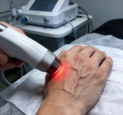
الملف الشخصي للمريض والبيانات السريرية الأساسية
- Case Identifier: HAND-RHIZ-2026-0412
- Patient Age / Sex: 61-Year-Old Female
- Primary Diagnosis: Advanced Thumb Basal Joint Osteoarthritis (Eaton-Littler Stage III) with Dorsal Subluxation, Marked Trapezial Sclerosis, and Secondary Adductor Pollicis Spasm, confirmed via Stress Radiographs and Musculoskeletal Ultrasound.
- History: 48 months of worsening right thumb base pain, loss of key pinch strength, and dropping household objects. Failed conservative management including custom thermoplastic splints, topical NSAIDs, oral analgesics, and three cortisone injections. Patient unable to hold dental instruments for more than ten minutes without severe pain.
- Baseline Metrics: Numerical Pain Rating Scale (NPRS) at 8/10 during key pinch tasks; Functional Index for Hand Osteoarthritis (FIHOA) at 24/30; Key Pinch Strength measured at 2.4 kg (contralateral unaffected thumb measured at 6.8 kg); Palmar Abduction restricted to 28°.
Multi-Stage Treatment Regimen
The treatment course comprised ten clinical sessions scheduled over four weeks, progressing from early pain management and lymphatic drainage to deep joint biostimulation and functional grip integration.
| رقم الجلسة. | Clinical Phase & Objective | Wavelength Setup | Operating Power (W) | تردد النبض (هرتز) | دورة التشغيل (%) | الطاقة المسلمة (J) | Cumulative Dose (J) |
| الجلسة 01 | Nociceptive Gating & Radial Styloid Drainage | 980nm (65%) + 1064nm (35%) | 8.0 واط | 8,000 Hz | 35% | 2,400 J | 2,400 J |
| Session 02 | Thenar Muscular Decompression | 980nm (60%) + 1064nm (40%) | 9.0 واط | 4,000 Hz | 45% | 2,700 J | 5,100 J |
| الجلسة 03 | Palmar Oblique Ligament Capillary Flush | 980nm (50%) + 1064nm (50%) | 10.0 واط | 2,000 هرتز | 50% | 3,000 J | 8,100 J |
| Session 04 | Dorsoradial Capsular Saturation | 980nm (40%) + 1064nm (60%) | 11.0 واط | 500 هرتز | 60% | 3,300 J | 11,400 J |
| Session 05 | Deep Trapezial Bed Photon Infusion | 980nm (35%) + 1064nm (65%) | 12.0 واط | 250 هرتز | 65% | 3,600 J | 15,000 J |
| الجلسة 06 | تثبيط السيتوكينات في الخلايا الليفية الزلالية | 980 نانومتر (30%) + 1064 نانومتر (70%) | 13.0 واط | 100 هرتز | 70% | 3,900 جول | 18,900 ج |
| الجلسة 07 | دفع عملية التنفس في الخلايا الغضروفية عالية التدفق | 980 نانومتر (20%) + 1064 نانومتر (80%) | 14.0 واط | مستمر (CW) | 100% | 4,200 جول | 23,100 ج |
| الجلسة 08 | تشبع الصفيحة شبه الغضروفية شبه المنحرفة | 980 نانومتر (20%) + 1064 نانومتر (80%) | 14.0 واط | مستمر (CW) | 100% | 4,200 جول | 27,300 ج |
| الجلسة 09 | تحميل ديناميكي لمقاومة الانضغاط | 980 نانومتر (30%) + 1064 نانومتر (70%) | 12.0 واط | 150 هرتز | 70% | 3,600 J | 30,900 ج |
| الجلسة 10 | التثبيت النهائي للأنسجة والخروج من المستشفى | 980nm (40%) + 1064nm (60%) | 10.0 واط | 400 هرتز | 60% | 3,000 J | 33,900 ج |
مؤشرات التطور السريري والنتائج
الأسبوع الأول (الجلسات 1-3):
- أدى التلامس اللطيف الذي يمر فوق منطقة «صندوق التبغ» التشريحية ونتوء الثنار إلى تخفيف التوتر حول المفصل.
- خفت آلام الراحة الليلية؛ وانخفضت درجة الألم على مقياس NPRS من 8/10 إلى 4/10.
- نام المريض طوال الليل دون أن يستيقظ بسبب الخفقان في قاعدة الإبهام بعد الجلسة 2.
- استرخى تشنج العضلة المقربة للإبهام بشكل ملحوظ أثناء الجس اليدوي.

الأسبوع 2 (الجلسات 4-6):
- اتسع نطاق الإبعاد الراحي من 28° إلى 42°؛ وتخفف تقلص الفراغ بين الأصابع الأول.
- تمكن المريض من فتح مقابض الأبواب وإمساك أدوات تناول الطعام دون الشعور بوخز حاد.
- تحسنت درجة FIHOA الوظيفية من 24/30 إلى 12/30.
- زادت قوة القبض على المفتاح من 2.4 كجم إلى 4.2 كجم.

الأسبوع 3 (الجلسات 7-8):
- تم تطبيق موجات مستمرة على خط المفصل CMC الأول باستخدام ملامس كوارتز مخروطي صلب.
- تمكن المريض من حمل أدوات خفيفة لتنظيف الأسنان لمدة 30 دقيقة دون حدوث التهاب بعد النشاط.
- أظهر التقييم بالموجات فوق الصوتية انخفاضًا في سماكة المحفظة وتلاشي الاحتقان الزليلي الموضعي.

الأسبوع 4 (الجلستان 9-10):
- استعادة الانحراف الراحي النشط الكامل (48°)، بما يتطابق مع المستوى الأساسي لليد المقابلة.
- بلغت قوة الضغط بالأصابع 6.2 كجم (في نطاق 90% من اليد السليمة).
- سُجل مؤشر NPRS النهائي عند 1/10 أثناء الضغط القوي والمستمر و0/10 في حالة الراحة.
- بلغت درجة FIHOA 2/30، مما مكن المريض من استئناف مهامه المهنية في مجال صحة الأسنان بدوام كامل.
تحليل مقارن: الليزر عالي الكثافة مقابل الأساليب التقليدية
يُظهر تقييم الضوء عالي الكثافة من الفئة الرابعة جنبًا إلى جنب مع الخيارات السريرية التقليدية السبب وراء قيام مراكز الطب الفيزيائي الحديثة بدمج أجهزة الليزر متعددة الأطوال الموجية في العلاج اليومي لليد:
حقن الكورتيكوستيرويدات داخل المفصل
في حين أن حقن الستيرويدات توفر راحة مؤقتة من الالتهاب، فإن الاستخدام المتكرر لها داخل المفصل الكاربوميتاكارصالي الصغير يثبط عملية التمثيل الغذائي للخلايا الغضروفية، ويضعف الأربطة الكبسولية المُثبِتة، ويُسرّع من حدوث خلع جزئي تدريجي للعظم الكعبري الأول. وفي المقابل، يعمل العلاج بالليزر عالي الكثافة متعدد الأطوال الموجية بطريقة غير جراحية. وبدلاً من إتلاف الغضروف، تعمل الفوتونات المركزة على تحفيز الدورة الدموية في الأوعية الدموية الدقيقة، وتعزيز تخليق ATP الخلوي، وتخفيف الألم في المفصل دون أي سمية كيميائية.
استئصال العضلة شبه المنحرفة مع إعادة بناء الأربطة (جراحة LRTI)
يؤدي الاستئصال الجراحي لعظم الترابيزيوم إلى القضاء على التلامس بين العظام، لكن هذه العملية تتطلب استئصال الأوتار، وفترات طويلة من تثبيت المفصل، وغالبًا ما تؤدي إلى ضعف مستمر في قوة القبضة. ويُعد العلاج بالليزر من الفئة الرابعة علاجًا فعالًا غير جراحي يحافظ على التشريح الطبيعي للمفصل، ويقلل من التهاب المحفظة، ويعيد القدرة على الحركة دون التعرض لصدمة جراحية.
الأدوية المضادة للالتهاب غير الستيرويدية (NSAIDs) التي تُؤخذ عن طريق الفم
تنتشر الأدوية المضادة للالتهابات الجهازية في جميع أنحاء الجسم، مما يثبط عمل البروستاجلاندينات الواقية في بطانة المعدة والكلى، في حين تصل تركيزات منخفضة من الدواء إلى أربطة اليد الكثيفة ذات الأوعية الدموية القليلة. وينطوي الاستخدام المزمن لمضادات الالتهاب غير الستيرويدية (NSAID) على مخاطر موثقة جيدًا تتمثل في الإصابة بقرحة المعدة وارتفاع ضغط الدم والضعف الكلوي. يستهدف العلاج بالليزر عالي الكثافة توجيه الطاقة العلاجية مباشرةً إلى كبسولة المفصل المتضررة، مما يخفف آلام المفاصل وتيبسها محليًّا دون آثار جانبية جهازية.
التطبيق السريري العملي والدروس المستفادة على الصعيد التشغيلي
يتطلب تحقيق نتائج سريرية متسقة عند استخدام أنظمة الليزر عالية الكثافة لعلاج تنكس المفصل القاعدي للإبهام اتباع نهج منظم:
- الضغط الديناميكي باستخدام مجسات الكوارتز المخروطية: يجب على الأطباء ممارسة ضغط قوي وثابت باستخدام مجس التلامس المخروطي المصنوع من الكوارتز مباشرةً على «صندوق التبغ» التشريحي والحواف الراحية لقاعدة عظم المشط الأول. يؤدي الضغط المادي إلى إزاحة الماء السطحي والدم الوريدي بعيدًا عن مسار الحزمة الضوئية، مما يخلق مسارًا بصريًّا مثاليًّا يوفر عددًا أكبر من الفوتونات إلى الهياكل العميقة للمحفظة.
- التطهير اللمفاوي الإقليمي: يجب دائمًا معالجة المسارات اللمفاوية القريبة أولاً. إن مسح العظم الشوكي الكعبري، والجزء البعيد من الساعد، والفضاء بين أصابع اليد (الثينار) قبل توجيه الطاقة إلى كبسولة المفصل، يؤدي إلى تطهير مسارات السوائل وتسريع تعافي الأنسجة.
- بروتوكول التدرج الترددي: ابدأ بترددات نبضية عالية (4,000 هرتز إلى 8,000 هرتز) خلال الزيارات الأولية لتهدئة المسارات العصبية الطرفية شديدة الحساسية، ثم انتقل إلى نبضات معتدلة (100 هرتز إلى 500 هرتز) لتحسين التروية المحلية وتخفيف تصلب المحفظة، واختتم العلاج بإرسال موجة مستمرة (CW) لتحفيز التمثيل الغذائي للخلايا الغضروفية وإعادة تشكيلها الهيكلي بمجرد أن يهدأ الألم الحاد.
يتيح تطبيق العلاج بالليزر متعدد الأطوال الموجية من الفئة الرابعة، القائم على فيزياء الأنسجة البصرية، لعيادات العلاج العظمي وعلاج اليد في العيادات الخارجية معالجة التهاب المفاصل العظمي المستعصي في المفاصل القاعدية ومساعدة المرضى على استعادة وظائف اليد اليومية بدقة وبدون ألم.