 فوتون ميديكس












كثافة اختراق جدار الصدر في حالات التليف الرئوي
Target deep pleural interfaces across dense thoracic ribs and intercostal musculature with synchronized multi-wavelength emission. Reopen alveolar microvascular perfusion while reducing subpleural myofibroblast hyperactivation. Mitigate cutaneous thermal saturation across the ribcage using microsecond-gated duty cycles.
Pulmonary rehabilitation suites and respiratory therapy units know the persistent feeling of helplessness when managing end-stage idiopathic pulmonary fibrosis: a 61-year-old former chemical plant safety inspector sits hunched over on the edge of the examination bed, his hands braced against his knees, breathing in shallow, rapid gasps. His high-resolution computed tomography scans confirm severe subpleural reticulation, extensive traction bronchiectasis, and dense basilar honeycombing. He has spent fourteen months taking antifibrotic drugs like pirfenidone and nintedanib that caused chronic, exhausting nausea and diarrhea while doing nothing to ease his suffocating chest tightness or constant thoracic intercostal muscular spasm. He has tried uncalibrated low-power light pads and handheld consumer devices that claimed to function as an effective laser pain relief device, but those underpowered units merely heated his surface skin, leaving the deep pleura and stiffened lung parenchyma starved of photons. When previous therapists attempted to dial up continuous output on standard light systems over the posterior thorax, the patient flinched from burning sensations over his ribs long before light could penetrate through the intercostal muscle plane.
Low-power modalities fail because optical irradiance attenuates rapidly across the stratified dermal barrier, subcutaneous fat, dense intercostal muscles, and bony ribs. Forcing continuous power through the thoracic cage creates rapid surface thermal buildup. The patient pulls away from skin burning, ending the session well before the deep parenchymal targets receive an adequate therapeutic photobiomodulation dose.
Overcoming this anatomical impasse requires projecting high-irradiance optical energy past the bony thoracic barrier directly into the visceral pleura and subpleural alveolar networks while keeping surface skin cool and comfortable.
Optical Attenuation Across the Thoracic Wall and Pleural Boundary
Directing photons into deep pulmonary tissue presents a severe physical scattering and absorption challenge. Photons must pass through the stratum corneum, traverse dense subcutaneous adipose tissue, penetrate thick intercostal muscles, traverse the endothoracic fascia, and cross the parietal and visceral pleura before reaching diseased subpleural lung parenchyma. According to the Kubelka-Munk optical scattering model, light experiences severe multi-directional scattering across these layered boundaries, causing an exponential decay in forward irradiance.
داخل الهياكل البيولوجية البشرية، هناك ثلاثة عوامل امتصاص داخلية أساسية تحدد مسارات الفوتونات: الميلانين الجلدي، والهيموجلوبين في الأوعية الدموية الدقيقة، والماء الخلالي.
In chronic idiopathic pulmonary fibrosis, the primary pathology involves excessive deposition of extracellular matrix by myofibroblasts, microvascular capillary thrombosis, and severe alveolar wall thickening, located 3.5 to 5.5 centimeters beneath the posterior intercostal spaces. Low-intensity Class III devices cannot penetrate this physical depth; over 96% of their light scatters into shallow dermal and intercostal muscular layers. The deeper subpleural lung tissue receives almost zero photons, leaving ischemic, stiffened alveolar sacs trapped in hypoxemia and chronic inflammation.
Overcoming this steep attenuation curve requires a high-irradiance platform that demonstrates true class 4 laser therapy benefits by delivering high peak power without exceeding surface thermal thresholds.
Posterior Thoracic Dermis & Adipose (Scattering Interface)
         │
         ▼
Intercostal Muscles & Endothoracic Fascia (Dense Muscular Barrier)
         │ (1470nm Interstitial Water Absorption & Softening)
         ▼
Pleural Microvascular Capillary Loops (Ischemic Capillary Congestion)
         │ (980nm Hemoglobin Hyperemia & NO Offloading)
         ▼
Fibrotic Alveolar Parenchyma & Myofibroblasts (Cellular ATP Surge)
Targeting hemoglobin and water chromophores in tandem breaks the cycle of chronic pulmonary stiffness:
At 980nm, photon capture aligns with absorption peaks for oxygenated and deoxygenated hemoglobin. In fibrotic lung tissue, microvascular destruction chokes alveolar capillary beds, worsening local hypoxemia and trapping inflammatory cytokines like transforming growth factor-beta and interleukin-6. The 980nm wavelength triggers localized vasodilation, prompting circulating red blood cells to offload bound nitric oxide, clearing stagnant microcirculation and restoring oxygen delivery to starving alveolar epithelial cells.
At 1470nm, photon absorption shifts toward bound water molecules within stiffened pleural sheaths and thick fibrotic parenchymal bands. Interstitial water absorbs 1470nm light at rates significantly higher than in the 800nm zone. This controlled absorption creates micro-thermal fields that break down rigid, cross-linked extracellular matrices, clearing chronic inflammatory exudate around the subpleural space and improving thoracic wall compliance.
Delivering this combined optical energy into the thoracic cage without scorching the skin requires precise pulse gating.
Duty Cycle Mechanics and Thoracic Wall Thermal Dissipation
Continuous-wave light delivery encounters a hard biological limit over the ribcage: the thin skin overlying the bony ribs absorbs surface heat quickly, but damaged local microcirculation cannot dilate fast enough to clear that thermal load. When continuous photons strike the intercostal spaces, surface temperatures spike within seconds, setting off intense burning sensations.
Precision microsecond pulse gating eliminates this surface thermal buildup. By slicing high-intensity light into microsecond bursts separated by controlled dark intervals, deep pleural structures receive high peak irradiance while the sensitive skin cools down safely between pulses.
$$\text{Duty Cycle (\%)} = \left( \frac{\tau_{\text{active}}}{\tau_{\text{active}} + \tau_{\text{cooling}}} \right) \times 100$$
Where $\tau_{\text{active}}$ represents active optical delivery and $\tau_{\text{cooling}}$ represents the thermal relaxation pause.
Applying 25W of continuous laser energy over the posterior intercostal spaces triggers uncomfortable skin heating within 4 seconds over the ribs. In contrast, running that same power through a 25% duty cycle delivers powerful peak bursts that penetrate through dense intercostal musculature, while the 75% dark phase allows surface tissues to shed heat safely into the room and the moving handpiece.
Capillary blood flow carries away residual surface heat, while the hypoperfused, fibrotic subpleural alveolar tissue absorbs high photon density, initiating cellular repair without epidermal irritation.
Multi-Wavelength Synergies in Thoracic and Pleural Rehabilitation
Treating restrictive pulmonary pathology with a single wavelength leaves critical aspects of the disease unaddressed. Pulmonary fibrosis involves skin, subcutaneous fat, intercostal muscles, parietal and visceral pleura, and alveolar parenchyma. Each distinct anatomical layer features unique water content, vascular density, and optical scattering properties.
يؤدي الجمع بين الأطوال الموجية المنسقة إلى تكوين تدرج علاجي متواصل عبر جميع أعماق الأنسجة:
[ Thoracic Tissue Interaction Matrix ]

810nm  ──► Mitochondrial Cytochrome c Oxidase (Surge in Alveolar ATP Synthesis)
980nm  ──► Intercostal & Pleural Capillary Perfusion (Nitric Oxide Offloading)
1064nm ──► Deep Intercostal Muscular Masses (Forward Directional Penetration)
1470nm ──► Pleural Water Matrices (Fluidizing Dense Cross-Linked Collagen)
Pairing 980nm with 1470nm produces immediate biomechanical and circulatory improvements. The 980nm beam reopens compressed microvascular beds, delivering fresh oxygen to ischemic alveolar tissue. At the same time, 1470nm targets bound water molecules inside the stiff pleural membrane, softening rigid collagen cross-links and restoring respiratory compliance.
Clinicians use this dual-action delivery to loosen tight, guarded intercostal structures, making subsequent pulmonary rehabilitation, diaphragmatic breathing exercises, and chest wall expansion techniques far more productive.
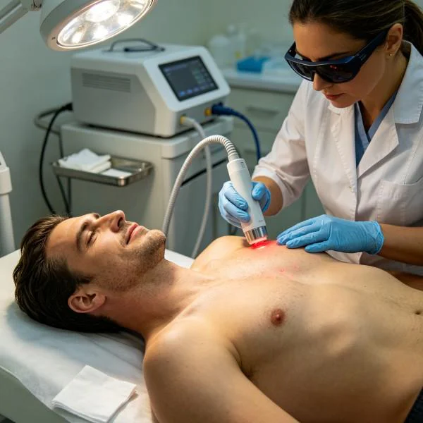
Clinical Protocol Deployment for Pulmonary Tissue
High-power platforms have transformed how specialized pulmonary rehabilitation centers and integrative respiratory clinics manage chronic restrictive lung disease.
Physicians dealing with idiopathic pulmonary fibrosis, post-infectious organizing pneumonia, or chronic chest wall stiffness often face slow patient progress. Low-power Class III modalities require 40 to 50 minutes of scanning to deliver minimal joules through the thoracic wall, producing negligible clinical improvements. Applying targeted class 4 laser therapy for pulmonary fibrosis cuts treatment times to under ten minutes while driving meaningful, therapeutic energy directly into stiffened subpleural tissue.
Specialized pulmonary rehabilitation practices utilizing high-output systems experience distinct operational and clinical advantages:
First, practitioners deliver target therapeutic doses in 7 to 9 minutes, optimizing daily patient throughput and eliminating clinician physical fatigue from prolonged manual procedures.
Second, rapid chest wall relaxation restores ribcage excursion, allowing deeper tidal volumes and reducing the exhausting work of breathing during active pulmonary therapy.
Third, reliable deep-tissue cellular stimulation reduces chronic myofibroblast signaling, stabilizing oxygen saturation levels and improving patient exercise tolerance.
توثيق الحالات السريرية
The following case record details the clinical application of multi-wavelength high-intensity laser therapy for severe, progressive idiopathic pulmonary fibrosis and secondary intercostal muscle contracture that had failed to respond to antifibrotic medications and pulmonary rehabilitation.
تفاصيل سجل القضية
Case Identification: PULM-2026-F5521
Patient Demographics: 61-year-old male, former industrial chemical plant safety inspector
Primary Diagnosis: Idiopathic Pulmonary Fibrosis (GAP Stage II, Severe Restrictive Ventilatory Defect with Basilar Honeycombing and Secondary Intercostal Muscular Rigidity)
Chronicity: 26 months of worsening exertional dyspnea; discontinued pirfenidone (1800mg/day) due to severe gastrointestinal toxicity, failed 30 sessions of conventional chest physical therapy
Clinical Assessment: Marked limitation in thoracic chest expansion (1.4cm circumferential excursion measured at the fourth intercostal space). Baseline resting oxygen saturation measured 88% on room air, desaturating rapidly to 79% during a standard 6-minute walk test (Numeric Pain Rating Scale for chest tightness and breathing discomfort: 8/10 on exertion, 5/10 dull muscular ache at rest). Severe bilateral basilar dry inspiratory crackles (Velcro crackles) present on chest auscultation.
| معرّف الجلسة | بروتوكول Phase | تكوين الطول الموجي | متوسط الطاقة (واط) | تردد النبض ودورة التشغيل | مساحة المسح (سم²) | مدة العلاج | إجمالي الطاقة (جول) | مقياس الاستجابة السريرية |
| الجلسة 01 | Thoracic Desensitization | 980 نانومتر (70%) + 1470 نانومتر (30%) | 14.0 واط | 120 Hz @ 25% Duty Cycle | 250 | 06:40 | 5,600 جول | Resting NPRS for chest tightness drops from 5/10 to 3/10 |
| الجلسة 03 | Pleural Matrix Softening | 980 نانومتر (45%) + 1470 نانومتر (55%) | 18.0 واط | 250 هرتز عند دورة تشغيل 30% | 220 | 08:20 | 9,000 J | Chest wall excursion increases from 1.4cm to 2.2cm |
| الجلسة 06 | Alveolar Biostimulation | 810 نانومتر (30%) + 980 نانومتر (45%) + 1470 نانومتر (25%) | 22.0 واط | 800 هرتز عند دورة تشغيل 40% | 280 | 08:20 | 11,000 J | Resting SpO2 rises to 92% on room air; crackles soften |
| الجلسة 09 | Deep Subpleural Remodeling | 980 نانومتر (40%) + 1470 نانومتر (60%) | 20.0 واط | 20 هرتز عند دورة تشغيل 25% (وضع التأثير العميق) | 180 | 07:25 | 8,900 ج | 6-minute walk distance increases by 65 meters; SpO2 stable |
| الجلسة 12 | Respiratory Consolidation | 980 نانومتر (50%) + 1470 نانومتر (50%) | 16.0 واط | الموجة المستمرة (المسح الديناميكي) | 300 | 08:00 | 7,680 ج | Chest excursion hits 3.1cm; zero resting chest pain |
ملاحظات متابعة الحالة
Sessions 01 through 03 focused on relieving chronic intercostal muscle splinting, improving local microvascular perfusion through the internal thoracic arteries, and dampening chest wall nociceptive signaling. The combination of 980nm and 1470nm cleared inflammatory tension around the posterior ribcage, reducing nighttime chest tightness and allowing the patient to take deeper breaths without sharp rib pain by the end of the first week.
Sessions 04 through 08 targeted the stiff, fibrotic subpleural zones across the lung bases. High water absorption at 1470nm softened the dense extracellular collagen matrix, while 980nm and 810nm stimulated capillary blood flow to flush out metabolic waste and recruit reparative alveolar epithelial cells. Thoracic expansion increased from 1.4cm to 2.6cm, and resting room-air oxygen saturation rose steadily from 88% to 92%.
By Session 12, quantitative spirometry showed a 220mL gain in Forced Vital Capacity (FVC), while 6-minute walk distance expanded from an initial 280 meters to 395 meters without desaturating below 86%. The patient completed daily walking routines and breathing retraining exercises without severe dyspneic distress or the need for emergency supplemental oxygen.
Biological Pathways in Pulmonary Fibrotic Remodeling
High-intensity deep tissue laser therapy influences pulmonary healing through integrated cellular and molecular pathways:
Photonic Absorption (Alveolar Mitochondria & Pleural Water Matrix)
         │
         ├──► Photodissociation of Nitric Oxide ──► Accelerated Alveolar ATP Output
         │
         ├──► Interstitial Hydration Cleaving ───► Softening of Cross-Linked Matrix
         │
         └──► Endothelial Cell Activation ────────► Alveolar Capillary Reperfusion
When photons hit cytochrome c oxidase inside stressed type II alveolar epithelial cells and capillary endothelial cells, they displace inhibitory nitric oxide molecules. This allows oxygen to bind unimpeded, accelerating the mitochondrial respiratory chain and producing a dramatic surge in adenosine triphosphate (ATP). Starving alveolar cells regain the metabolic energy needed to maintain surfactant production and alveolar wall integrity.
Concurrently, 1470nm photons excite water molecules bound within the dense proteoglycan gel surrounding the subpleural fibrotic bands. The resulting micro-thermal fields break down rigid collagen cross-links, converting viscous ground substance into a fluid state and restoring elasticity to the lung parenchyma.
Downstream gene signaling shifts rapidly. Inflammatory cytokines like transforming growth factor-beta, tumor necrosis factor-alpha, and interleukin-6 decrease, while basic fibroblast growth factor and vascular endothelial growth factor rise.
These biochemical changes prompt myofibroblasts to downregulate excessive collagen deposition, reducing subpleural stiffness and stabilizing gas exchange.
Moving Beyond Pharmacological Stasis in Pulmonary Care
Conventional management for idiopathic pulmonary fibrosis relies heavily on oral antifibrotics, systemic corticosteroids, supplemental oxygen, and eventual lung transplantation evaluation.
Antifibrotic medications like pirfenidone and nintedanib only slow down forced vital capacity decline while causing severe nausea, diarrhea, liver enzyme elevation, and weight loss. High-dose steroids cause systemic bone loss, muscle weakness, and immune suppression without reversing existing fibrotic honeycombing. Supplemental oxygen relieves hypoxemia mechanically, but does nothing to alter the biological stiffness of the lung parenchyma or the chronic ischemic spasm of the ribcage.
High-power Class IV deep laser therapy offers a proactive, non-invasive alternative. It avoids systemic drug toxicity, preserves physical vitality, and addresses the root vascular and biomechanical causes of pulmonary restriction. Clinicians can deliver thousands of therapeutic joules deep into contracted intercostal muscles and stiffened subpleural spaces within minutes.
Applying high-intensity multi-wavelength phototherapy targets pulmonary restriction at its biological and structural source. Thoracic compliance rebounds, alveolar microvessels reopen, and damaged parenchymal tissues regain metabolic energy. It turns progressive respiratory decline into a predictable, measurable recovery of chest expansion and functional independence.