 포톤메딕스




중증 무릎 관절염에서 연골하 골 허혈 극복하기
Dual-spectral coherent photons bypass synovial fluid attenuation, dynamic pulse duty cycles prevent superficial thermal boundary accumulation, and targeted near-infrared energy restores chondrocyte cytochrome c oxidase function while clearing periarticular microvascular congestion.
A 66-year-old retired landscape contractor sits in an outpatient consultation room, bracing both forearms against his knees before attempting to stand. Every forward step produces a deep, grinding ache behind the patellar tendon, accompanied by an unstable lateral wobble during the loading response. His right knee exhibits chronic swelling, with an indurated suprapatellar pouch and a warm infrapatellar fat pad that flinches under minimal thumb pressure. Over the previous four years, the patient underwent periodic intra-articular corticosteroid injections, three rounds of high-molecular-weight hyaluronic acid viscosupplementation, and ongoing prescription meloxicam. While the early injections provided temporary relief, the therapeutic duration steadily declined from months to mere days, leaving him with chronic gastritis and the standing recommendation for total knee replacement surgery. The primary clinical bottleneck in treating advanced articular degeneration without operative joint reconstruction is soft-tissue photon attenuation: the degenerated tibiofemoral cartilage, meniscus, and subchondral bone lie four to six centimeters below the skin surface, shielded by dense fibrous capsular ligaments, subcutaneous adipose tissue, and thick, protein-rich synovial fluid pockets. Low-power modalities and superficial infrared devices scatter completely within the dermal layer, failing to deliver therapeutic doses to the hypoxic joint interior. Reversing severe articular pain demands delivering verified photon densities through thick joint structures to suppress local inflammatory signaling while reactivating cellular ATP production inside starving chondrocytes.
Optical Penetration Physics Across Fibrotic Synovial Barriers
Delivering therapeutic photon densities into an osteoarthritic joint space requires managing significant energy loss caused by tissue scatter and fluid absorption. As photons traverse the stratum corneum, subcutaneous fat, and dense collateral ligaments, optical attenuation follows an exponential decay curve governed by the specific absorption and scattering coefficients of each biological layer.
Wavelength-Targeted Chromophore Saturation in Degenerate Joints
The physiological success of high-intensity class IV light depends on selecting wavelengths that penetrate deep tissues while activating targeted chromophores in the damaged capsule. Using a single emission wavelength creates clinical compromises, whereas combining complementary near-infrared bands enables concurrent vascular and cellular remodeling:
- Microvascular recruitment and capillary flushing at 980nm: The 980nm wavelength matches the optical absorption characteristics of oxygenated hemoglobin and local water. In osteoarthritic synovium, microvascular networks become chronically compressed and fibrotic, causing tissue hypoxia and trapping acidic metabolic waste products. Delivering 980nm energy creates localized microthermal gradients along capillary walls, stimulating endothelial nitric oxide release, opening closed microvascular sphincters, and jumpstarting lymphatic reabsorption within congested periarticular tissues.
- Mitochondrial cytochrome c oxidase saturation at 1064nm: The 1064nm wavelength displays the lowest scattering coefficient among near-infrared therapeutic spectra in dense human connective tissue. Photons at this wavelength pass through thick capsular walls and joint fluid to reach the subchondral bone plate and articular cartilage. Inside dormant chondrocytes and subchondral osteoblasts, 1064nm light binds to cytochrome c oxidase, displacing inhibitory nitric oxide, rebuilding the mitochondrial proton gradient, and driving adenosine triphosphate (ATP) production required for cellular maintenance.
듀티 사이클 조절을 통한 피부 열 축적 방지
Directing twenty to thirty Watts of continuous laser energy toward a joint capsule generates significant surface heat. Continuous wave (CW) delivery quickly exceeds the thermal relaxation threshold of human skin, triggering cutaneous pain receptors and risking epidermal blistering long before the deep joint receives an adequate biostimulative dose.
Modern high-intensity class IV therapy platforms address this challenge using pulsed duty cycle gating. By modulating beam delivery into microsecond bursts (ranging from 20 Hz to 20,000 Hz) with controlled pulse durations and rest intervals, high peak power reaches deep tissues during the active phase, while superficial capillaries conduct heat away during the rest phase. This thermal management keeps skin temperatures safely below 42 degrees Celsius, enabling clinicians to perform sustained arthritis laser therapy without risking photothermal burns.
+--------------------------------------------------------------------------------------------------+
|                            ARTICULAR CAPSULE PHOTON PROPAGATION                                  |
+--------------------------------------------------------------------------------------------------+
| Quartz Glass Massage Handpiece Contact Compression                                              |
|   |                                                                                              |
|   v                                                                                              |
| [Epidermis & Dermis]        ---> Surface heat dissipated via microsecond duty cycle pauses       |
|   |                                                                                              |
|   v                                                                                              |
| [Prepatellar Adipose]       ---> Mechanical probe pressure clears interstitial water barrier      |
|   |                                                                                              |
|   v                                                                                              |
| [Capsular Ligament Layer]   ---> 980nm dilates compressed vessels, clearing inflammatory waste   |
|   |                                                                                              |
|   v                                                                                              |
| [Synovial Cavity Margin]    ---> Downregulation of IL-1beta, TNF-alpha, and COX-2 enzymes         |
|   |                                                                                              |
|   v                                                                                              |
| [Articular Cartilage & Bone]---> 1064nm activates Cytochrome C Oxidase to restore ATP production  |
+--------------------------------------------------------------------------------------------------+
Biomolecular Cascades: Suppressing Synovial Catabolism and Peripheral Pain
Managing advanced articular degeneration requires addressing structural cartilage loss, chronic biochemical inflammation, and persistent peripheral nerve sensitization. Degenerated chondrocytes and inflamed synovial cells produce catabolic cytokines, including interleukin-1 beta (IL-1β), tumor necrosis factor-alpha (TNF-α), and matrix metalloproteinases (MMPs), which break down type II collagen and aggrecan while sensitizing articular nociceptors.
Applying focused high-intensity light interrupts this destructive cycle. Targeted near-infrared irradiation downregulates nuclear factor kappa B (NF-κB) transcription inside synovial fibroblasts, decreasing the production of inflammatory mediators like prostaglandin E2 (PGE2) and cyclooxygenase-2 (COX-2) to help protect remaining cartilage integrity. This biological modulation represents one of the primary arthritis laser therapy benefits for patients with persistent joint effusion.
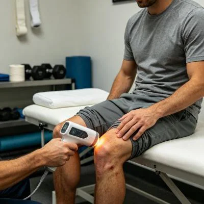
Simultaneously, implementing structured laser therapy for arthritis pain acts directly on sensory nerve pathways. Delivering high photon doses hyperpolarizes unmyelinated C-fibers and thinly myelinated A-delta fibers within the articular capsule. Research by Bjordal and colleagues in physical rehabilitation literature demonstrates that high-energy near-infrared illumination reduces peripheral nerve conduction velocity and curbs substance P accumulation in periarticular tissues, providing rapid and lasting pain relief without systemic side effects.
임상 사례 기록: 외래 정형외과 재활
The documented case below outlines the outpatient rehabilitation course of a patient with advanced knee osteoarthritis and secondary capsular flexion contracture, treated using a multi-wavelength Class IV rehabilitation laser platform equipped with a quartz massage applicator.
환자 프로필 및 임상적 기저 상태
- Case Identifier: GONARTH-OSTEO-2026-0315
- Patient Age / Sex: 66-Year-Old Male
- Primary Diagnosis: Advanced Medial Compartment Knee Osteoarthritis (Kellgren-Lawrence Grade III) with Significant Joint Space Loss, Subchondral Sclerosis, Infrapatellar Fat Pad Hypertrophy, and Secondary Flexion Contracture, confirmed via Weight-Bearing Radiographs and Diagnostic Musculoskeletal Ultrasound.
- History: 5 years of progressive right knee pain, joint stiffness, and mobility loss. Failed conservative measures including physical therapy, oral NSAIDs, hyaluronic acid injections, and four cortisone injections. Patient experiences severe pain during weight-bearing, cannot walk beyond 150 meters without resting, and walks with an antalgic limp.
- Baseline Metrics: Numerical Pain Rating Scale (NPRS) at 8/10 during weight-bearing movement; Western Ontario and McMaster Universities Osteoarthritis Index (WOMAC) at 71/96; Knee Active Range of Motion (AROM) Flexion restricted to 88°, Extension lacking 14° (flexion contracture); Mid-patellar joint circumference measured at 43.2 cm.
다단계 치료 요법
이 치료 과정은 4주에 걸쳐 진행된 10회의 임상 세션으로 구성되었으며, 초기 통증 관리와 림프 배농부터 심부 관절 생체 자극 및 기능적 가동성 향상 단계로 단계적으로 진행되었습니다.
| 세션 번호. | 임상 단계 및 목적 | 파장 설정 | 작동 전력 (W) | 펄스 주파수 (Hz) | 듀티 사이클(%) | 전달되는 에너지(J) | 누적 선량 (J) |
| 세션 01 | 통각 게이트 현상 및 슬와부 배액 | 980nm (65%) + 1064nm (35%) | 12.0 W | 8,000 Hz | 40% | 3,600 J | 3,600 J |
| 제2회 세션 | 내측 구획의 부종 완화 | 980nm (60%) + 1064nm (40%) | 14.0 W | 4,000 Hz | 50% | 4,200 J | 7,800 J |
| 세션 03 | 슬개골 상부 활막 세척 | 980nm (50%) + 1064nm (50%) | 16.0 W | 2,000 Hz | 60% | 4,800 J | 12,600 J |
| 제4회 세션 | 슬개골하 지방 패드 감작 감소 | 980nm (40%) + 1064nm (60%) | 18.0 W | 500Hz | 65% | 5,400 J | 18,000 J |
| 제5회 세션 | 심부 캡슐 섬유화성 연화 | 980nm (35%) + 1064nm (65%) | 20.0 W | 250 Hz | 70% | 6,000 J | 24,000 J |
| 제6회 세션 | 활막 섬유아세포의 사이토카인 억제 | 980nm (30%) + 1064nm (70%) | 22.0 W | 100Hz | 75% | 6,600 J | 30,600 J |
| 제7회 세션 | 고플럭스 연골세포 호흡 연구 추진 | 980nm (20%) + 1064nm (80%) | 24.0 W | 연속(CW) | 100% | 7,200 J | 37,800 J |
| 제8회 세션 | 연골하 골 기질의 포화도 | 980nm (20%) + 1064nm (80%) | 24.0 W | 연속(CW) | 100% | 7,200 J | 45,000 J |
| 제9회 세션 | 동적 확장 생체역학적 통합 | 980nm (30%) + 1064nm (70%) | 20.0 W | 150 Hz | 75% | 6,000 J | 51,000 J |
| 세션 10 | 최종 조직 통합 및 퇴원 | 980nm (40%) + 1064nm (60%) | 16.0 W | 400 Hz | 65% | 4,800 J | 55,800 J |
임상 경과 및 결과 지표
WEEK 1 (Sessions 1-3):
- Deep contact sweeping over the popliteal drainage basin and medial joint line reduced periarticular tension.
- Resting nocturnal pain subsided; NPRS dropped from 8/10 to 4/10.
- Patient slept through the night without waking from positional joint aching after Session 2.
- Mid-patellar joint circumference decreased from 43.2 cm to 41.0 cm as effusion drained.

WEEK 2 (Sessions 4-6):
- Knee active flexion expanded from 88° to 116°; extension deficit reduced from 14° to 5°.
- Patient climbed household stairs hand-over-hand without requiring pauses on intermediate steps.
- Ultrasound evaluation showed suprapatellar fluid pocket thickness decreased from 13 mm to 3.5 mm.
- WOMAC functional score improved from 71 to 38.

WEEK 3 (Sessions 7-8):
- Continuous wave delivery applied over the patellar retinaculum and tibiofemoral margins with firm compression.
- Patient walked 1.2 kilometers outdoors without joint catching, sudden giving way, or post-activity swelling.
- Crepitus during active knee flexion softened considerably as synovial membrane congestion subsided.

WEEK 4 (Sessions 9-10):
- Full active knee extension achieved (0° neutral); active flexion reached 128°.
- Mid-patellar circumference stabilized at 39.1 cm, matching the unaffected contralateral limb.
- Final NPRS recorded at 1/10 during extended walking and 0/10 at rest.
- WOMAC score reached 8/96, restoring daily mobility and deferring surgical total knee replacement.
비교 분석: 고강도 레이저 대 기존 치료법
고강도 IV급 레이저를 기존의 임상 치료법과 함께 평가해 보면, 현대적인 물리치료 센터들이 왜 다파장 레이저를 일상적인 치료에 도입하고 있는지 알 수 있습니다:
관절 내 코르티코스테로이드 주사
스테로이드 주사는 광범위한 화학적 면역억제제 역할을 하여 관절 염증을 일시적으로 완화시킵니다. 그러나 관절 내로 반복적으로 스테로이드가 투여되면 연골 손실이 가속화되고, 본래의 연골세포 생존력이 손상되며, 시간이 지남에 따라 관절 간격이 좁아질 위험이 있습니다. 고강도 다파장 레이저 치료는 비침습적이고 비파괴적인 대안을 제공합니다. 집중된 광자는 연골세포를 손상시키지 않으면서 미세혈관 순환을 촉진하고, 세포 내 ATP 합성을 증진시키며, 화학적 독성 없이 관절 통증을 완화합니다.
전치환 무릎 관절 치환술 (TKA)
관절 치환술은 원래의 관절 표면을 제거하고 금속 및 폴리에틸렌 임플란트로 대체하는 수술입니다. 심한 해부학적 손상에 대해서는 효과적이지만, 이 수술은 수술 전후 감염, 심부 정맥 혈전증, 임플란트 이완, 그리고 긴 재활 기간 등의 위험을 수반합니다. 4등급 레이저 치료는 본래의 관절 해부학적 구조를 보존하고, 관절낭 염증을 완화하며, 수술로 인한 외상 없이 관절 가동 범위를 회복시키는 효과적인 비수술적 치료법입니다.
경구용 비스테로이드성 항염증제(NSAIDs)
전신성 항염증제는 신체 전체로 순환하며, 위 점막과 신장에서 보호 작용을 하는 프로스타글란딘을 억제하는 한편, 혈관이 잘 발달되지 않은 관절 연골에는 제한된 농도의 약물만 전달됩니다. 비스테로이드성 항염증제(NSAID)를 장기간 복용할 경우 위궤양, 고혈압, 신장 기능 저하와 같은 위험이 있다는 사실은 이미 잘 알려져 있습니다. 고강도 레이저 치료는 손상된 관절낭에 치료 에너지를 직접 전달하여, 전신적인 부작용 없이 국소적으로 관절 통증과 뻣뻣함을 완화합니다.
실무적 임상 적용 및 운영상 시사점
만성 관절 퇴행성 질환 치료에 고강도 레이저 시스템을 사용할 때 일관된 임상 결과를 얻으려면 체계적인 시술이 필요합니다:
- 석영 마사지 프로브를 이용한 동적 압박: 의료진은 석영 접촉 프로브를 사용하여 슬개건과 경골-대퇴골 관절선을 둘러싼 연조직 경계에 단단하고 일정한 압력을 가해야 합니다. 물리적 압박은 표층의 수분과 정맥혈을 빔 경로에서 멀리 밀어내어, 깊은 캡슐 구조로 더 많은 광자를 전달할 수 있는 최적의 광학 경로를 형성합니다.
- 국소 림프 순환 촉진: 항상 근위부 림프 경로를 먼저 치료해야 합니다. 관절낭에 에너지를 집중하기 전에 슬와부, 대퇴삼각부, 종아리 근위부 근육을 훑어주면 체액이 흐르는 경로를 원활하게 하고 조직 회복을 촉진합니다.
- 주파수 단계적 증가 프로토콜: 초기 내원 시에는 과민해진 말초 신경 경로를 진정시키기 위해 높은 펄스 주파수(4,000 Hz~8,000 Hz)로 시작하고, 국소 혈류를 개선하고 관절낭의 경직을 완화하기 위해 중간 강도의 펄스(100 Hz~500 Hz)로 전환한 후, 급성 통증이 가라앉은 후에는 연속파(CW)를 적용하여 연골세포의 대사 및 구조적 재형성을 촉진합니다.
조직 광물리학을 기반으로 한 4등급 다파장 레이저 치료를 적용함으로써, 외래 정형외과 및 물리치료 클리닉은 완치하기 어려운 만성 관절 퇴행성 질환을 해결하고, 환자들이 통증 없이 활동적인 일상으로 돌아갈 수 있도록 돕습니다.