 포톤메딕스
포톤메딕스












퇴행성 무릎 반월상 연골 파열에서 심부 저산소증 해결
Penetrate avascular fibrocartilaginous zones using synchronized multi-wavelength delivery. Stimulate microvascular synovial margins while fluidizing dense collagenous proteoglycan matrices. Eliminate anterior cutaneous overheating across the patellar retinaculum using sub-millisecond gated duty cycles.
A specialized sports medicine consultation room sees this exact clinical deadlock every afternoon: a 49-year-old high-performance tennis instructor sits with his hands locked around his right knee, unable to squat or pivot without sharp, sickening clicks along the medial joint line. Diagnostic magnetic resonance imaging confirms a Grade II degenerative horizontal cleavage tear of the medial meniscus posterior horn with secondary parameniscal cyst formation and subchondral marrow edema. Over seven months of escalating pain, he has endured multiple hyaluronic acid injections, taken high-dose oral meloxicam that triggered severe acid reflux, and tested various low-power optical gadgets that claimed to deliver laser knee therapy but barely produced enough warmth to penetrate his jeans. His joint feels stiff, spongy, and mechanically blocked. When previous operators pushed continuous output on basic units to drive energy past the thick patellar tendon, the patient flinched from sharp, burning sensations across his skin long before therapeutic photons reached the deep fibrocartilage.
Low-power modalities fail because optical irradiance drops off rapidly inside the thick stratum corneum, infrapatellar fat pad, and dense fibrous joint capsule. Forcing continuous power through this barrier causes surface thermal energy to accumulate immediately. The patient pulls away from dermal burning, ending the session well before the avascular meniscal core receives an adequate photobiomodulation dose.
Overcoming this joint space bottleneck requires driving high-irradiance optical energy deep into the posterior horn and subchondral plate while keeping the thin skin over the patellar border cool and protected.
Photonic Attenuation Curves Across Dense Meniscal Architecture
Delivering light into a degenerative meniscal tear presents an intricate optical challenge. Photons must pass through stratified epidermis, dense subcutaneous adipose tissue, the tough fibrous joint capsule, and the thick infrapatellar fat pad before reaching the fibrocartilaginous meniscus. Under the Kubelka-Munk optical scattering model, light experiences severe multi-directional scattering across these collagenous boundaries, causing an exponential decay in forward irradiance.
Inside human knee structures, three primary endogenous absorbers dictate photon pathways: cutaneous melanin, microvascular hemoglobin, and interstitial water.
In degenerative meniscal pathology, the underlying injury sits primarily in the avascular white-white or red-white zone, located 3.5 to 5.0 centimeters beneath the anterior joint line. Low-intensity Class III devices cannot penetrate this physical depth; over 94% of their light scatters into shallow dermal and bursal tissues. The damaged fibrocartilage receives virtually zero energy, leaving torn collagen fibrils trapped in hypoperfusion and chronic mechanical shearing.
Overcoming this steep decay curve requires a high-irradiance laser treatment for knee pain capable of delivering high peak power without exceeding surface thermal thresholds.
Anterior Dermal Layer & Subcutaneous Fat (Scattering Trap)
         │
         ▼
Infrapatellar Fat Pad & Joint Capsule (Dense Fibrous Sheet)
         │ (1470nm Aqueous Fluidization & Cleaving)
         ▼
Perimeniscal Capillary Loops (Ischemic Microvascular Zone)
         │ (980nm Hemoglobin Hyperemia & NO Offloading)
         ▼
Degenerative Meniscal Horn & Subchondral Plate (Cellular ATP Surge)
Targeting hemoglobin and water chromophores in tandem transforms how degenerated fibrocartilage responds:
At 980nm, photon capture aligns with absorption peaks for oxygenated and deoxygenated hemoglobin. In degenerative meniscal tears, the peripheral red-red zone suffers from microvascular congestion, creating localized hypoxia and trapping inflammatory cytokines like interleukin-1-beta and tumor necrosis factor-alpha. The 980nm wavelength triggers localized vasodilation, prompting circulating red blood cells to offload bound nitric oxide, clearing sluggish microcirculation and delivering oxygen to starving fibrochondrocytes.
At 1470nm, photon absorption shifts toward bound water molecules within the thickened synovial lining and proteoglycan ground substance. Interstitial water absorbs 1470nm light at rates significantly higher than in the 800nm zone. This controlled absorption creates micro-thermal fields that break down rigid, polymerized proteoglycan matrices, clearing chronic inflammatory fluid from the parameniscal space and restoring joint sliding mechanics.
피부에 화상을 입히지 않으면서 이 결합된 광출력을 신경이 밀집된 좁은 관절 공간에 전달하려면 정밀한 펄스 게이트 제어가 필요합니다.
Duty Cycle Mechanics and Periarticular Thermal Dissipation
Continuous-wave light delivery encounters a hard biological limit around the knee: the thin skin overlying the joint margin absorbs surface heat quickly, but damaged local autonomic tone prevents adequate microvascular flushing of that thermal load. When continuous photons strike the joint line, surface temperatures spike within seconds, setting off intense burning sensations.
Precision microsecond pulse gating eliminates this surface thermal buildup. By slicing high-intensity light into microsecond bursts separated by controlled dark intervals, deep cartilaginous structures receive high peak irradiance while the sensitive skin cools down safely between pulses.
$$\text{듀티 사이클 (\%)} = \left( \frac{\tau_{\text{펄스}}}{\tau_{\text{펄스}} + \tau_{\text{간격}}} \right) \times 100\TP4T$
여기서 $\tau_{\text{pulse}}$는 능동적 광 전달을 나타내고, $\tau_{\text{interval}}$는 열 이완 일시 정지 시간을 나타낸다.
Applying 24W of continuous laser energy over the medial joint space triggers uncomfortable skin heating within 4 seconds over the medial collateral ligament. In contrast, running that same power through a 20% duty cycle delivers powerful peak bursts that penetrate through the thick capsule, while the 80% dark phase allows surface tissues to shed heat safely into the room and the moving handpiece.
Blood flow in viable collateral vessels carries away residual surface heat, while the hypoperfused, torn fibrocartilage absorbs high photon density, initiating cellular healing without epidermal irritation.
Multi-Wavelength Synergies in Knee Joint Connective Tissue
Treating complex meniscal tears with a single wavelength leaves critical parts of the anatomy unaddressed. Meniscal pathology involves skin, subcutaneous fat, capsular ligaments, synovial fluid, and dense fibrocartilaginous wedges. Each distinct anatomical layer features unique water content, vascular density, and optical scattering properties.
조화를 이룬 파장들을 결합함으로써 모든 조직 깊이에 걸쳐 끊김 없는 치료 농도 구배를 형성합니다:
[ Articular Knee Tissue Interaction Matrix ]

810nm  ──► Mitochondrial Cytochrome c Oxidase (Surge in Chondrocyte ATP Synthesis)
980nm  ──► Synovial Capillary Perfusion & NO Release (Flushing Inflammatory Fluid)
1064nm ──► Deep Patellar Tendon & Collateral Ligament (Forward Directional Penetration)
1470nm ──► Synovial & Meniscal Hydration Shells (Fluidizing Rigid Proteoglycan Chains)
Pairing 980nm with 1470nm produces immediate biomechanical and circulatory improvements. The 980nm beam opens compressed microvascular beds, delivering fresh oxygen to ischemic tissue. At the same time, 1470nm targets bound water molecules inside the stiff synovial capsule, softening rigid collagen cross-links and draining localized fluid.
Clinicians use this dual-action delivery to loosen tight, guarded joint envelopes, making subsequent quadriceps strengthening, closed-chain loading, and unweighted range-of-motion drills far more productive.
Clinical Protocol Deployment and Surgical Laser Integration
High-power Class IV platforms have transformed how specialized orthopedic centers and sports injury clinics manage degenerative knee pathology.
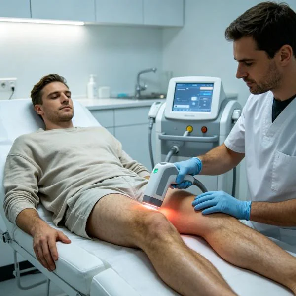
Physicians and physiotherapists managing chronic meniscal tears, chondromalacia patellae, or post-surgical joint contractures often face limited options between failed conservative modalities and arthroscopy. Low-intensity Class III modalities require 35 to 45 minutes of scanning to deliver minimal joules through the knee joint, producing negligible clinical improvements. While advanced surgical laser technology and specialized surgical lasers serve critical roles in operating suites for precise, non-contact partial meniscectomies, ablation of torn flap margins, and targeted hemostasis without mechanical crushing, an external non-invasive high-power Class IV platform delivers therapeutic photonic flux through intact skin to decompress and regenerate damaged joint tissues without incisions or anesthesia.
Specialized knee rehabilitation departments utilizing high-output systems experience distinct operational and clinical advantages:
첫째, 시술자는 6~8분 만에 적절한 치료용 에너지량을 투여할 수 있어, 일일 환자 처리량을 극대화하고 과도한 수동 마찰로 인한 시술자의 신체적 피로를 해소할 수 있습니다.
Second, rapid effusion reduction restores joint kinematics, allowing earlier participation in functional closed-chain rehabilitation.
Third, reliable deep-tissue cellular repair restores microvascular blood flow, clearing resting joint ache and slowing progressive cartilage breakdown.
임상 사례 기록
The following case record details the clinical application of multi-wavelength high-intensity laser therapy for a severe, treatment-resistant degenerative medial meniscal tear that had failed to respond to corticosteroid injections, viscosupplementation, and conventional rehabilitation.
사건 기록 상세 정보
Case Identification: KNEE-2026-M5012
Patient Demographics: 49-year-old male, competitive tennis coach
Primary Diagnosis: Degenerative Complex Horizontal Cleavage Tear of the Medial Meniscus Posterior Horn (Outerbridge Grade II Meniscal Cleavage with Parameniscal Edema and Subchondral Bone Bruising)
Chronicity: 7 months of persistent knee catching and aching; failed two intra-articular cross-linked hyaluronic acid injections, failed 16 sessions of conventional physical therapy involving ice and ultrasound
Clinical Assessment: McMurray test positive on the medial joint line with an audible click and sharp catching pain. Active knee flexion restricted to 102 degrees due to mechanical fullness and discomfort (Numeric Pain Rating Scale: 8/10 during squatting or pivoting, 5/10 constant dull ache at rest). Significant joint line effusion (1+ on the stroke test) with visible quadriceps inhibition (2.5cm circumference deficit compared to the unaffected left leg).
| 세션 ID | Phase 프로토콜 | 파장 구성 | 평균 전력(W) | 펄스 주파수 및 듀티 사이클 | 스캔 영역 (cm²) | 치료 기간 | 총 에너지(줄) | 임상 반응 지표 |
| 세션 01 | 낭 내 부종 완화 | 980nm (70%) + 1470nm (30%) | 12.0W | 100 Hz @ 20% 듀티 사이클 | 150 | 06:40 | 4,800 J | Resting NPRS drops from 5/10 to 3/10; night throbbing eases |
| 세션 03 | 활막 기질의 연화 | 980nm (45%) + 1470nm (55%) | 16.0W | 250 Hz @ 25% 듀티 사이클 | 120 | 08:20 | 8,000 J | Flexion increases to 115°; stroke test down to trace effusion |
| 제6회 세션 | Fibrocartilage Biostim | 810nm (30%) + 980nm (45%) + 1470nm (25%) | 20.0W | 800 Hz @ 35% 듀티 사이클 | 160 | 08:20 | 10,000 J | McMurray test triggers faint click without pain; NPRS: 2/10 |
| 제9회 세션 | Deep Subchondral Remodeling | 980nm (40%) + 1470nm (60%) | 18.0W | 20 Hz @ 25% 듀티 사이클 (딥 임팩트 모드) | 100 | 07:25 | 8,010 J | Active knee flexion reaches 130°; bodyweight squat tolerated |
| 제12회 | 동적 압밀 | 980nm (50%) + 1470nm (50%) | 15.0W | 연속파 (동적 스윕) | 180 | 08:00 | 7,200 J | Zero resting or exertion pain; full tennis coaching resumed |
사건 진행 기록
Sessions 01 through 03 focused on clearing periarticular vascular congestion, reducing suprapatellar effusion, and dampening unmyelinated nociceptive firing along the saphenous nerve branches. The combination of 980nm and 1470nm cleared inflammatory edema around the posterior medial joint line, reducing nocturnal throbbing and allowing the knee to bend past 110 degrees by the end of the first week.
Sessions 04 through 08 targeted the thick, fibrotic scar tissue within the coronary ligaments and the ischemic red-white meniscal zone. High water absorption at 1470nm softened the rigid proteoglycan gel, while 980nm and 810nm stimulated capillary blood flow to flush out metabolic waste and recruit reparative cells. Passive knee flexion expanded from 102° to 128° without sharp joint line catching.
By Session 12, McMurray testing was completely negative for mechanical catching or pain. Follow-up diagnostic ultrasound confirmed complete reabsorption of the parameniscal fluid collection, with stable, regular meniscal margins. The patient resumed competitive tennis coaching and rotational court drills without an antalgic limp, joint swelling, or post-activity soreness.
Biological Pathways in Fibrocartilage Regeneration
고강도 심부 조직 레이저 치료는 세포 내 및 세포 외 기전을 통합하여 치유를 촉진합니다:
Photonic Absorption (Fibrochondrocytes & Synovial Water Matrix)
         │
         ├──► Dissociation of Inhibitory Nitric Oxide ──► Accelerated Chondrocyte ATP Output
         │
         ├──► Interstitial Hydration Cleaving ────────► Clearance of Parameniscal Fluid Stasis
         │
         └──► Endothelial Cell Activation ─────────────► Perimeniscal Capillary Reperfusion
When photons hit cytochrome c oxidase inside stressed fibrochondrocytes and synoviocytes, they displace inhibitory nitric oxide molecules. This allows oxygen to bind unimpeded, accelerating the mitochondrial respiratory chain and producing a dramatic surge in adenosine triphosphate (ATP). Starving fibrochondrocytes regain the metabolic energy needed to maintain extracellular matrix balance and synthesize healthy aggrecan and type II collagen.
Concurrently, 1470nm photons excite water molecules bound within the dense proteoglycan gel surrounding the meniscal tear. The resulting micro-thermal fields break down rigid collagen cross-links, converting viscous ground substance into a fluid state and opening pathways for synovial fluid exchange and waste clearance.
하류 유전자 신호전달이 급격히 변화한다. 인터루킨-1-베타, 종양괴사인자-알파, 매트릭스 메탈로프로테이나제와 같은 염증성 사이토카인은 감소하는 반면, 변형성장인자-베타와 루브리신의 합성은 증가한다.
These biochemical changes prompt fibrochondrocytes to stabilize remaining torn margins and restore native joint fluid lubricin, protecting the articular cartilage from accelerated wear.
Moving Beyond Arthroscopic Resection and Injections
Conventional management for degenerative meniscal tears relies heavily on oral NSAIDs, repeated corticosteroid injections, and arthroscopic partial meniscectomy.
Systemic anti-inflammatory medications cause gastrointestinal bleeding and kidney strain while doing nothing to resolve the physical ischemia or mechanical shearing of the torn meniscus. Repeated cortisone injections provide short-term symptom relief, but accelerate articular cartilage loss and weaken adjacent capsular ligaments over time. Arthroscopic partial meniscectomy trims away shock-absorbing fibrocartilage, permanently altering joint contact stress and dramatically increasing the risk of rapid secondary osteoarthritis.
High-power Class IV deep laser therapy provides a focused, non-invasive alternative. It avoids systemic drug toxicity, preserves native meniscal volume, and eliminates surgical trauma. Clinicians can deliver thousands of therapeutic joules deep into contracted capsular folds and ischemic meniscal margins within minutes.
Applying high-intensity multi-wavelength phototherapy targets meniscal degeneration at its biological and vascular source. Swollen synovial tissues soften, microvascular circulation rebounds, and inflamed periarticular structures heal cleanly. It turns months of agonizing knee catching into a predictable, lasting return to full joint mobility and high-level athletic performance.