 FotonMedix












Surmonter la dégénérescence méniscale avasculaire dans l'arthrose du genou
Targeted intra-articular photonic flux, dual-target water and hemoglobin absorption, and duty cycle gating stimulate avascular fibrocartilage without thermal injury.
Orthopedic rehabilitation practices face severe treatment limitations when managing complex degenerative meniscus tears complicated by medial compartment knee osteoarthritis. Patients present with chronic weight-bearing joint line pain, mechanical clicking, and progressive effusion that fail to resolve through physical exercise, unloader bracing, or oral pharmacotherapy. Physical therapists attempting to treat this pathology with an ordinary medical grade cold laser therapy device quickly encounter an unyielding anatomical barrier: low-power light completely dissipates within the quadriceps tendon, thick infrapatellar fat pads, and fibrous joint capsules. Before light energy reaches 15 millimeters into the joint space, photon density drops below the minimal photobiomodulation threshold, leaving the avascular white-white zone of the degenerate fibrocartilage untouched. Conversely, relying on continuous-wave high-intensity output causes rapid epidermal heat spikes over the patellar margins, forcing operators to abort sessions before adequate biostimulation occurs. Overcoming this barrier requires using a multi-wavelength therapy laser capable of maintaining high photon penetration past dense synovial tissue, utilizing 980 nm and 1470 nm chromophore targets, and managing thermal relaxation with gated pulsing.
Photonic Transmission Barriers Across Multi-Tissue Articular Envelopes
Directing coherent light into the deep fibrocartilage of the medial meniscus demands traversing an optical obstacle course: thick epidermis, dense subcutaneous adipose layers, fibrous capsular ligaments, and viscous synovial fluid. Photons moving across these heterogeneous tissue interfaces undergo severe spatial dispersion and multi-directional elastic scattering, as characterized in tissue optics models formulated by researchers like Steven Jacques and Valery Tuchin.
In dense fibrous connective tissue, anisotropic scattering rapidly degrades directional beam coherence. Low-power units lose clinical efficacy because their superficial photon intensity drops below the biological activation threshold of 0.02 W per square centimeter long before clearing the anterior synovial lining. To reach the degenerate inner meniscus margin situated 30 to 45 millimeters beneath the skin line, clinical practices must utilize an advanced laser therapy device engineered for high-intensity Class IV output. Sustained high photon flux ensures that after accounting for the logarithmic scattering losses within periarticular tissues, a sufficient residual photon density enters the joint space to activate chondrocyte and fibrochondrocyte repair pathways.
Chromophore Synergy: Synovial Perfusion and Fibrocartilage Hydration
Restoring a degenerative joint environment requires addressing active synovial inflammation alongside the structural breakdown of avascular fibrocartilage. Deploying a dual-wavelength platform targets these physiological factors simultaneously:
The 980 nm wavelength interacts strongly with tissue hemoglobin, targeting the hyperemic, inflamed synovial lining and peri-meniscal capillary networks in the outer red-red vascular zone. Chronic knee osteoarthritis leads to impaired synovial microcirculation, sustained capillary congestion, and local accumulation of degradative matrix metalloproteinases. Irradiating this vascularized area with 980 nm light causes immediate photodissociation of nitric oxide from cytochrome c oxidase in cellular mitochondria. This bioenergetic cascade stimulates local arteriolar dilation, clears pro-inflammatory exudates, lowers joint effusion, and enhances aerobic ATP production in surrounding tissues.
The 1470 nm wavelength matches a dominant resonance absorption peak of water, which constitutes over 70% of healthy meniscal tissue and synovial fluid. Degenerative meniscal tears disrupt the glycosaminoglycan-water matrix, causing structural brittleness and fibrillar splitting. Delivering 1470 nm radiation introduces controlled, non-destructive photothermal resonance into interstitial water molecules. This localized energy transfer reorganizes disrupted collagen cross-links, enhances extracellular matrix permeability, and promotes nutrient diffusion into the avascular white-white meniscal zone where natural vascular supply is absent. Working with a dependable laser equipment supplier ensures clinical access to stable, calibrated dual-wavelength platforms capable of tailoring optical emission to specific joint conditions.
Thermal Dissipation Dynamics and Gated Duty Cycle Delivery
Delivering elevated energy dosages into tight joint spaces risks thermal stacking within superficial cutaneous layers overlying the patella and tibial plateau. Safe clinical execution requires managing the thermal relaxation time of human skin and subcutaneous tissue, which spans 20 to 45 milliseconds.
Implementing pulsed duty-cycle modulation resolves this surface-heat constraint. Delivering intense peak wattage during narrow micro-pulses followed by calculated off-times allows superficial capillaries to conduct excess heat away through normal thermal dissipation. Meanwhile, coherent photon packets continue penetrating through deeper capsular layers into the joint cavity. Regulating the duty cycle between 25% and 50% allows clinicians to saturate deep articular tissues with therapeutic energy levels while maintaining superficial skin temperatures well below the threshold of thermal discomfort.
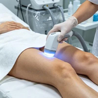
Clinical Protocol: Class IV Laser Photobiomodulation in Degenerative Meniscal Tears
The following clinical data represents an outpatient rehabilitation protocol applied to a patient presenting with medial compartment knee osteoarthritis complicated by a degenerative medial meniscus tear.
Profil des patients et caractéristiques cliniques initiales
- Case Identifier: FTM-ORTHO-2026-0943
- Patient Age: 54
- Sexe : Femme
- Primary Diagnosis: Grade III degenerative tear of the posterior horn of the medial meniscus with Kellgren-Lawrence Grade 2 medial compartment knee osteoarthritis, symptom duration 9 months
- Prior Interventions: Oral non-steroidal anti-inflammatory drugs, unloader knee bracing, two intra-articular hyaluronic acid injections (temporary lubrication with persistent joint line tenderness), and physical therapy focused on quadriceps strengthening
- Baseline Diagnostics: Magnetic Resonance Imaging (MRI) confirmed an intrameniscal high-signal line communicating with the inferior articular surface of the posterior horn of the medial meniscus, accompanied by moderate joint effusion and localized medial joint-space narrowing (2.8 mm vs. 4.6 mm in the lateral compartment). The patient presented with pain during weight-bearing knee flexion, nocturnal aching, and recurrent joint effusion. Baseline Visual Analog Scale (VAS) pain score registered 7.8/10 during stair descent. Western Ontario and McMaster Universities Osteoarthritis Index (WOMAC) score measured 68.5%.
Paramètres thérapeutiques et schéma posologique technique
The patient completed a four-week clinical protocol comprising twelve sessions scheduled three times per week. Treatments utilized contact scanning with deep tissue compression around the medial joint line to displace superficial venous blood, combined with non-contact passes across the popliteal fossa to target the posterior horn.
| Plage de sessions | Optical Wavelength Mix | Puissance de sortie de crête | Fréquence de déclenchement des impulsions | Cycle de service effectif | Durée de la session | Exposition rayonnante appliquée | Total de l'énergie livrée |
| Séances 1 à 3 | 75% 980 nm, 25% 1 470 nm | 9,0 W | 20 Hz | 30% | 600 s | 16.0 J/cm² | 1,620 J |
| Séances 4 à 6 | 65% 980 nm, 35% 1 470 nm | 11.0 W | 40 Hz | 35% | 540 s | 23.0 J/cm² | 2,079 J |
| Séances 7 à 9 | 55% 980 nm, 45% 1 470 nm | 13.0 W | 70 Hz | 40% | 480 s | 31.0 J/cm² | 2,496 J |
| Séances 10 à 12 | 45% 980 nm, 55% 1470 nm | 14,0 W | Continuous / 10 Hz mix | 60% | 420 s | 35,0 J/cm² | 3,528 J |
Indicateurs objectifs de progression clinique
Treatments were delivered without local anesthetics, skin chilling sprays, or concomitant oral analgesics. Cutaneous temperatures were tracked in real time using non-contact infrared sensors, maintaining surface levels below 41.6 degrees Celsius during every pass.
| Paramètre clinique | Évaluation initiale | Après la session 3 | Post-session 6 | Après la 9e séance | Fin (Séance 12) | Suivi à 90 jours |
| Stair Descent Pain (VAS 0–10) | 7.8 | 5.2 | 3.1 | 1.4 | 0.4 | 0.0 |
| WOMAC Disability Index (%) | 68.5% | 51.0% | 34.5% | 19.0% | 7.5% | 5.0% |
| Amplitude de flexion passive du genou (deg) | 104° | 112° | 124° | 134° | 138° | 140° |
| Medial Joint Line Tenderness | Sévère | Modéré | Doux | Trace | Négatif | Négatif |
| Suprapatellar Effusion Depth (mm) | 12.4 | 9.8 | 5.2 | 2.1 | Trace (<1.0) | Résolu |
Tissue Recovery and Joint Remodeling Mechanisms
Initial treatment sessions focused primarily on the 980 nm wavelength to down-regulate pro-inflammatory cytokines such as interleukin-1 beta and matrix metalloproteinase-13 within the synovial lining. Within the first three sessions, suprapatellar effusion decreased from 12.4 mm to 9.8 mm, providing rapid decompression of the joint capsule and reducing resting pain from 7.8 to 5.2 on the VAS scale.
As the protocol advanced into weeks two and three, raising the 1470 nm ratio delivered targeted photothermal energy into the water-dense extracellular ground substance of the damaged meniscus. This interaction softened rigid fibrotic adhesions and stimulated proteoglycan production by viable fibrochondrocytes at the tear margin. By session nine, joint line tenderness shifted from severe to trace, and passive flexion reached 134 degrees without joint jamming. At the 90-day follow-up, the patient reported pain-free ambulation, zero joint effusion, and full participation in daily activities without pharmaceutical reliance or surgical referral.
High-Power Laser Therapy Compared to Standard Knee Interventions
Managing degenerative meniscal tears and early osteoarthritis using conventional medical options carries notable limitations. Repeated intra-articular corticosteroid injections temporarily reduce synovitis but accelerate articular cartilage loss, weaken subchondral bone, and increase long-term joint degradation risks. Oral non-steroidal anti-inflammatory drugs provide minor pain blunting while exposing older patients to renal, cardiovascular, and gastrointestinal complications.
Arthroscopic partial meniscectomy surgically excises torn meniscal fragments, but removing fibrocartilage permanently decreases joint contact area and triples contact stresses on tibial plateau cartilage, accelerating osteoarthritis progression. Physical therapy alone often struggles when intense joint effusion and pain inhibit basic quadriceps recruitment.
High-intensity Class IV laser therapy offers a distinct, non-invasive alternative. By combining 980 nm and 1470 nm wavelengths with thermal relaxation duty gating, this method projects high photon density through periarticular soft-tissue barriers straight into the intra-articular space. Clinicians can resolve synovial inflammation, clear chronic joint effusions, and stimulate natural fibrocartilage matrix maintenance without mechanical excision, systemic chemical exposure, or surgical recovery periods. High-output therapeutic platforms deliver dependable, long-term functional recovery for patients suffering from degenerative knee joint conditions.