 FotonMedix












Overcoming Chronic Canine Wound Dehiscence Breakdown
Multi-wavelength Class IV photon deposition accelerates capillary budding in ischemic wound beds, clears extracellular wound exudate via water resonance, and prevents thermal damage across granulating margins using microsecond duty cycle regulation.
A 7-year-old Boxer lies depressed on the examination mat, wearing an oversized Elizabethan collar while foul-smelling, blood-tinged serosanguinous fluid soaks through heavy gauze wraps. Twelve days ago, the dog underwent surgical resection of a subcutaneous mast cell tumor over the lateral thigh, leaving a tension-heavy ten-centimeter incision. Yesterday evening, the distal two-thirds of the surgical closure suffered catastrophic breakdown: sutures pulled through friable skin margins, wound edges retracted by nearly three centimeters, and raw, yellowish slough exposed the underlying fascia. The dog whines with every attempt to adjust its hind leg, panting heavily from sharp nociceptive spikes. The owner is traumatized by the sight of open muscle, facing an unexpected quote for revision surgery with tension-relieving flaps under general anesthesia. Meanwhile, high-dose oral tramadol and gabapentin leave the dog heavily sedated and anorexic, while oral cephalexin fails to halt wound margins from macerating under persistent tissue edema.
Managing severe surgical wound breakdown and persistent pain presents a tough physical dilemma. Dehisced surgical wounds in tension zones are hypovascular, ischemic at retracted margins, and inundated with inflammatory fluid that degrades newly formed extracellular matrix. Conventional low-level therapy probes fail completely here: their low milliwatt output cannot deposit sufficient photons through thick wound slough and fibrin layers. Technicians spend twenty minutes hovering a tiny wand over an open lesion without reaching the cellular energy threshold needed to stimulate wound contracture. Turning around a stalling open wound requires an industrial-grade wound healing laser treatment platform, capable of driving high photon density into ischemic wound beds to shut down infection, accelerate granulation, and clear deep-seated tissue inflammation.
Optical Penetration Gradients and Multi-Spectrum Chromophore Targeting
Photons aimed at an open, dehisced surgical site encounter drastic optical resistance. The wound bed is a chaotic mixture of organizing fibrin slough, exposed fascia, capillary pooling, and hyperemic, edematous skin margins. Single-wavelength, low-output light loses up to eighty-five percent of its irradiance within the first two millimeters of serosanguinous exudate, scattering harmlessly before reaching the basal fibroblastic layers where collagen remodeling occurs.
Reactivating cellular repair in an open wound bed demands synchronized multi-wavelength targeting across biological optical windows:
- 980 nm Hemoglobin Resonance and Microvascular Recanalization: Ischemic wound margins suffer from local venous stasis, creating tissue acidosis and tissue hypoxia that halt cellular migration. Photons at 980 nm match the optical absorption peak of oxygenated and deoxygenated hemoglobin. This produces mild, localized photothermal expansion within spastic microcapillaries, opening up shut-down vascular loops, accelerating the delivery of circulating leukocytes to clear bioburden, and establishing healthy capillary buds across the wound base.
- 1064 nm Deep Fascial Penetration: Demonstrating low scattering coefficients and minimal absorption across water and melanin, the 1064 nm wavelength travels straight through dense fibrin slough and underlying deep fascial planes. It deposits photon energy directly into damaged myofibroblasts and tenocytes, stimulating mitochondrial cytochrome c oxidase activity and boosting cellular adenosine triphosphate synthesis to drive active wound contracture.
- 1470 nm High-Affinity Water Modulation and Edema Evacuation: Severe wound dehiscence creates heavy interstitial fluid pooling that macerates wound borders and stretches local nociceptive nerve terminals. Light at 1470 nm exhibits strong affinity for water molecules, mobilizing stagnant interstitial fluid pockets and accelerating fluid evacuation through local dermal lymphatics to relieve wound tension.
This coordinated multi-spectral array delivers direct relief in clinical settings. Managing acute tissue distress with a calibrated platform provides genuine laser therapy for post-surgical pain, suppressing substance P and local inflammatory bradykinins within minutes. In parallel, incorporating these biological mechanisms into postoperative rehabilitation workflows elevates standard laser therapy physiotherapy, transitioning an open, non-healing incision into a rapidly contracting, granulating tissue bed.
Eliminating Margin Thermal Injury via Microsecond Duty Cycle Pulsing
Delivering high-power Class IV photomedicine across fragile, denuded wound margins presents a clear thermal management challenge: granulating tissue lacks protective stratum corneum and sebaceous insulation. Applying continuous-wave high-power light directly to an open wound bed risks overheating surface capillaries, causing microvascular coagulation, cell death, or acute patient discomfort.
Microsecond duty cycle regulation eliminates this risk by dividing continuous photon emission into controlled micro-bursts separated by thermal relaxation intervals:
Continuous Wave: [================ Continuous Heat Accumulation ================] -> Capillary Coagulation Risk
Pulsed 35% Duty: [== Pulse ON ==] [ Thermal Dissipation ] [== Pulse ON ==]      -> Deep Granulation Saturation
During the active pulse phase, high peak irradiance drives photons deep into the wound base, overcoming tissue scatter and fibrin barriers. During the dark phase, capillary blood flow in the adjacent healthy tissue whisks away surface heat. Because deep fibroblasts retain photonic stimulation while surface tissues cool rapidly, the wound bed receives full therapeutic fluence while fragile healing margins remain safe, comfortable, and protected against thermal injury.
This precise thermal management allows veterinary teams to deliver comprehensive treatments in five to eight minutes of continuous non-contact sweeping. The patient rests calmly without tight physical restraint, eliminating clinic anxiety and keeping daily appointments running smoothly.
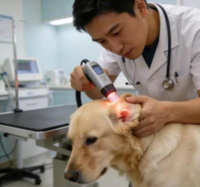
Clinical Protocol and Objective Healing Metrics
According to photobiological wound repair frameworks documented by Hamblin and supported in veterinary surgical trials published in Veterinary Surgery, near-infrared photobiomodulation downregulates pro-inflammatory cytokines such as interleukin-1 beta and tumor necrosis factor-alpha, while upregulating transforming growth factor-beta and vascular endothelial growth factor to accelerate neoangiogenesis, fibroblast proliferation, and epidermal re-epithelialization.
The following clinical case details this multi-wavelength protocol in a severe case of full-thickness surgical wound dehiscence managed without revision surgery.
Case Record: Canine Full-Thickness Surgical Wound Dehiscence
- Référence du dossier : SURG-2026-CANINE-1674
- Profil du patient : 7-year-old male intact Boxer
- Masse corporelle : 33.4 kg
- Diagnostic : Full-thickness distal surgical wound dehiscence over the lateral left thigh following wide mast cell tumor excision; open defect measuring 9.5 cm in length by 3.2 cm in width at widest margin; exposed biceps femoris fascia with heavy fibrinous slough, marked margin maceration, and active serosanguinous discharge; Colorado State University Canine Acute Pain Scale score: 3/4; reluctance to bear weight on the left hindlimb
- Traitement conservateur antérieur : Oral cephalexin at 25 mg/kg every 12 hours for seven days, tramadol at 3 mg/kg every 8 hours, hydrogel wet-to-dry bandage changes every 48 hours; minimal progress with ongoing suture pull-out and worsening margin necrosis
Paramètres du protocole de traitement complet
| Numéro de session | Phase of Tissue Repair | Puissance de sortie (W) | Mode de fonctionnement et rapport cyclique | Distribution spectrale ciblée | Fréquence (Hz) | Énergie totale (Joules) | Fluence (J/cm²) |
| 01 | Acute Pain & Edema Clearance | 8,0 W | En mode pulsé (rapport cyclique 30%) | High 980 nm / Capillary Focus | 3,500 Hz | 1 440 J | 4.8 J/cm² |
| 02 | Interstitial Fluid Mobilization | 10,0 W | En mode pulsé (rapport cyclique 35%) | High 1470 nm / Water Focus | 2 000 Hz | 2,100 J | 7,0 J/cm² |
| 03 | Microvascular Recanalization | 12,0 W | En mode pulsé (rapport cyclique 35%) | Mélange équilibré 980 nm / 1064 nm | 1 200 Hz | 2,520 J | 8.4 J/cm² |
| 04 | Granulation Bed Induction | 15,0 W | Superpulsed (40% Duty Cycle) | Penetration Dominant (1064 nm) | 500 Hz | 3,600 J | 12,0 J/cm² |
| 05 | Myofibroblast Contracture Trigger | 16,0 W | En mode pulsé (rapport cyclique 40%) | Quad-wavelength balanced array | 200 Hz | 3,840 J | 12.8 J/cm² |
| 06 | Collagen Cross-Linking Phase | 16,0 W | En mode pulsé (rapport cyclique 40%) | Deep Extracellular Matrix Focus | 100 Hz | 3,840 J | 12.8 J/cm² |
| 07 | Epithelial Margin Migration | 14,0 W | Continuous Dynamic Sweep | Multi-spectral Regenerative Mix | CW / Dynamic | 3 360 J | 11.2 J/cm² |
| 08 | Epidermal Maturation Consolidation | 10,0 W | En mode pulsé (rapport cyclique 30%) | Balanced Maintenance Profile | 30 Hz | 2,100 J | 7,0 J/cm² |
Longitudinal Wound Area and Acute Pain Score Evolution
Wound Surface Defect Area (cm²):
Pre-Treatment: [==================================] 30.4 cm²
Session 03:    [==========================] 21.0 cm²
Session 05:    [================] 10.5 cm²
Session 08:    [=] 1.2 cm² (Near-complete epithelial closure)

CSU Canine Acute Pain Score (0 to 4 Scale, Lower = Comfortable):
Pre-Treatment: [==============================] 3.0 (Severe pain, vocal on palpation)
Session 03:    [================] 1.5
Session 05:    [=======] 0.5
Session 08:    [] 0.0 (Pain-free, full weight-bearing gait)
By the third session, the foul-smelling serosanguinous oozing ceased entirely, replaced by a clean, vascularized red bed of budding granulation tissue without fibrinous slough. By session five, active myofibroblast contracture pulled the wound borders inward, reducing defect surface area from 30.4 cm² down to 10.5 cm², with advancing pink epithelial borders. At the conclusion of eight treatments over four weeks, the surgical wound achieved complete closure without revision surgery, leaving a pliable, flat linear scar that preserved full stifle and hip range of motion, allowing systemic analgesics to be discontinued completely.
Practice Efficiency and Economic Value in Surgical Complication Management
Managing major surgical wound dehiscence conventionally drains hospital resources and strains client relationships. Taking a dehisced patient back to the operating room requires another round of general anesthesia, extensive tissue undermining, secondary surgical trauma, and unpredictable tension sutures that often fail again. The alternative of indefinite wet-to-dry bandage changes means weekly sedation charges, expensive dressing materials, and weeks of messy home care that exhaust pet owners.
Integrating high-power Class IV photomedicine establishes concrete advantages across the practice:
1. Completely Non-Surgical Defect Closure
Providing an effective, scalpel-free wound healing protocol spares medically compromised or senior pets the physiological risks of repeat anesthesia. Practices avoid the frustration of revision surgery failures while delivering a clean, functional scar that avoids tension-induced contracture.
2. Rapid In-Clinic Workflow Turnaround
Surgical wound revision ties up an operating room and surgical nurses for ninety minutes, followed by weeks of anxious rechecks. A high-output Class IV system delivers targeted doses in six to eight minutes of non-contact sweeping in a regular exam room, allowing technicians to manage wound appointments efficiently between routine patient check-ins.
3. Tangible Visual Progress That Reassures Pet Owners
Pet owners are deeply unsettled by open, draining surgical wounds. Seeing active bleeding stop, healthy red granulation tissue form, and wound margins visibly pull together within the first week restores confidence in the hospital’s clinical care, ensuring consistent compliance with bandage management and activity restrictions.
High-power Class IV photomedicine provides a reliable, high-throughput solution for severe surgical complications and chronic open wounds. By delivering balanced multi-wavelength photon energy directly into hypoxic, damaged tissue beds without surface thermal risks, veterinary practices can heal difficult surgical wounds, eliminate reliance on heavy pain medications, and guide recovering pets back to comfortable, active lives.