 FotonMedix




Superare l'attenuazione fotonica nei tessuti profondi nella radicolopatia cervicale
Three synchronized multi-wavelength delivery modes bypass superficial subcutaneous scatter, eliminate thermal epidermal accumulation through sub-millisecond duty cycle gating, and deposit therapeutic photon density directly into sub-axial facet capsular interfaces and compressed nerve root corridors.
The typical clinical morning in an outpatient spine center begins with an all-too-familiar dilemma. A patient sits on the examination table clutching their posterior cervical spine, radiating numbness tracking down the C6 dermatome into the thumb and index finger. Standard physical therapy has reached an impasse; cervical traction yields only transient decompression, and high-dose nonsteroidal anti-inflammatory regimens are causing epigastric distress. When clinicians attempt intervention using conventional modality equipment, the photon energy disperses harmlessly across the trapezius and deep cervical fascia without altering the inflammatory cascade surrounding the intervertebral foramen. Delivering meaningful therapeutic photon fluence through dense cervical paraspinal musculature to unseat refractory neuroinflammation requires abandoning underpowered modalities and addressing photon scatter, target chromophore affinity, and tissue thermal relaxation times directly at the biological site of pathology.
Biological Barriers and Photonic Decay in Sub-Axial Spine Pathology
Targeting the neural exit pathways of the cervical spine represents one of the most demanding biophysical challenges in musculoskeletal rehabilitation. The target anatomy—specifically the disc-osteophyte complexes, the posterior longitudinal ligament, the facet joint capsules, and the irritated dorsal root ganglion—resides beneath multiple tissue planes. Photons directed from a cutaneous handpiece must traverse the epidermis, dermis, subcutaneous adipose tissue, the superficial investing fascia, splenius capitis, semispinalis capitis, and the deep multifidus musculature before encountering the targeted neural interfaces at depths ranging from 35 to 55 millimeters.
Each biological layer presents a distinct extinction coefficient driven by tissue-specific scatter and absorption characteristics. In biological tissues, Rayleigh and Mie scattering dominate the light-tissue interaction within the near-infrared window. Scattering shifts photon paths off axis, rapidly dispersing energy laterally and causing an exponential drop in forward irradiance. When relying on low-level light devices operating below 1 Watt of continuous output, forward photon density plummets to biologically undetectable levels well before penetrating the superficial epimysium. High-energy Class 4 systems overcome this boundary by utilizing sufficient incident irradiance to saturate superficial scattering centers, establishing a viable forward-propagating photon flux that reaches the deep degenerative margins of the sub-axial cervical spine.
Achieving meaningful clinical outcomes with a high-power therapeutic laser machine requires strategic balancing of primary tissue chromophores. Biological tissues display three primary optical absorbing agents in the red and near-infrared spectrum: water, melanin, and hemoglobin derivatives (both oxygenated and deoxygenated). Epidermal melanin forms the initial gatekeeper; its absorption curve declines progressively from visible wavelengths into the near-infrared window. Once photons clear the epidermal layer, the targeted deployment of specific photon bands dictates the physiological fate of the transmitted energy:
Tissue Depth & Extinction Mechanism Across Cervical Soft Tissue:
[Surface Epidermis]      --> Melanin absorption threshold (mitigated by moving handpiece technique)
[Adipose & Fascia]       --> High Mie scattering; requires sufficient incident photon density
[Deep Paraspinal Muscle] --> Hemoglobin absorption band (980nm photothermal conversion & perfusion)
[Spinal Nerve Corridor]  --> Water absorption band (1470nm direct interstitial edema suppression)
The 980nm wavelength matches the peak absorption spectrum of deoxygenated and oxygenated hemoglobin. While this absorption profile limits its penetration relative to 810nm or 1064nm, it serves a distinct clinical function: local photothermal stimulation of microvascular perfusion, temporary localized vasodilation, and rapid washout of algogenic chemical mediators such as bradykinin, substance P, and tumor necrosis factor-alpha. Conversely, the 1470nm wavelength interacts aggressively with interstitial water. In severe cervical spondylosis, nerve root irritation triggers localized intraneural edema, venous congestion, and axonal ischemia within the neural canal. Because free water molecules absorb 1470nm energy with exceptional selectivity, this wavelength heats and mobilizes localized fluid accumulation within inflammatory pockets, resolving intraneural pressure without relying on mechanical tissue distraction.
Delivering high photon flux into biological tissue inevitably creates the risk of superficial thermal injury. When photons are absorbed by water and hemoglobin, radiant energy converts directly into kinetic thermal energy. Biological tissue possesses a characteristic thermal relaxation time—the duration required for target tissue to dissipate 50 percent of its absorbed heat via passive conduction. If incident photon delivery outpaces this thermal dissipation rate, cutaneous temperature escalates past 43 degrees Celsius, initiating nociceptor activation, patient discomfort, and potential epidermal blistering.
Thermal Dissipation Dynamics During Pulsed Delivery:
Laser Emission Window (ON)   : [==== High Peak Power Pulse ====] (Cellular Chromophore Saturation)
                                |<-------- Pulse Width -------->|
Inter-Pulse Quench (OFF)     : [ . . . . Thermal Relaxation Phase . . . . ] (Heat Sinks to Deep Tissues)
                                |<--------- Quench Interval --------->|
Net Result                   : Maximum depth of penetration without cutaneous thermal ceiling breach
Pulsed duty cycle control provides the definitive clinical solution to this physiological bottleneck. By operating the delivery system in gated pulse frequencies ranging from 10Hz to 20,000Hz, peak power can be elevated dramatically to guarantee deep forward scattering while keeping mean power strictly regulated. A 20W peak pulse delivered at a 20 percent duty cycle drives photons deep into the facet capsule while giving the cutaneous surface four times the emission duration to cool through vascular micro-convection and structural heat conduction. Through this precise temporal modulation, therapeutic laser therapy deposits high-density photonic doses into the cervical spine without exceeding cutaneous thermal thresholds.
Cellular Bioenergetics and Neuropathic Cascade Interruption
The clinical efficacy of laser treatment for cervical spondylosis depends upon triggering intracellular biostimulatory responses within mechanically compressed and biochemically compromised cells. Cytochrome c oxidase, the terminal enzyme (Complex IV) of the mitochondrial electron transport chain, acts as the primary mitochondrial photoacceptor within the near-infrared spectrum. Under conditions of prolonged ischemic compression caused by cervical canal narrowing, swollen disc margins, or uncovertebral joint hypertrophy, the microenvironment suffers profound hypoxic stress. Mitochondrial respiration stalls as nitric oxide binds competitively to the binuclear heme a3/copper B active center of Cytochrome c oxidase, displacing oxygen and shutting down adenosine triphosphate synthesis.
Irradiating this hypoxic tissue with resonant photons dislodges nitric oxide through light-induced photolysis. Once nitric oxide is cleared from the enzyme’s respiratory binding pocket, oxygen binds without hindrance, restarting the mitochondrial proton pump across the inner mitochondrial membrane. This immediate re-establishment of the transmembrane electrochemical gradient drives a rapid upsurge in cellular adenosine triphosphate generation.
Downstream biological cascades follow this metabolic reactivation:
- Transient Reactive Oxygen Species Modulation: Controlled, low-amplitude generation of reactive oxygen species activates endogenous cellular defense systems, activating the Nrf2-ARE antioxidant pathway and triggering the transcription of superoxide dismutase, catalase, and glutathione peroxidase.
- Membrane Hyperpolarization and Calcium Influx: Gated photonic energy stimulates transient receptor potential cation channels, normalizing the resting membrane potential of damaged dorsal root axons and dampening ectopic, spontaneous discharge from irritable neuromas.
- Suppression of Pro-Inflammatory Cyclooxygenase Transcripts: Elevated mitochondrial output downregulates NF-kappa-B nuclear translocation, reducing gene transcription of cyclooxygenase-2, inducible nitric oxide synthase, and interleukin-1 beta within the deep cervical ligaments and intervertebral joint capsules.
- Schwann Cell Proliferation and Axonal Repair Signaling: Photonic stimulation upregulates cyclic AMP and activates brain-derived neurotrophic factor, accelerating axonal sprout elongation, promoting Schwann cell remyelination, and restoring compound muscle action potentials along the cervical nerve distribution.
These bioenergetic mechanisms explain why simply applying heat via superficial infrared lamps, microwave diathermy, or hot packs cannot replicate high-intensity Class 4 outcomes. Superficial conductive heat merely alters local skin surface vascular tone. Coherent, high-intensity near-infrared photon flux penetrates deep enough to initiate photobiomodulatory responses within the uncinate processes, the nerve root sheath, and the compressed cervical cord parenchyma.
Diagnostic Staging and Patient Stratification Parameters
Not every presentation of neck pain responds equally to photonic intervention. Accurate patient stratification ensures that therapeutic energy is applied to correct clinical targets while identifying cases requiring neurosurgical consultation. Cervical spondylosis encompasses four distinct yet often overlapping clinical phenotypes:
- Cervical Spondylotic Myelopathy: Involves progressive compression of the spinal cord itself, clinically marked by hyperreflexia, lower extremity spasticity, positive Hoffmann and Babinski signs, and gait ataxia. Patients displaying these advanced compressive myelopathy symptoms must be referred immediately for neurosurgical decompression; laser therapy cannot substitute for urgent structural decompression when cord signal changes appear on T2-weighted MRI.
- Radicolopatia spondilotica cervicale: The classic candidate for deep photon delivery. Patients present with unilateral or bilateral radiating arm pain, paresthesias, sensory loss in distinct dermatomal bands (typically C5, C6, or C7), and motor weakness of the biceps, triceps, or wrist extensors. The primary pathology involves disc protrusion, uncinate spurring, and neurovascular bundle entrapment at the neural exit foramen.
- Cervical Facet Arthropathy: Localized axial neck pain exacerbated by cervical extension, rotation, and lateral bending. Tenderness localizes over the articular pillars. The therapeutic target is the inflamed synovial lining of the zygapophyseal joints, located approximately 25 to 35 millimeters beneath the skin.
- Cervicogenic Myofascial Pain Syndrome: Chronic secondary spasm of the upper trapezius, levator scapulae, and splenius cervicis, frequently housing hyperirritable trigger points that maintain noxious sensory feedback to the spinal trigeminal nucleus.
Prior to powering the clinical laser unit, practitioners must calculate total target volumetric joules rather than relying on arbitrary treatment durations. Dosage in deep spinal care is governed by the Arndt-Schulz rule: insufficient energy fails to elicit a biological response, whereas excessive energy risks tissue inhibition or thermal damage. In deep cervical pathology, due to tissue scatter and depth attenuation, approximately 95 to 97 percent of surface-delivered photon energy attenuates within overlying tissues. Therefore, to deliver an effective threshold dose of 4 to 8 Joules per square centimeter to the damaged nerve root interface at a depth of 4 centimeters, the surface-applied dosage must reach 100 to 150 Joules per square centimeter across the clinical projection field.
Comprehensive Clinical Case History: Outpatient Spine Center
The following case history demonstrates the therapeutic parameters, diagnostic markers, and clinical trajectory observed during multi-wavelength Class 4 laser therapy.
Profilo del paziente e esami diagnostici iniziali
- ID pratica del dipartimento: FTM-RAD-2026-0884
- Età del paziente: 53 years
- Sesso biologico: Maschio
- Occupazione: Systems Software Architect (prolonged static desk posture: 10 to 12 hours daily)
- Condizione attuale: Severe right-sided cervicobrachial pain radiating across the right shoulder, lateral arm, and into the dorsoradial aspect of the hand, index finger, and thumb.
- Durata dei sintomi: 16 weeks of progressive deterioration; unresponsive to oral meloxicam (15mg daily), gabapentin (300mg three times daily), and 12 sessions of standard manual physical therapy.
- Livello funzionale di riferimento:
- Visual Analog Scale for Axial Neck Pain: 6 / 10
- Visual Analog Scale for Right Arm Radicular Pain: 8.5 / 10
- Neck Disability Index: 58% (severe functional disability)
- Active Cervical Spine Range of Motion: Right lateral flexion restricted to 14 degrees (normal: 45); extension restricted to 8 degrees with immediate radicular exacerbation.
- Esame neurologico:
- Sensory: Hypoesthesia to light touch and pinprick mapping specifically across the right C6 dermatome.
- Motor: Right biceps and brachioradialis strength graded at 4/5 on the Medical Research Council scale; wrist extensor weakness graded at 4-/5.
- Deep Tendon Reflexes: Right biceps reflex diminished (1+) compared to contralateral side (2+).
- Provocative Testing: Positive Spurling’s maneuver to the right, reproducing lancinating brachialgia; positive Shoulder Abduction (Bakody’s) relief sign.
- Magnetic Resonance Imaging (Cervical Spine, 3.0T):
- Multilevel spondylotic changes maximal at C5-C6.
- Right paracentral and foraminal soft disc protrusion measuring 3.8mm, directly effacing the ventral subarachnoid space and causing moderate-to-severe right neural foraminal stenosis with right C6 exiting nerve root impingement.
- Surrounding T2 hyperintensity consistent with perineural and capsular inflammatory edema. No cord myelomalacia.
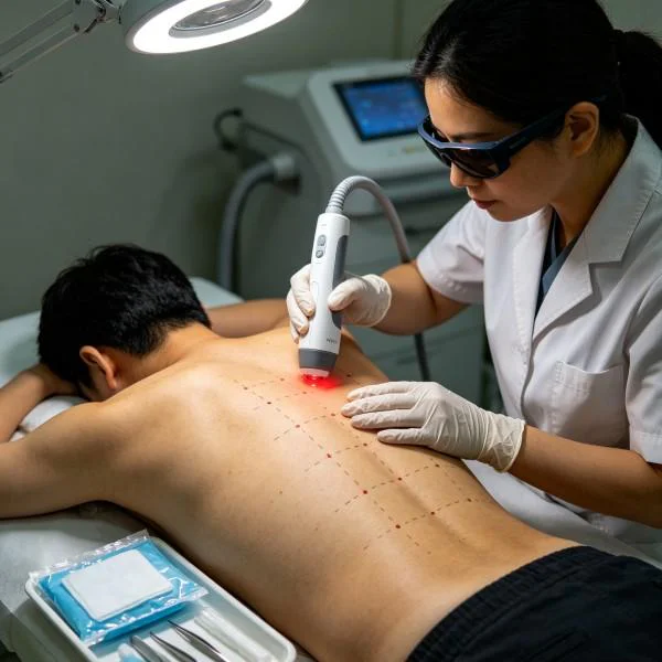
Protocollo terapeutico standardizzato e matrice dell'energia cinetica
The patient underwent a course of 12 treatment sessions scheduled across 4 consecutive weeks (3 sessions per week for 3 weeks, tapering to 2 sessions in the final week). The protocol was delivered using a multi-wavelength Class 4 medical laser system featuring synchronized output across 810nm, 980nm, and 1470nm, equipped with a large-aperture optical collimation handpiece with integrated surface thermal monitoring.
+-------------------------------------------------------------------------------------------------------+
| Week | Session | Wavelength Ratio   | Power (W) | Frequency (Hz) | Duty Cycle | Energy (J) | Area (cm2) |
+-------------------------------------------------------------------------------------------------------+
| Wk 1 | S-01    | 810nm (70%) 980 (30)| 8.0 W CW  | Continuous     | 100%       | 3,840 J    | 80 cm2     |
| Wk 1 | S-02    | 810/980/1470 (5:3:2)| 12.0 W P  | 100 Hz         | 60%        | 5,184 J    | 80 cm2     |
| Wk 1 | S-03    | 810/980/1470 (4:3:3)| 14.0 W P  | 250 Hz         | 50%        | 5,880 J    | 100 cm2    |
| Wk 2 | S-04    | 810/980/1470 (4:3:3)| 16.0 W P  | 500 Hz         | 50%        | 6,720 J    | 100 cm2    |
| Wk 2 | S-05    | 810/980/1470 (4:3:3)| 18.0 W P  | 1,000 Hz       | 50%        | 7,560 J    | 100 cm2    |
| Wk 2 | S-06    | 810/980/1470 (3:3:4)| 20.0 W P  | 2,500 Hz       | 40%        | 7,680 J    | 120 cm2    |
| Wk 3 | S-07    | 810/980/1470 (3:3:4)| 22.0 W P  | 5,000 Hz       | 40%        | 8,448 J    | 120 cm2    |
| Wk 3 | S-08    | 810/980/1470 (3:3:4)| 22.0 W P  | 5,000 Hz       | 40%        | 8,448 J    | 120 cm2    |
| Wk 3 | S-09    | 810/980/1470 (3:2:5)| 24.0 W P  | 10,000 Hz      | 35%        | 8,064 J    | 120 cm2    |
| Wk 4 | S-10    | 810/980/1470 (4:3:3)| 20.0 W P  | 2,000 Hz       | 50%        | 8,400 J    | 120 cm2    |
| Wk 4 | S-11    | 810/980/1470 (4:3:3)| 18.0 W P  | 1,000 Hz       | 50%        | 7,560 J    | 100 cm2    |
| Wk 4 | S-12    | 810/980 (60% : 40%) | 12.0 W CW | Continuous     | 100%       | 5,760 J    | 80 cm2     |
+-------------------------------------------------------------------------------------------------------+
Progressione clinica cumulativa
+----------------------------------------------------------------------------------------------------+
| Checkpoint    | Neck VAS | Arm VAS | NDI (%) | C6 Motor Grade | Biceps Reflex | Spurling's Sign     |
+----------------------------------------------------------------------------------------------------+
| Pre-Treatment | 6.0 / 10 | 8.5 / 10| 58 %    | 4- / 5         | 1+ (Diminished)| Positive (Instant) |
| Post-S-03     | 4.5 / 10 | 6.0 / 10| 46 %    | 4 / 5          | 1+ (Diminished)| Positive (Delayed) |
| Post-S-06     | 3.0 / 10 | 3.5 / 10| 28 %    | 4+ / 5         | 2- (Emerging)  | Mild Discomfort    |
| Post-S-09     | 1.5 / 10 | 1.5 / 10| 14 %    | 5 / 5          | 2  (Normal)    | Negative           |
| Post-S-12     | 0.5 / 10 | 0.0 / 10| 6 %     | 5 / 5          | 2  (Normal)    | Negative           |
| 90-Day Follow | 0.0 / 10 | 0.0 / 10| 4 %     | 5 / 5          | 2  (Normal)    | Negative           |
+----------------------------------------------------------------------------------------------------+
Commento clinico sulle fasi di avanzamento
During Sessions 1 through 3, therapy focused on establishing microvascular perfusion and dampening peripheral nociceptive signaling without overheating sensitive paraspinal tissues. Continuous-wave delivery at 8.0 Watts delivered energy to the superficial paracervical musculature, clearing local myofascial tension. By Session 2, integrating the 1470nm wavelength activated fluid mobilization within the edematous neuroforaminal corridor. The patient experienced a 2.5-point drop on the arm Visual Analog Scale by the conclusion of Session 3, reporting that sharp lancinating pains had transitioned into a dull, intermittent ache localized primarily above the elbow.
Sessions 4 through 8 transitioned to higher peak outputs, running up to 22.0 Watts. Operating at high frequencies (500Hz to 5,000Hz) with a 40 to 50 percent duty cycle maintained deep forward photon penetration to the uncinate and facet joints while keeping epidermal temperatures under 40.5 degrees Celsius. By Session 6, right wrist extensor strength improved to 4+/5, and sensory dysesthesia along the thumb and index finger receded. The patient was able to discontinue gabapentin completely under medical supervision.
Sessions 9 through 12 focused on consolidating neural recovery and stimulating long-term tissue remodeling. High-frequency pulse trains at 10,000Hz stimulated deep axonal repair pathways while minimizing surface thermal accumulation. By the twelfth session, radicular pain was resolved, the Neck Disability Index had fallen from an initial 58 percent down to 6 percent, and active cervical rotation expanded symmetrically to 42 degrees without producing brachial radiation. At the 90-day follow-up examination, the clinical gains remained stable, with no return of neuroforaminal irritation or motor weakness.
Handpiece Delivery Dynamics and Epidermal Thermal Management
The physical execution of Class 4 therapy requires precise operational discipline. Unlike low-power devices applied using stationary skin contact, high-power systems cannot remain static over biological tissue. Delivering 20 Watts of continuous or pulsed multi-wavelength energy through a static optical probe would exceed the thermal threshold of epidermal melanin and dermal water within seconds, triggering tissue burns.
Transverse Handpiece Trajectory across Cervical Landmarks:
[Occipital Protuberance]  <--- Sweep Path Zone 1: Suboccipital Insertion & Ligamentum Nuchae
         |
[C3-C5 Motion Segments]   <--- Sweep Path Zone 2: Bilateral Zygapophyseal Facet Joints
         |
[C5-C7 Foraminal Gate]    <--- Sweep Path Zone 3: Direct Uncinate Process & Nerve Root Exit
         |
[T1-T3 Cervicothoracic]   <--- Sweep Path Zone 4: Trapezius and Levator Scapulae Drainage Line
Therapy is administered using a continuous dual-vector scanning technique:
- Scan Rate Velocity: The handpiece optic must maintain a uniform scanning velocity between 3 and 5 centimeters per second across the designated treatment zone. This speed allows sufficient photonic accumulation in target tissues while staying ahead of the cutaneous thermal conduction curve.
- Contact Pressure vs. Non-Contact Standoff: When treating the posterior paraspinal musculature, a mechanical contact handpiece with a curved quartz spacer compresses superficial venous blood out of the local path. Because hemoglobin acts as an active near-infrared absorber, mechanical tissue blanching lowers superficial absorption, allowing photons to penetrate deeper into underlying fascia. Over bony prominences or irritated spinous processes, clinicians transition to an open collimated standoff distance of 2 to 3 centimeters, using sweeping transverse strokes to avoid localized hot spots.
- Feedback termico: Treatment protocols utilize integrated infrared thermal sensing built directly into the delivery handpiece. Surface cutaneous temperatures are monitored in real time, keeping the skin within the therapeutic target window of 39.5 to 41.5 degrees Celsius. If surface temperatures reach 42.5 degrees Celsius, the operator increases sweep velocity or shifts to a lower duty cycle pulse mode, preventing tissue overheating while maintaining overall treatment dosage.
High Intensity Laser Integration in Spine Care
Managing degenerative spine pathology has long been complicated by the limitations of conventional modalities. High-intensity Class 4 laser therapy provides a targeted non-invasive approach that works across several clinical applications:
+------------------------------------------------------------------------------------------------------+
| Modality/Approach       | Tissue Target Depth | Mechanism of Action       | Major Limitations        |
+------------------------------------------------------------------------------------------------------+
| Low-Level Light Therapy | 2 mm to 8 mm        | Superficial Photochemistry| Fails to reach deep root |
| Cervical Traction       | Macroscopic Joint   | Mechanical Distraction    | Rebounds upon standing   |
| NSAID Pharmacotherapy   | Systemic Vascular   | COX Enzyme Inhibition     | GI, renal, cardiac risk  |
| Epidural Injections     | Targeted Foraminal  | Steroid Receptor Blockade | Invasive; structural wear|
| Surgical Decompression  | Direct Bone/Disc    | Anatomical Excision       | Adjacent segment decline |
| Class 4 Laser Therapy   | 30 mm to 55 mm      | Photobiomodulation & Edema| Requires high power unit |
+------------------------------------------------------------------------------------------------------+
Low-Level Light Therapy operates at milliwatt power levels. While effective for superficial conditions such as epicondylitis or minor carpal tunnel entrapment, it cannot penetrate the dense connective tissue of the cervical spine. High-power systems generate the irradiance required to maintain forward-propagating photon flux across deep tissue interfaces, delivering meaningful doses directly to damaged nerve roots.
Mechanical cervical traction offers transient relief by separating the neural exit foramina, but its mechanical benefits diminish once the patient stands and axial loading resumes. It does not alter local inflammatory cascades or stimulate mitochondrial energy production. High-intensity laser therapy, by contrast, targets the biochemical environment of the injured joint capsule, reducing perineural edema and downregulating pro-inflammatory cytokine activity.
Systemic NSAID therapies carry documented gastrointestinal, renal, and cardiovascular risks when used long-term, and their systemic distribution delivers only a fraction of the drug to poorly vascularized degenerative discs. Epidural steroid injections introduce anti-inflammatory agents into the spinal canal, but repeated exposure can weaken surrounding ligamentous structures and carries risks of dural puncture or infection. Surgical intervention remains necessary for progressive motor deficits and unstable spinal columns, but decompression procedures often accelerate adjacent-segment wear and require lengthy recovery periods.
High-power multi-wavelength Class 4 laser therapy bridges the gap between passive physical therapy and invasive interventions. By combining deep penetration, multi-wavelength tissue targeting, and controlled thermal delivery, this approach gives clinicians a reproducible, non-surgical tool to relieve nerve root compression and treat the structural drivers of chronic cervical spine pain.