 FotonMedix



Overcoming Hip Capsular Fibrosis and Subchondral Hypoxia
High-flux near-infrared photons penetrate dense acetabular fibrocartilage, downregulate articular nociceptive cascades via microsecond thermal relaxation gating, and stimulate subchondral microvascular perfusion without cutaneous heat accumulation.
A 58-year-old retired groundskeeper struggles into the orthopedic consultation room, leaning heavily on a cane while dragging the right leg in an antalgic gait. Passive internal rotation of the right hip elicits immediate groin pain that radiates down the anterior thigh into the suprapatellar retinaculum. The patient has endured three years of escalating joint degeneration. Two ultrasound-guided fluoroscopic steroid injections provided brief relief, but each subsequent intervention yielded shorter benefit windows, culminating in recurring morning stiffness lasting over an hour and severe nocturnal pain that interrupts sleep. Oral celecoxib and acetaminophen barely take the edge off, leaving the patient with gastrointestinal reflux and the immediate recommendation of total hip arthroplasty (THA). The core technical obstacle in managing advanced coxarthrosis without surgical replacement centers on optical biophysics: the human hip joint is the deepest major articulation in the body, buried beneath four to six centimeters of dense adipose tissue, the tensor fasciae latae, and the thick gluteus medius muscle mass. Low-power modalities and superficial phototherapy disperse within the first ten millimeters of skin and subcutaneous fat. Delivering adequate photon counts past this massive physical barrier to stimulate hypoxic subchondral osteocytes and relieve capsular tension requires overcoming soft-tissue attenuation, preventing cutaneous overheating, and maintaining sustained photon delivery.
Optical Penetration Across Thick Pelvic Soft Tissue Barriers
Driving photons into the acetabular labrum requires understanding how coherent light behaves across dense biological layers. As light travels through skin, subcutaneous adipose, fascia lata, and deep gluteal musculature, it experiences significant energy reduction caused by molecular absorption and optical scattering.
Wavelength-Selective Coupling in Deep Articular Envelopes
The clinical effectiveness of high-intensity class IV light depends on selecting wavelengths that bypass superficial tissue absorption while targeting specific biological components within the joint capsule. Combining distinct wavelengths enables structural and cellular modulation deep inside the acetabular cavity:
- Microvascular recruitment and capillary dilation at 980nm: The 980nm wavelength coincides with the optical absorption peak of oxygenated hemoglobin and local water. In osteoarthritic hip capsules, chronic capsular contracture causes local capillary beds to become compressed and hypoxic. Photons operating at 980nm induce localized microvascular warming, triggering endothelial nitric oxide release, relaxing vascular smooth muscle, and promoting local fluid drainage in congested periarticular tissues.
- Mitochondrial cytochrome c oxidase saturation at 1064nm: The 1064nm wavelength features the lowest scattering coefficient among near-infrared therapeutic bands in dense skeletal tissue. These photons penetrate thick muscular layers to reach the deep acetabular rim and femoral head margin. Inside chondrocytes and subchondral osteoblasts, they bind to cytochrome c oxidase, displacing inhibitory nitric oxide, elevating mitochondrial membrane potentials, and restoring adenosine triphosphate (ATP) production needed for cellular repair.
Preventing Cutaneous Overheating via Duty Cycle Modulation
Directing twenty to thirty Watts of continuous laser energy toward a deep joint creates substantial heat accumulation in superficial tissues. Continuous wave (CW) emission rapidly exceeds the thermal relaxation threshold of human skin, triggering thermal nociceptors and risking skin injury long before deep acetabular structures receive an adequate dose.
Modern high-intensity class IV therapy platforms address this challenge using pulsed duty cycle gating. By modulating beam delivery into microsecond bursts (ranging from 20 Hz to 15,000 Hz) with controlled pulse durations and rest intervals, high peak power reaches deep tissues during the active phase, while superficial capillaries conduct heat away during the rest phase. This thermal management keeps skin temperatures safely below 42 degrees Celsius, enabling clinicians to perform sustained laser therapy for pain management without risking photothermal burns.
+--------------------------------------------------------------------------------------------------+
|                              DEEP HIP CAPSULE PHOTON PROPAGATION                                 |
+--------------------------------------------------------------------------------------------------+
| Quartz Glass Massage Handpiece Contact Compression                                              |
|   |                                                                                              |
|   v                                                                                              |
| [Epidermis & Dermis]        ---> Surface heat dissipated via microsecond duty cycle pauses       |
|   |                                                                                              |
|   v                                                                                              |
| [Gluteal Adipose Layer]     ---> Mechanical probe pressure clears interstitial fluids            |
|   |                                                                                              |
|   v                                                                                              |
| [Fascia Lata & Musculature] ---> 980nm dilates compressed vessels, clearing inflammatory waste   |
|   |                                                                                              |
|   v                                                                                              |
| [Iliofemoral Ligament]      ---> 1064nm penetrates dense collagen fibrils with minimal scatter  |
|   |                                                                                              |
|   v                                                                                              |
| [Acetabular Labrum & Bone]  ---> Chondrocyte Cytochrome C Oxidase activation restores ATP flow   |
+--------------------------------------------------------------------------------------------------+
Biological Pathways in Arthritic Degeneration: Synovial Cytokines and Sensory Nociception
Managing advanced degenerative joint disease requires addressing joint wear, inflammatory cytokines, and persistent peripheral nerve irritation. Damaged synovial cells and subchondral bone release inflammatory mediators like interleukin-1 beta (IL-1β), tumor necrosis factor-alpha (TNF-α), and matrix metalloproteinases (MMPs), which break down proteoglycans in articular cartilage and sensitize local C-fiber nociceptors.
Applying high-intensity laser therapy for joint pain interrupts this biochemical cascade. Focused near-infrared irradiation downregulates nuclear factor kappa B (NF-κB) transcription inside synovial lining cells, decreasing the production of inflammatory mediators like prostaglandin E2 (PGE2) and cyclooxygenase-2 (COX-2) to help protect cartilage integrity.
Simultaneously, using high-intensity laser therapy for joint pain relief acts directly on sensory nerve pathways. Delivering high photon doses hyperpolarizes unmyelinated nociceptive fibers within the articular capsule. Research by Bjordal and colleagues in physical rehabilitation literature demonstrates that high-energy near-infrared illumination reduces peripheral nerve conduction velocity and curbs substance P accumulation in periarticular tissues, providing lasting pain relief without reliance on pharmaceuticals.
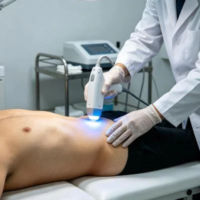
Clinical Case Documentation: Outpatient Orthopedic Rehabilitation
The documented case below details the outpatient rehabilitation course of a patient with advanced degenerative hip osteoarthritis, treated using a multi-wavelength Class IV rehabilitation laser platform equipped with a quartz massage applicator.
Patient Profile and Clinical Baseline
- Case Identifier: HIP-OSTEO-2026-0732
- Patient Age / Sex: 58-Year-Old Male
- Primary Diagnosis: Severe Primary Hip Osteoarthritis (Kellgren-Lawrence Grade III) with Superolateral Joint Space Narrowing, Subchondral Sclerosis, and Secondary Capsular Fibrosis, confirmed via Weight-Bearing Pelvic Radiographs and Diagnostic Musculoskeletal Ultrasound.
- History: 36 months of progressive right hip stiffness, groin pain, and functional impairment. Failed conservative measures including physical therapy, aquatic exercise, NSAIDs, and two intra-articular corticosteroid injections. Patient experiences severe pain during weight-bearing, is unable to put on socks independently, and walks with an antalgic gait.
- Baseline Metrics: Numerical Pain Rating Scale (NPRS) at 8/10 during weight-bearing; Harris Hip Score (HHS) at 41.5/100; Hip Active Range of Motion (AROM) Flexion restricted to 70°, Internal Rotation restricted to 5°; Timed Up and Go (TUG) test measured at 16.8 seconds.
Multi-Stage Treatment Regimen
The treatment course comprised ten clinical sessions scheduled over four weeks, progressing from early pain management and lymphatic drainage to deep joint biostimulation and functional mobility.
| Session No. | Clinical Phase & Objective | Wavelength Setup | Operating Power (W) | Pulse Frequency (Hz) | Duty Cycle (%) | Energy Delivered (J) | Cumulative Dose (J) |
| Session 01 | Nociceptive Gating & Inguinal Drainage | 980nm (65%) + 1064nm (35%) | 14.0 W | 8,000 Hz | 40% | 4,200 J | 4,200 J |
| Session 02 | Periarticular Myofascial Decompression | 980nm (60%) + 1064nm (40%) | 16.0 W | 4,000 Hz | 50% | 4,800 J | 9,000 J |
| Session 03 | Tensor Fasciae Latae Capillary Flush | 980nm (50%) + 1064nm (50%) | 18.0 W | 2,000 Hz | 60% | 5,400 J | 14,400 J |
| Session 04 | Iliofemoral Ligament Saturation | 980nm (40%) + 1064nm (60%) | 20.0 W | 500 Hz | 65% | 6,000 J | 20,400 J |
| Session 05 | Deep Acetabular Rim Photon Infusion | 980nm (35%) + 1064nm (65%) | 22.0 W | 250 Hz | 70% | 6,600 J | 27,000 J |
| Session 06 | Subchondral Bone Matrix Perfusion | 980nm (30%) + 1064nm (70%) | 24.0 W | 100 Hz | 75% | 7,200 J | 34,200 J |
| Session 07 | High-Flux Chondrocyte Respiration Push | 980nm (20%) + 1064nm (80%) | 26.0 W | Continuous (CW) | 100% | 7,800 J | 42,000 J |
| Session 08 | Dense Fibrocartilage Matrix Remodeling | 980nm (20%) + 1064nm (80%) | 26.0 W | Continuous (CW) | 100% | 7,800 J | 49,800 J |
| Session 09 | Weight-Bearing Joint Stance Integration | 980nm (30%) + 1064nm (70%) | 22.0 W | 150 Hz | 75% | 6,600 J | 56,400 J |
| Session 10 | Final Tissue Consolidation & Discharge | 980nm (40%) + 1064nm (60%) | 18.0 W | 400 Hz | 65% | 5,400 J | 61,800 J |
Clinical Progression and Outcome Metrics
WEEK 1 (Sessions 1-3):
- Deep contact application over the femoral triangle and greater trochanter relieved periarticular tension.
- Resting groin pain decreased; NPRS dropped from 8/10 to 4/10.
- Patient slept through the night without waking from position-related hip pain after Session 2.
- Antalgic limp softened as muscular splinting in the gluteus medius decreased.

WEEK 2 (Sessions 4-6):
- Hip flexion active range increased from 70° to 98°; internal rotation expanded to 14°.
- Patient put on footwear independently without requiring assistive shoe horns.
- Timed Up and Go (TUG) test improved from 16.8 seconds to 11.2 seconds.
- Harris Hip Score rose from 41.5 to 63.8.

WEEK 3 (Sessions 7-8):
- Continuous wave delivery applied over the anterior and posterior joint lines with firm quartz contact.
- Patient completed 20 minutes of treadmill walking without groin pinching or joint flare-up.
- Diagnostic ultrasound showed reduced capsular thickening and increased local microvascular flow.

WEEK 4 (Sessions 9-10):
- Hip flexion reached 115°; internal rotation reached 22°.
- TUG test clocked at 8.4 seconds, matching normal functional levels for age.
- Final NPRS recorded at 1/10 during extended walking and 0/10 at rest.
- Harris Hip Score reached 84.6/100, restoring independent mobility and deferring joint replacement.
Comparative Analysis: High-Intensity Laser vs. Conventional Modalities
Comparing high-intensity class IV light with traditional treatment paths clarifies why outpatient orthopedic clinics incorporate this technology into daily care:
Intra-Articular Corticosteroid Injections
Fluoroscopically guided steroid injections suppress local inflammation temporarily, but they do not address tissue hypoxia or mechanical capsule tightness. Repeated intra-articular steroid injections suppress chondrocyte viability, accelerate cartilage degradation, and increase the risk of rapid joint space loss. In contrast, multi-wavelength high-intensity laser therapy works non-invasively. Rather than damaging cartilage, concentrated photons stimulate microvascular circulation, boost cellular ATP synthesis, and reduce joint discomfort without chemical toxicity.
Total Hip Arthroplasty (THA)
Total hip replacement removes the native joint and replaces it with prosthetic titanium, ceramic, or polyethylene components. While effective for end-stage joint destruction, surgery involves substantial blood loss, risks of periprosthetic infection, dislocation hazards, and lengthy rehabilitation timelines. Class IV laser therapy offers an effective non-surgical treatment that preserves native joint anatomy, reduces capsular inflammation, and restores mobility without operative trauma.
Oral Non-Steroidal Anti-Inflammatory Drugs (NSAIDs)
Systemic anti-inflammatory medications circulate throughout the body, inhibiting protective prostaglandins in the gastric lining and kidneys while providing limited drug concentration to deep, avascular joint capsules. Long-term NSAID use carries well-documented risks of gastrointestinal ulceration, hypertension, and renal impairment. High-intensity laser therapy targets therapeutic energy directly into the deep hip capsule, relieving joint pain and stiffness locally without systemic adverse effects.
Practical Clinical Application and Operational Takeaways
Achieving dependable clinical results when using high-intensity laser systems for deep joint pain requires systematic clinical execution:
- Dynamic compression with quartz massage probes: Clinicians should apply firm, steady pressure with the quartz contact probe into the soft-tissue boundaries surrounding the greater trochanter and anterior hip capsule. Physical compression displaces superficial water and venous blood away from the beam path, creating an optimal optical pathway that delivers higher photon counts into deep capsule structures.
- Regional lymphatic clearing: Always treat proximal lymphatic pathways first. Sweeping the inguinal crease, iliac crest border, and proximal gluteal folds before directing energy into the deep joint capsule clears fluid pathways and accelerates tissue recovery.
- Frequency progression protocol: Begin with high pulse frequencies (4,000 Hz to 8,000 Hz) during the initial visits to calm hypersensitive peripheral nerve pathways, transition to moderate pulsing (100 Hz to 500 Hz) to improve local perfusion and clear capsular stiffness, and finish with continuous wave (CW) delivery to stimulate chondrocyte metabolism and structural remodeling once acute pain has subsided.
Grounding clinical protocols in the optical principles of human tissue allows physical medicine clinics and orthopedic centers to resolve deep, chronic joint pain and help patients regain active, independent lives.