 FotonMedix


Canine Elbow Arthrosis Capsule Scattering Bottlenecks
Targeted water and hemoglobin resonance channels clear chronic periarticular effusion while gated duty cycle emission protects thin collateral ligaments.
A nine-year-old Bernese Mountain Dog hesitates at the clinic entryway, its forelimbs trembling under unequal load distribution before collapsing into a rigid chest lay. Palpation of the left elbow triggers immediate retraction, localized vocalization, and intense spasm across the triceps brachii and extensor carpi radialis groups. Bilateral elbow dysplasia with secondary medial coronoid process disease and severe periarticular osteophytosis has left the joint capsule thick, fibrosed, and chronically distended with inflammatory fluid. The owner brings bloodwork showing rising blood urea nitrogen and creatinine after months of daily NSAID administration, forcing an immediate cessation of systemic anti-inflammatories. When non-surgical phototherapy is introduced, the primary clinical barrier centers on whether laser treatment for canine arthritis can penetrate thickened synovial envelopes, dense collateral ligaments, and subchondral bone without causing thermal spikes on heavily pigmented, fur-dense skin.
Overcoming deep elbow joint pathology requires calculating the optical attenuation curve across dense fibrous structures. In chronic canine elbow osteoarthrosis, incident light encounters dense epidermal fur layers, pigmented basal dermis, thickened subcutaneous fibrous tissue, hyperplastic synovial membranes, and sclerotic bone margins. Standard superficial illumination scatters within the first few millimeters of collagenous joint capsules, failing to deliver the required photon fluence to deep subchondral nociceptors and ischemic articular cartilage. Implementing high-intensity Class 4 multi-wavelength protocols bridges this anatomical barrier, ensuring adequate energy density reaches deep intra-articular structures to relieve pain and stimulate cellular repair.
Stratified Tissue Extinction Across The Canine Forelimb
Treating chronic forelimb joint degeneration involves navigating complex optical decay curves. The canine elbow is tightly wrapped in dense connective tissue: thick outer skin with dense hair follicles, followed by the deep antebrachial fascia, medial and lateral collateral ligaments, and hypertrophied joint capsules lined with fibrotic synovial folds.
[Incident High-Intensity Dual Photon Beam]
                     │
                     ▼ (Melanin Absorption & Fur Follicle Scatter)
        [Dense Dermal & Subdermal Fascia]
                     │
                     ▼ (Mie Scattering in Thick Collateral Ligaments)
        [Hyperplastic Synovial Joint Capsule]
                     │
                     ▼ (Water Extinction in Stagnant Synovial Effusion)
        [Periarticular Osteophytes & Sclerotic Bone]
                     │
                     ▼ (Target Fluence > Chondrocyte Activation Threshold)
        [Medial Coronoid Margin & Subchondral Nociceptive Fibers]
Low-level modalities disperse within superficial soft tissue, losing coherence before reaching the joint cavity. To stimulate cellular healing, photon density must remain above the biological threshold at the level of the medial coronoid process and articular cartilage. Choosing effective canine laser treatment for arthritis requires matching tissue-specific chromophores to deep joint targets rather than relying on unfocused surface illumination.
Bypassing these stratified barriers demands simultaneous delivery across complementary spectral peaks. Synchronizing these wavelengths allows clinicians to address vascular constriction, soft tissue spasm, and intra-capsular effusion within the same treatment session.
Target Tissue Stratum          Optimal Spectrum Band      Primary Physiological Dynamic
-----------------------------------------------------------------------------------------
Spastic Triceps Musculature    980 nm                     Microvascular dilation, NO induction
Effusive Joint Hydrops         1470 nm                    Selective water drainage, decompression
Subchondral Articular Margin   980 nm / 1470 nm Ratio     Mitochondrial ATP synthesis, pain block
The 980nm Hemoglobin Profile And Periarticular Reperfusion
The 980nm wavelength matches the peak absorption profiles of oxygenated and deoxygenated hemoglobin within the periarticular microvasculature. In chronic elbow dysplasia, sustained joint instability and compensatory muscle splinting constrict local capillary loops, starving articular tissues of oxygen and trapping lactic acid within the triceps and biceps brachii muscles.
Die Absorption von 980-nm-Photonen löst eine lokalisierte photothermische Erweiterung in den tiefen Kapillarbetten rund um das Gelenk aus. Endothelzellen setzen Stickstoffmonoxid frei, wodurch sich die glatten Muskel-Sphinkter entspannen und die mikrovaskuläre Durchblutung wiederhergestellt wird. Dieser Zufluss von sauerstoffreichem Blut spült Entzündungsmediatoren aus dem periartikulären Gewebe und stimuliert die Cytochrom-c-Oxidase in den Mitochondrien der Chondrozyten und Tenozyten. Die Wiederherstellung der Adenosintriphosphat-Produktion liefert die metabolische Energie, die erforderlich ist, um ischämische Muskelkrämpfe zu beseitigen und die Kollagenneuausrichtung in den geschädigten Gelenkstrukturen zu fördern.
The 1470nm Water Absorption Pathway In Joint Capsule Hydrops
Chronic elbow arthrosis leads to persistent synovial fluid pooling within the joint capsule, generating hydrostatic pressure that compresses sensory nerve endings against osteophytic margins. The 1470nm spectrum specifically targets free and bound water molecules in this inflammatory fluid, providing an absorption coefficient significantly higher than standard near-infrared bands.
When 1470nm photons strike edematous capsular tissues, targeted energy absorption alters interstitial fluid pressure gradients. Trapped fluid drains into collateral lymphatic networks, decompressing the pinched articular capsule. Combining 1470nm fluid mobilization with 980nm microvascular stimulation delivers rapid pain relief. This coordinated action makes laser therapy for dog arthritis a dependable modality, cutting the total number of clinical sessions needed to restore functional limb movement.
Duty Cycle Engineering For Periarticular Bone Safety
Delivering high-power photons through thin soft tissue over the elbow carries a real risk of thermal buildup. Cortical bone on the medial epicondyle and olecranon absorbs continuous wave energy rapidly. If an unmodulated beam stays focused over these bony landmarks, thermal accumulation triggers periosteal pain and potential tissue irritation, forcing clinicians to stop treatment before delivering an effective dose to the joint core.
Durch gesteuerte dynamische Tastverhältnisse werden diese thermischen Spitzen verhindert. Bei der Modulation des Strahls werden Mikrosekunden-Impulsfolgen mit kalibrierten thermischen Entspannungsintervallen abgewechselt.
Continuous Wave (Thermal Runaway Over Epicondyles):
Power ────────────────────────────────────────── (Periosteal Thermal Risk & Pain)

Gated Dynamic Duty Cycle (Structured Thermal Dissipation):
Power ┌───┐    ┌───┐    ┌───┐    ┌───┐    ┌───┐
      │   │    │   │    │   │    │   │    │   │   (Deep Articular Penetration / Zero Burn)
Base  ┘   └────┘   └────┘   └────┘   └────┘   └────
      |Ton|    Toff     |
Because dense collateral ligaments and bone dissipate heat faster than they absorb peak energy pulses, dynamic duty cycles let clinicians deploy high peak irradiance through the joint capsule while maintaining safe surface temperatures. Operating between 35 percent and 45 percent duty cycles enables safe, comfortable treatment directly over the medial and lateral elbow margins.
Durch die Anpassung der Impulsfrequenzen zwischen 15 Hertz und 800 Hertz lässt sich die biologische Wirkung gezielt steuern: Niedrige Frequenzen unterdrücken die Übertragung von Substanz P und lindern so Gelenkschmerzen, während höhere Frequenzen in späteren Rehabilitationsphasen die Organisation der Tenozyten und die Synthese der extrazellulären Matrix des Knorpels anregen.
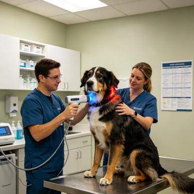
Klinischer Fallbericht
The data below comes from Veterinary Orthopedic and Mobility Rehabilitation unit records, documenting non-surgical recovery metrics in a large-breed canine with severe elbow osteoarthrosis.
Patientenprofil und diagnostische Einstufung
Case File Number: VOM-2026-4892
Subject: Canine, Bernese Mountain Dog
Alter: 9 Jahre und 4 Monate
Geschlecht: Männlich (kastriert)
Weight: 44.5 Kilograms
Presenting Complaint: Severe left thoracic limb lameness, painful weight bearing, pronounced head bob at walk, refusal to descend stairs, bilateral periarticular elbow thickening.
Orthopedic Examination: Left elbow flexion limited to 45 degrees of total arc; severe crepitus and painful response during terminal extension; palpable joint effusion along the caudolateral joint pouch; compensatory spasm across the left trapezius and triceps brachii.
Radiographic Findings: Severe bilateral elbow arthrosis with fragmented medial coronoid process remodeling, extensive osteophytosis exceeding 6 mm along the anconeal process, and pronounced subchondral sclerosis. Staged at International Elbow Working Group Grade 3 (Severe). Pre-treatment Canine Brief Pain Inventory Pain Severity Score: 8.2 out of 10. Pain Interference Score: 8.6 out of 10.
| Sitzungs-ID | Tag | Behandlungsort | Wellenlängenverteilung | Spitzenleistung (W) | Einschaltdauer (%) | Impulsfrequenz (Hz) | Gesamtenergie (J) | Klinische Beobachtungen |
| S-01 | D-01 | Left Elbow & Triceps | 80% 980 nm / 20% 1470 nm | 10.0 | 30% | 15 | 2,800 | Marked joint guarding; off-contact circular sweeps applied around collateral margins. |
| S-02 | D-03 | Left Elbow & Shoulder Girdle | 75% 980 nm / 25% 1470 nm | 12.0 | 35% | 30 | 3,600 | Triceps muscle spasm softens; dog tolerates joint palpation without pulling limb back. |
| S-03 | D-06 | Elbow Joint Capsule Complex | 70% 980 nm / 30% 1470 nm | 14.0 | 35% | 60 | 4,400 | Caudolateral joint effusion visibly recedes; standing weight-bearing shifts toward neutral. |
| S-04 | D-09 | Medial Coronoid & Epicondyle | 65% 980 nm / 35% 1470 nm | 16.0 | 40% | 120 | 5,200 | Elbow extension arc gains 12 degrees; head bob at walk visibly decreases. |
| S-05 | D-13 | Whole Forelimb Kinetic Chain | 60% 980 nm / 40% 1470 nm | 18.0 | 45% | 250 | 6,200 | Dog navigates entry steps unassisted; symmetric limb loading observed in stance. |
| S-06 | D-17 | Left Elbow Capsule & Brachium | 50% 980 nm / 50% 1470 nm | 20.0 | 45% | 500 | 7,000 | Passive range of motion improves by 22 degrees overall; joint crepitus softens. |
| S-07 | D-22 | Regionale Muskel- und Gelenkerkrankungen | 50% 980 nm / 50% 1470 nm | 18.0 | 40% | Dynamisch | 6,400 | Gait symmetry improves on hard floors; Canine Brief Pain Inventory scores decline. |
| S-08 | D-28 | Biomechanische Erhaltungszone | 50% 980 nm / 50% 1470 nm | 16.0 | 35% | Dynamisch | 5,600 | Die volle Bewegungsfähigkeit ist wiederhergestellt; der Patient geht täglich 30 Minuten ohne Hinken. |
Objektive Messgrößen für die funktionelle Genesung
Day 1 (Intake Baseline)
[Pain Severity: 8.2/10]      ████████████████░░░░
[Elbow Flexion Deficit: 45°] ████████████████████

Day 13 (Midway Status)
[Pain Severity: 3.8/10]      ████████░░░░░░░░░░░░
[Elbow Flexion Deficit: 22°] ██████████░░░░░░░░░░

Day 28 (Protocol Completion)
[Pain Severity: 1.4/10]      ███░░░░░░░░░░░░░░░░░
[Elbow Flexion Deficit: 6°]  ███░░░░░░░░░░░░░░░░░
By session eight on Day 28, the patient demonstrated major clinical recovery. The Canine Brief Pain Inventory Pain Severity Score dropped from an intake baseline of 8.2 down to 1.4, while the Pain Interference Score fell from 8.6 to 1.8. Total elbow range of motion expanded by 39 degrees, bringing the flexion-extension arc close to physiological norms and resolving the pronounced compensatory head bob during walking.
Digital circumference measurements around the left elbow decreased by 1.6 centimeters, confirming the drainage of chronic synovial hydrops. Follow-up renal blood panels remained stable, confirming that non-invasive photobiomodulation relieved severe chronic pain without placing metabolic stress on the kidneys or liver.
Praktische Vorteile gegenüber herkömmlichen Verfahren
Managing severe canine elbow arthrosis presents ongoing operational and therapeutic hurdles in veterinary practice. Long-term administration of systemic NSAIDs or corticosteroids carries high risks of renal and hepatic toxicity, especially in older large-breed dogs, requiring recurring blood panels that increase client expenses and force sudden medication stops. Surgical options, including arthroscopic debridement or joint resurfacing, require general anesthesia, significant financial commitments, and prolonged post-operative confinement that can be difficult for aging dogs.
Underpowered low-level phototherapy units worsen these clinical delays. Low-wattage hardware requires prolonged session times of thirty to forty minutes to deliver minimal photon quantities through thick joint capsules and dense musculature. This hands-on demand ties up technicians and examination suites, driving up clinic labor overhead while delivering slow, inconsistent clinical progress.
High-power Class 4 multi-wavelength protocols eliminate these clinical bottlenecks. Delivering up to twenty watts of peak power through balanced 980nm and 1470nm wavelengths cuts hands-on treatment times down to under ten minutes per joint. This delivery speed allows veterinary practices to treat deep periarticular structures efficiently, tripling daily room capacity while keeping large-breed patients relaxed on the treatment mat.
Dynamic duty cycle pulsing protects sensitive tissues over the medial epicondyle and olecranon, preventing thermal spikes on dense, dark-coated limbs. Pet owners see noticeable reductions in lameness and stiffness within the first three visits, validating their commitment and keeping compliance high across the complete rehabilitation plan. For veterinary practices, combining deep intra-capsular penetration with rapid treatment times turns complex canine osteoarthritis cases into a dependable, non-invasive clinical service.