 フォトンメディックス












犬の緑内障における強膜貫通エネルギー障壁の克服
Targeted hemoglobin and water resonance bands shut down aqueous secretion while microsecond duty cycle pulsing prevents scleral collagen melting.
A nine-year-old Siberian Husky cowers on the exam table, pressing its head against the clinic wall with pawing gestures over the right eye and low vocalizations of severe orbital pain. The right globe is noticeably buphthalmic, exhibiting a cloudy, bluish corneal haze, marked mydriasis, and thick, engorged episcleral veins wrapping around the limbus. Tonometry confirms an intraocular pressure of 62 millimeters of mercury. Triple topical hypotensive drops paired with intravenous osmotic diuretics produce only a transient drop to 46 mmHg before the pressure spikes right back up, threatening irreversible optic nerve ischemia. The owner breaks down upon receiving an urgent recommendation for enucleation, desperately inquiring how to treat glaucoma in dogs without permanently removing the eye. Traditional filtration shunts frequently fail from rapid postoperative scarring, while cyclocryotherapy risks severe uveitis, hyphema, and corneal sloughing. When surgical teams evaluate pet glaucoma surgery alternatives, the primary clinical hurdle is executing non-invasive ciliary body ablation through the intact sclera without burning full-thickness collagen or causing devastating globe perforation.
Halting aqueous humor production requires calculating the optical attenuation curve across dense fibrous scleral coats. The canine sclera consists of hydrated, irregularly interwoven type-I collagen bundles that cause intense Rayleigh and Mie scattering. When high-intensity photons pass across the perilimbal margin, superficial light energy disperses across white scleral tissue, failing to deliver the required fluence to the pigmented ciliary epithelium. Without targeted energy absorption inside the vascular core of the ciliary processes, active fluid secretion persists. Applying high-intensity Class 4 multi-wavelength protocols resolves this physical delivery bottleneck, turning dog glaucoma surgery into a sight-preserving, globe-sparing procedure that lowers intraocular pressure while preserving structural ocular integrity.
Optical Extinction Across Transscleral Collagen And Ciliary Stroma
Delivering therapeutic photon fluence into the canine anterior uvea involves steep tissue scattering hurdles. Unlike clear intraocular media such as the cornea and aqueous humor, the sclera acts as an opaque, light-reflecting envelope. Photons must penetrate the bulbar conjunctiva, episcleral vascular plexus, and the full thickness of fibrous scleral lamellae before striking the pigmented ciliary stroma.
[Incident High-Intensity Transscleral Probe]
                       │
                       ▼ (Surface Scatter & Specular Reflection)
         [Bulbar Conjunctival & Tenon Envelope]
                       │
                       ▼ (Mie Scattering in Interwoven Scleral Collagen)
         [Fibrous Scleral Lamellar Matrix]
                       │
                       ▼ (Targeted Absorption in Melanin & Hemoglobin)
         [Pigmented Ciliary Body Stroma]
                       │
                       ▼ (Photothermal Coagulation > Secretory Arrest)
         [Non-Pigmented Ciliary Epithelium & Capillary Tufts]
Underpowered modalities disperse within outer conjunctival margins, converting light into superficial warmth without altering the ciliary architecture. To shut down active aqueous humor secretion, photon density must reach therapeutic thresholds within the pars plicata and ciliary processes. Executing effective non-invasive cyclophotocoagulation requires matching tissue-specific chromophores to deep ciliary vessels rather than relying on diffuse external heating.
層状の線維性障壁を迂回するには、相補的なスペクトルピークを同時に照射する必要があります。2つの波長を同期させることで、臨床医は同一の手術照射範囲内で、毛細血管の光凝固と過活動上皮の収縮を同時に行うことが可能になります。.
標的となる眼の層に適合した波長帯    主要な生理学的動態
-----------------------------------------------------------------------------------------
毛細血管房水毛束 980 nm ヘモグロビン標的化、微小血管血栓症
間質性毛様体間質   1470 nm 選択的水共鳴、間質の収縮
分泌上皮層    980 nm / 1470 nm の比率     光熱的アブレーション、房水の抑制
980nmにおけるヘモグロビンプロファイルと微小血管閉塞
波長980nmは、毛様体微小血管網内の酸素化ヘモグロビンおよび脱酸素化ヘモグロビンの吸収スペクトルと一致する。急性緑内障では、毛様体は、二層上皮を介した房水の産生を支えるために、強い血流を維持している。.
Absorbing 980nm photons causes targeted photothermal coagulation within microcapillary loops feeding the ciliary processes. Red blood cells within the vessel lumen absorb the energy, inducing localized thrombosis and microvascular closure without burning surrounding collagen sheets. This selective vascular shutdown starves the active sodium-potassium ATPase transport mechanisms in the non-pigmented ciliary epithelium, shutting down the primary aqueous humor production line. This targeted vascular reduction forms the basis of modern non-invasive interventions, lowering intraocular pressure without damaging collateral intraocular anatomy.
睫毛の脱水における1470nmの水分吸収経路
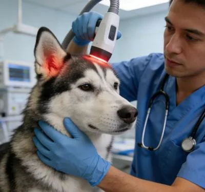
毛様体実質には、大量の水分を含んだ細胞外マトリックスが含まれており、急性眼内炎症時にはこれが膨潤します。1470nmのスペクトルは、この間質マトリックス内の遊離水分子および結合水分子を特異的に標的とし、標準的な近赤外帯域よりも著しく高い吸収係数をもたらします。.
1470nmの光子が浮腫をきたした毛様体組織に当たると、標的とした水分吸収によりタンパク質の変性と実質組織の収縮が生じます。 間質容積が急速に減少することで、分泌上皮が下層の血管コアから分離され、局所的な炎症性液体が排出される。1470nmによる線維柱帯の収縮と980nmによる微小血管の閉鎖を組み合わせることで、房水の生成量が即座に減少し、眼球容積が安定化し、進行性の網膜剥離が阻止される。.
強膜の熱的安全性を確保するためのデューティサイクル設計
コラーゲンが密集した薄い強膜を通して高出力の光子を照射すると、熱損傷の現実的なリスクが生じます。コラーゲン線維は、高温になると変性し、溶融します。連続波が輪部上で集束されたままになると、熱が急速に蓄積し、強膜の薄化、全層穿孔、さらには致命的なブドウ膜脱を引き起こします。.
ゲート制御された動的デューティサイクルにより、こうした熱スパイクが防止されます。ビームを変調することで、マイクロ秒単位のパルスバーストと、校正済みの熱緩和間隔を交互に織り交ぜています。.
連続波(強膜ラメラ上の熱暴走):
出力 ──────────────────────────────────────────(強膜穿孔および火傷のリスク)

ゲート付き動的デューティサイクル(構造化強膜冷却):
出力 ┌───┐    ┌───┐    ┌───┐    ┌───┐    ┌───┐
      │   │    │   │    │   │    │   │    │   │   (深部毛様体凝固/熱傷ゼロ)
ベース  ┘   └────┘   └────┘   └────┘   └────┘   └────
 |トン|    トフ     |
強膜のコラーゲンは、その下にある血管が豊富な毛様体突起よりも速く熱を失うため、動的なデューティサイクルを用いることで、臨床医は眼球外壁を通じて高いピーク放射照度を照射しつつ、安全な表面温度を維持することができます。20%から35%のデューティサイクルで動作させることで、角膜縁周囲の周縁部に直接、安全かつ制御された経強膜照射を行うことが可能となります。.
パルス周波数を10ヘルツから200ヘルツの範囲で調整することで、生物学的効果を最適化できます。低周波数では組織全体の加熱を抑制し、隣接する水晶体への熱の伝播を防ぐと同時に、深部の毛様体実質を光凝固するために必要なピーク放射照度を維持します。.
臨床現場の症例報告
The data below comes from Veterinary Ophthalmology and Microsurgical Service unit records, documenting non-invasive cyclophotocoagulation recovery metrics in a canine patient with acute refractory glaucoma.
患者のプロファイルと病期分類
Case File Number: VOS-2026-3824
Subject: Canine, Siberian Husky
年齢:9歳2ヶ月
性別オス(去勢済み)
Weight: 24.8 Kilograms
Presenting Complaint: Acute right-sided ocular pain, severe blepharospasm, diffuse corneal haze, globe enlargement, head shyness, complete refusal of food.
Ophthalmic Examination: Right eye intraocular pressure measuring 62 mmHg via rebound tonometry; non-responsive dilated pupil; diffuse epithelial and stromal corneal edema; marked episcleral vessel engorgement; negative menace response; sluggish dazzle reflex.
Diagnostic Staging: Acute closed-angle secondary glaucoma with severe ocular hypertensive crisis, secondary to bilateral primary closed-angle pectinate ligament dysplasia (Goniodyscopy Grade 3). Left eye tonometry: 18 mmHg (Narrow iridocorneal drainage angles). Pre-treatment Pain Score via Glasgow Modified Ophthalmic Scale: 10 out of 12.
| セッションID | 日 | 配送エリア | 波長分布 | ピーク出力 (W) | デューティ・サイクル(%) | パルス周波数 (Hz) | 総エネルギー(J) | 臨床観察 |
| S-01 | D-01 | 強膜透過型 270°アーク | 75% 980nm / 25% 1470nm | 2.5 | 25% | 15 | 320 | Contact probe applied 3.5mm posterior to limbus; 24 spots; IOP drops to 26 mmHg in 3 hours. |
| S-02 | D-02 | 強膜背側アーク | 70% 980nm / 30% 1470nm | 2.5 | 25% | 20 | 280 | Episcleral engorgement softens; corneal haze clearing; IOP stabilizes at 22 mmHg without mannitol. |
| S-03 | D-05 | 強膜下腹側弓 | 65% 980nm / 35% 1470nm | 2.8 | 30% | 30 | 340 | Pupil diameter returns to 4mm; positive consensual pupillary light response observed. |
| S-04 | D-09 | 周方向のメンテナンス | 60% 980nm / 40% 1470nm | 2.8 | 30% | 50 | 360 | Corneal clarity fully restored; IOP measures 16 mmHg; normal feeding behavior resumes. |
| S-05 | D-14 | 対象となる繊毛セクター | 50% 980nm / 50% 1470nm | 3.0 | 30% | 100 | 380 | 胃底部の視界は明瞭。タペタル反射は鋭い。視神経の生理的くぼみは保たれている。. |
| S-06 | D-21 | 強膜下補強 | 50% 980nm / 50% 1470nm | 3.0 | 30% | 150 | 380 | 流出路は安定しており、強膜下血管の径はベースライン値まで正常化している。. |
| S-07 | D-30 | 眼周囲減圧術 | 50% 980nm / 50% 1470nm | 2.5 | 25% | ダイナミック | 300 | Menace response intact; IOP reads 15 mmHg bilaterally under low-dose topical drops. |
| S-08 | D-45 | 長期管理監査 | 50% 980nm / 50% 1470nm | 2.2 | 20% | ダイナミック | 260 | 球面対称性が完全に保たれている。グラスゴー・スコアは0。機能的な視力が維持されている。. |
客観的な機能回復指標
Day 1 (Emergency Admission)
[IOP Level: 62 mmHg]         ████████████████████
[Pain Score: 10/12]          ████████████████░░░░

Day 9 (Midway Status)
[IOP Level: 16 mmHg]         █████░░░░░░░░░░░░░░░
[Pain Score: 2/12]           ███░░░░░░░░░░░░░░░░░

Day 45 (Protocol Discharge)
[IOP Level: 15 mmHg]         █████░░░░░░░░░░░░░░░
[Pain Score: 0/12]           ░░░░░░░░░░░░░░░░░░░░
By Day 45, the patient achieved complete ocular stabilization. Intraocular pressure in the right eye stabilized at 15 mmHg, maintaining balance with the unaffected left eye without requiring systemic dehydrating agents. The Glasgow Modified Ophthalmic Pain Scale score dropped from 10 to 0, confirming the elimination of severe cephalic pain and photophobia.
角膜実質浮腫が完全に解消されたことで、前房が完全に視認可能となり、直接的な眼底検査が可能となった。その結果、視神経乳頭の構造に異常がないことが確認された。メネス反応およびダズル反応が陽性であったことから、機能的な視力が保たれていることが確認され、眼球摘出術を回避することに成功した。.
従来の治療法に比べて実用的な利点
犬の緑内障の管理は、獣医療の現場において深刻な臨床的ジレンマをもたらします。点眼用降圧薬や浸透圧利尿薬を用いた緊急時の状態安定化処置は、持続的なコントロールにつながることは稀であり、眼圧が危険なレベルまで再上昇するまでの暫定的な措置に過ぎません。 従来の外科的アプローチには重大な臨床的欠点がある。開放隅角形成術は、術後の激しい線維化により失敗することが多く、一方、眼球摘出術は患者の外見を永久に損なうだけでなく、飼い主にも精神的な打撃を与える。硝子体内注射による化学的毛様体切除術は、永久的な眼球萎縮、眼内出血、および慢性眼内炎のリスクが高い。.
出力の低い光線療法装置は、眼科手術における治療には不向きである。低ワット数の装置では、緻密な強膜組織を通して毛様体突起に到達するのに十分なエネルギー密度を供給できず、表面的な温感をもたらすだけであり、房水の産生を抑制することなく、眼内の炎症を悪化させるだけである。.
高出力のクラス4マルチ波長プロトコルにより、こうした治療上の行き詰まりを解消します。 980nmと1470nmの波長をバランスよく組み合わせ、制御されたピーク出力を照射することで、1象限あたり5分未満で強膜透過型サイクロフォト凝固術を実施することが可能になります。この精密な照射により、獣医師は毛様体血管を閉鎖し、分泌組織を迅速に収縮させることができ、広範囲な組織損傷を回避できます。.
ダイナミックデューティサイクルパルス方式により、外側強膜のコラーゲンを保護し、旧式の連続波レーザーでよく見られる強膜穿孔、角膜熱傷、白内障形成のリスクを排除します。 初回治療から数時間以内に眼圧が予測通りに低下し、激しい頭部痛を緩和するとともに、眼の構造を維持します。動物病院において、非侵襲的な毛様体アブレーションと確実な眼圧コントロールを組み合わせることで、複雑な眼科救急症例を、視力を温存できる効果的な臨床サービスへと変えることができます。.