 FotonMedix


克服犬青光眼治疗中的巩膜外能量屏障
Targeted hemoglobin and water resonance bands shut down aqueous secretion while microsecond duty cycle pulsing prevents scleral collagen melting.
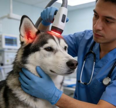
A nine-year-old Siberian Husky cowers on the exam table, pressing its head against the clinic wall with pawing gestures over the right eye and low vocalizations of severe orbital pain. The right globe is noticeably buphthalmic, exhibiting a cloudy, bluish corneal haze, marked mydriasis, and thick, engorged episcleral veins wrapping around the limbus. Tonometry confirms an intraocular pressure of 62 millimeters of mercury. Triple topical hypotensive drops paired with intravenous osmotic diuretics produce only a transient drop to 46 mmHg before the pressure spikes right back up, threatening irreversible optic nerve ischemia. The owner breaks down upon receiving an urgent recommendation for enucleation, desperately inquiring how to treat glaucoma in dogs without permanently removing the eye. Traditional filtration shunts frequently fail from rapid postoperative scarring, while cyclocryotherapy risks severe uveitis, hyphema, and corneal sloughing. When surgical teams evaluate pet glaucoma surgery alternatives, the primary clinical hurdle is executing non-invasive ciliary body ablation through the intact sclera without burning full-thickness collagen or causing devastating globe perforation.
Halting aqueous humor production requires calculating the optical attenuation curve across dense fibrous scleral coats. The canine sclera consists of hydrated, irregularly interwoven type-I collagen bundles that cause intense Rayleigh and Mie scattering. When high-intensity photons pass across the perilimbal margin, superficial light energy disperses across white scleral tissue, failing to deliver the required fluence to the pigmented ciliary epithelium. Without targeted energy absorption inside the vascular core of the ciliary processes, active fluid secretion persists. Applying high-intensity Class 4 multi-wavelength protocols resolves this physical delivery bottleneck, turning dog glaucoma surgery into a sight-preserving, globe-sparing procedure that lowers intraocular pressure while preserving structural ocular integrity.
Optical Extinction Across Transscleral Collagen And Ciliary Stroma
Delivering therapeutic photon fluence into the canine anterior uvea involves steep tissue scattering hurdles. Unlike clear intraocular media such as the cornea and aqueous humor, the sclera acts as an opaque, light-reflecting envelope. Photons must penetrate the bulbar conjunctiva, episcleral vascular plexus, and the full thickness of fibrous scleral lamellae before striking the pigmented ciliary stroma.
[Incident High-Intensity Transscleral Probe]
                       │
                       ▼ (Surface Scatter & Specular Reflection)
         [Bulbar Conjunctival & Tenon Envelope]
                       │
                       ▼ (Mie Scattering in Interwoven Scleral Collagen)
         [Fibrous Scleral Lamellar Matrix]
                       │
                       ▼ (Targeted Absorption in Melanin & Hemoglobin)
         [Pigmented Ciliary Body Stroma]
                       │
                       ▼ (Photothermal Coagulation > Secretory Arrest)
         [Non-Pigmented Ciliary Epithelium & Capillary Tufts]
Underpowered modalities disperse within outer conjunctival margins, converting light into superficial warmth without altering the ciliary architecture. To shut down active aqueous humor secretion, photon density must reach therapeutic thresholds within the pars plicata and ciliary processes. Executing effective non-invasive cyclophotocoagulation requires matching tissue-specific chromophores to deep ciliary vessels rather than relying on diffuse external heating.
要穿透分层纤维屏障,必须同时利用互补的光谱峰进行能量传递。通过同步双波长,临床医生可以在同一手术作用范围内,对睫状微血管进行光凝,并收缩过度活跃的上皮组织。.
Targeted Ocular Stratum       Matched Wavelength Band    Primary Physiological Dynamic
-----------------------------------------------------------------------------------------
Ciliary Capillary Tufts       980 nm                     Hemoglobin targeting, microvascular thrombosis
Interstitial Ciliary Stroma   1470 nm                    Selective water resonance, stromal shrinkage
Secretory Epithelium Layer    980 nm / 1470 nm Ratio     Photothermal ablation, aqueous suppression
980nm波长下的血红蛋白谱与微血管闭塞
980纳米波长与睫状体微血管网中含氧血红蛋白和脱氧血红蛋白的吸收光谱相吻合。在急性青光眼中,睫状体保持着强烈的血流,以支持房水在双层上皮中的生成。.
Absorbing 980nm photons causes targeted photothermal coagulation within microcapillary loops feeding the ciliary processes. Red blood cells within the vessel lumen absorb the energy, inducing localized thrombosis and microvascular closure without burning surrounding collagen sheets. This selective vascular shutdown starves the active sodium-potassium ATPase transport mechanisms in the non-pigmented ciliary epithelium, shutting down the primary aqueous humor production line. This targeted vascular reduction forms the basis of modern non-invasive interventions, lowering intraocular pressure without damaging collateral intraocular anatomy.
1470nm波长在睫状体脱水中的水吸收途径
睫状体基质中含有大量水合的细胞外基质,在急性眼内炎症期间会发生肿胀。1470nm波段的光谱专门针对该间质基质中的游离水分子和结合水分子,其吸收系数显著高于标准的近红外波段。.
当1470nm光子照射到水肿的睫状体组织时,靶向吸水会导致蛋白质变性及基质收缩。 间质体积迅速减少,使分泌上皮与下方的血管核心分离,并排出局部炎症液体。将1470nm引起的基质收缩与980nm引起的微血管闭合相结合,可立即使房水生成量下降,从而稳定眼球体积并阻止进行性视网膜脱离。.
用于巩膜热安全性的占空比设计
通过薄而富含胶原蛋白的巩膜传递高功率光子,存在造成热损伤的实际风险。胶原纤维在高温下会变性并熔化。如果连续波持续聚焦于角膜缘,热量会迅速积聚,导致巩膜变薄、全层穿孔以及灾难性的葡萄膜脱出。.
带门控的动态占空比可防止这些热峰值。通过调制光束,将微秒级脉冲群与经过校准的热松弛间隔交替进行。.
连续波(巩膜层上的热失控):
功率 ──────────────────────────────────────────(巩膜穿孔及烧伤风险)

门控动态占空比(结构化巩膜冷却):
功率 ┌───┐    ┌───┐    ┌───┐    ┌───┐    ┌───┐
      │   │    │   │    │   │    │   │    │   │   (深层睫状体凝固 / 零灼伤)
基底  ┘   └────┘   └────┘   └────┘   └────┘   └────
 |Ton|    Toff     |
由于巩膜胶原蛋白的散热速度快于其下方的血管丰富的睫状突,动态占空比使临床医生能够在保持安全表面温度的同时,通过眼球外壁施加高峰值辐照度。在20%至35%的占空比范围内运行,可实现直接作用于睫缘周缘区域的安全、可控的经巩膜给药。.
通过将脉冲频率调节在10赫兹至200赫兹之间,可以精准调控生物效应:较低的频率可限制组织整体升温,防止热量向相邻的晶状体扩散,同时保持光凝固深层睫状体基质所需的峰值辐照度。.
临床案例报告
The data below comes from Veterinary Ophthalmology and Microsurgical Service unit records, documenting non-invasive cyclophotocoagulation recovery metrics in a canine patient with acute refractory glaucoma.
患者概况与诊断分期
Case File Number: VOS-2026-3824
Subject: Canine, Siberian Husky
年龄:9岁2个月
性别雄性(绝育)
Weight: 24.8 Kilograms
Presenting Complaint: Acute right-sided ocular pain, severe blepharospasm, diffuse corneal haze, globe enlargement, head shyness, complete refusal of food.
Ophthalmic Examination: Right eye intraocular pressure measuring 62 mmHg via rebound tonometry; non-responsive dilated pupil; diffuse epithelial and stromal corneal edema; marked episcleral vessel engorgement; negative menace response; sluggish dazzle reflex.
Diagnostic Staging: Acute closed-angle secondary glaucoma with severe ocular hypertensive crisis, secondary to bilateral primary closed-angle pectinate ligament dysplasia (Goniodyscopy Grade 3). Left eye tonometry: 18 mmHg (Narrow iridocorneal drainage angles). Pre-treatment Pain Score via Glasgow Modified Ophthalmic Scale: 10 out of 12.
| 会话 ID | 日 | 配送区域 | 波长分布 | 峰值功率(瓦) | 占空比 (%) | 脉冲频率(赫兹) | 总能量(焦耳) | 临床观察 |
| S-01 | D-01 | 经巩膜270°弧形 | 75% 980纳米 / 25% 1470纳米 | 2.5 | 25% | 15 | 320 | Contact probe applied 3.5mm posterior to limbus; 24 spots; IOP drops to 26 mmHg in 3 hours. |
| S-02 | D-02 | Transscleral Dorsal Arc | 70% 980nm / 30% 1470nm | 2.5 | 25% | 20 | 280 | Episcleral engorgement softens; corneal haze clearing; IOP stabilizes at 22 mmHg without mannitol. |
| S-03 | D-05 | Transscleral Ventral Arc | 65% 980纳米 / 35% 1470纳米 | 2.8 | 30% | 30 | 340 | Pupil diameter returns to 4mm; positive consensual pupillary light response observed. |
| S-04 | D-09 | 周向维护 | 60% 980nm / 40% 1470nm | 2.8 | 30% | 50 | 360 | Corneal clarity fully restored; IOP measures 16 mmHg; normal feeding behavior resumes. |
| S-05 | D-14 | 目标纤毛区 | 50% 980nm / 50% 1470nm | 3.0 | 30% | 100 | 380 | Fundic view clear; tapetal reflex sharp; physiological cupping of optic nerve preserved. |
| S-06 | D-21 | 巩膜外加固 | 50% 980nm / 50% 1470nm | 3.0 | 30% | 150 | 380 | Outflow pathway stable; episcleral vessel caliber normalized to baseline. |
| S-07 | D-30 | 眼周减压术 | 50% 980nm / 50% 1470nm | 2.5 | 25% | 动态 | 300 | Menace response intact; IOP reads 15 mmHg bilaterally under low-dose topical drops. |
| S-08 | D-45 | 长期控制审计 | 50% 980nm / 50% 1470nm | 2.2 | 20% | 动态 | 260 | Globe symmetry fully maintained; Glasgow score 0; functional vision sustained. |
客观功能恢复指标
Day 1 (Emergency Admission)
[IOP Level: 62 mmHg]         ████████████████████
[Pain Score: 10/12]          ████████████████░░░░

Day 9 (Midway Status)
[IOP Level: 16 mmHg]         █████░░░░░░░░░░░░░░░
[Pain Score: 2/12]           ███░░░░░░░░░░░░░░░░░

Day 45 (Protocol Discharge)
[IOP Level: 15 mmHg]         █████░░░░░░░░░░░░░░░
[Pain Score: 0/12]           ░░░░░░░░░░░░░░░░░░░░
By Day 45, the patient achieved complete ocular stabilization. Intraocular pressure in the right eye stabilized at 15 mmHg, maintaining balance with the unaffected left eye without requiring systemic dehydrating agents. The Glasgow Modified Ophthalmic Pain Scale score dropped from 10 to 0, confirming the elimination of severe cephalic pain and photophobia.
Complete clearing of corneal stromal edema restored full anterior chamber visualization, permitting direct fundic inspection that confirmed intact optic disc architecture. Positive menace and dazzle responses confirmed the preservation of functional vision, successfully averting enucleation surgery.
相较于传统治疗方式的实际优势
Managing canine glaucoma presents severe clinical dilemmas in veterinary practice. Emergency medical stabilization with topical hypotensive drops and osmotic diuretics rarely provides lasting control, serving only as a temporary measure before intraocular pressure rebounds dangerously. Conventional surgical approaches carry heavy clinical drawbacks: open filtration surgeries frequently fail due to aggressive postoperative fibroplasia, while enucleation permanently disfigures the patient and devastates pet owners emotionally. Chemical ciliary ablation with intravitreal injections carries high risks of permanent phthisis bulbi, hyphema, and chronic endophthalmitis.
Underpowered phototherapy devices fail in ophthalmic surgical care. Low-wattage units cannot deliver sufficient energy density through dense scleral tissue to reach the ciliary processes, producing only superficial warmth that increases intraocular inflammation without stopping aqueous humor production.
High-power Class 4 multi-wavelength protocols resolve these therapeutic dead ends. Delivering controlled peak power through balanced 980nm and 1470nm wavelengths enables transscleral cyclophotocoagulation in under five minutes per quadrant. This precise delivery allows veterinary surgeons to close ciliary vessels and shrink secretory tissue rapidly, avoiding extensive tissue trauma.
Dynamic duty cycle pulsing protects the outer scleral collagen, eliminating the risks of scleral perforation, corneal thermal burns, and cataract formation common with older continuous-wave lasers. Intraocular pressure drops predictably within hours of the first session, relieving excruciating cephalic pain and preserving ocular architecture. For veterinary practices, combining non-invasive ciliary ablation with reliable intraocular pressure control turns complex ophthalmic emergencies into an effective, sight-sparing clinical service.