 FotonMedix











Managing Stifle Desmitis Economics And Photon Penetration
Selective water absorption targeting alongside microvascular perfusion pathways overcomes dense ligament scattering while slashing clinic operational overhead.
An eight-year-old Golden Retriever hobbles into the examination room with acute right stifle instability, toe-touching on hard tiles while the owner winces at every step. Palpation reveals medial buttress formation, joint effusion, and severe stifle hyperflexion pain following a partial cranial cruciate ligament tear with secondary patellar desmitis. The owner is staring down an estimate sheet with panic in their eyes: surgical stabilization runs well over four thousand dollars, while traditional post-operative physiotherapy stacks endless recurring visits. When conservative non-surgical management comes up, the immediate friction point in practice management is always transparency around dog laser therapy cost alongside whether non-invasive photon delivery can actually penetrate thick fibrous capsular scar tissue without burning skin coats.
Navigating dense periarticular collagen bundles requires calculating the optical attenuation curve across stratified joint capsules. When high-intensity photons pass through hyperplastic synovial membranes, irregular fibrillar networks scatter the incoming beam, turning focused light into diffuse, ineffective scatter. Without sufficient fluence reaching deep into the intra-articular origin of the cruciate ligaments, bio-stimulation stalls. Veterinary teams need an efficient protocol that bridges cellular healing kinetics with clinical viability, ensuring every single joule directly reduces patient recovery timelines.
Tissue Attenuation Barriers In Chronic Canine Stifles
Stifle joint pathologies present unique optical challenges compared to flat muscle planes. The canine knee is enveloped in dense connective tissues: the skin layer carries high concentrations of pheomelanin and eumelanin, immediately followed by the fascia lata, the dense fibrous joint capsule, and the infrapatellar fat pad.
[Incident High-Intensity Photon Beam]
                  │
                  ▼ (Epidermal Melanin Attenuation)
       [Dense Fascia Lata Barrier]
                  │
                  ▼ (Mie Scattering in Thick Collagen Fibrils)
     [Fibrous Stifle Joint Capsule]
                  │
                  ▼ (Intermolecular Water & Fat Scattering)
     [Infrapatellar Fat Pad / Synovium]
                  │
                  ▼ (Residual Targeted Flux > Cellular Threshold)
     [Cruciate Ligament Insertion & Meniscal Rims]
Standard near-infrared light scatters outward within the first four millimeters of fibrous connective tissue. Low-power modalities disperse across the dense collagenous network of the joint capsule, leaving intra-articular structures starved of regenerative photon density. Reaching therapeutic thresholds within deep synovial fluid pockets demands high-intensity Class 4 delivery configured with specific absorption dynamics. Clinics evaluating pet equipment often find that laser therapy for dogs hinges on matching tissue-specific chromophores to depth demands rather than relying on raw continuous power.
Balancing deep penetration against surface safety requires simultaneous emission across dual therapeutic spectra. By synchronizing precise wavelengths, practitioners manipulate absorption dynamics to clear anatomical barriers.
Targeted Anatomical Stratum    Matched Wavelength Band    Primary Physiological Dynamic
-----------------------------------------------------------------------------------------
Collateral Microcapillaries   980 nm                     Endothelial NO release, oxygenation
Deep Synovial Hydrops         1470 nm                    Targeted water clearance, decompression
Intra-Articular Stifle Core   980 nm / 1470 nm Ratio     Fibroblastic synthesis, ATP induction
The 980nm Chromophore Coupling In Periarticular Tissues
The 980nm wavelength coincides with defined absorption peaks for both oxygenated and deoxygenated hemoglobin within the periarticular microvasculature. In chronic cruciate desmitis, sustained joint laxity induces localized ischemia, leading to tissue hypoxia and accumulation of lactic acid along the patellar tendon attachment.
Absorbing 980nm photons triggers rapid photothermal warming within microscopic vascular networks, inducing localized vasodilation. Nitric oxide dissociates from hemoglobin carriers, relaxing smooth muscle sphincters across capillary beds. This targeted flush of oxygenated red blood cells into hypoxic periarticular zones accelerates cytochrome c oxidase activation inside fibroblast mitochondria. Elevating cellular adenosine triphosphate synthesis gives strained ligament fibers the bioenergetic fuel necessary to realign disorganized collagen fibrils.
The 1470nm Selective Water Absorption Profile In Effusion
Inflamed stifle joints collect excessive fluid within the infrapatellar region, generating painful capsular distension that restricts range of motion. The 1470nm wavelength interacts directly with free and structured water molecules within extracellular matrices. Its water absorption coefficient is substantially higher than standard infrared bands, enabling precise targeting of edema.
When 1470nm photons encounter localized effusive fluid, the targeted energy absorption alters interstitial fluid pressure gradients. This shifts stagnant, protein-rich exudate into surrounding lymphatic channels, decompressing sensory nociceptors embedded within the joint capsule. Combining 1470nm fluid mobilization with 980nm microvascular stimulation delivers rapid pain reduction, fundamentally shifting clinic unit economics and lowering the cumulative laser therapy for dogs cost by cutting the total number of visits needed to stabilize the joint.
Duty Cycle Engineering For Thermal Dissipation
Applying high average powers through dense canine stifle anatomy carries a significant risk of epidermal and periosteal overheating. Melanin in the dermis and compact bone surfaces in the patella absorb continuous energy rapidly. If a continuous wave remains engaged over bony landmarks, thermal accumulation triggers pain responses and thermal injury, forcing clinicians to pull the handpiece away before achieving an effective intra-articular dose.
Implementing gated dynamic duty cycles eliminates this thermal liability. Pulsing the laser beam intersperses microsecond pulse bursts with calibrated thermal relaxation intervals.
Continuous Wave (Rapid Thermal Spikes on Patella):
Power ────────────────────────────────────────── (Periosteal Pain & Thermal Risk)

Gated Dynamic Duty Cycle (Safe Peak Irradiance):
Power ┌───┐    ┌───┐    ┌───┐    ┌───┐    ┌───┐
      │   │    │   │    │   │    │   │    │   │   (Maximum Penetration / Zero Burn)
Base  ┘   └────┘   └────┘   └────┘   └────┘   └────
      |Ton|    Toff     |
Because dense fibrous tissues lose heat faster than they absorb peak irradiance pulses, a structured duty cycle allows clinicians to deploy high peak power to bypass dense connective layers while keeping average surface temperatures well within safe thresholds. Modulating duty cycles between 30 percent and 50 percent allows treatment directly over bony insertions, like the tibial tuberosity, without uncomfortable thermal spikes.
Adjusting pulsing frequencies between 20 Hertz and 1000 Hertz further aligns treatment goals. Low pulse frequencies suppress substance P transmission in peripheral nociceptors for immediate post-treatment pain relief, while higher frequencies stimulate tenocyte division and extracellular collagen remodeling during later rehabilitation phases.
Clinical Field Case Report
The data below reflects documented clinical records from the Department of Small Animal Orthopedic Rehabilitation, tracking conservative recovery metrics in a partial cranial cruciate ligament tear.
Patient Profile And Diagnostic Staging
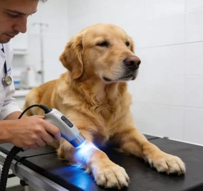
Case File Number: SAO-2026-4412
Subject: Canine, Golden Retriever
Age: 8 Years 6 Months
Sex: Male (Intact)
Weight: 34.2 Kilograms
Presenting Complaint: Grade 3 unilateral right pelvic limb lameness following a sudden pivot during exercise, audible joint clicking, refusal to bear weight during gait changes.
Orthopedic Findings: Positive cranial drawer sign under sedation, mild tibial thrust, palpable medial buttress swelling on the right stifle, extension pain score of 8 out of 10.
Imaging Diagnosis: High-resolution stifle ultrasonography confirms partial tear of the cranial cruciate ligament with concurrent grade 2 patellar desmitis and moderate joint effusion. Pre-treatment Liverpool Osteoarthritis in Dogs score: 38 out of 56.
| Session ID | Day | Treatment Site | Wavelength Distribution | Peak Power (W) | Duty Cycle (%) | Pulse Frequency (Hz) | Total Energy (J) | Clinical Observations |
| S-01 | D-01 | Medial & Lateral Stifle | 80% 980nm / 20% 1470nm | 10.0 | 35% | 20 | 2,400 | Intense joint guarding; contact sweeping technique applied around capsule margins. |
| S-02 | D-03 | Joint Capsule & Tendon | 75% 980nm / 25% 1470nm | 12.0 | 40% | 50 | 3,000 | Periarticular muscle tension down; patient tolerates palpation without vocalizing. |
| S-03 | D-06 | Stifle & Popliteal Bed | 70% 980nm / 30% 1470nm | 14.0 | 40% | 100 | 3,600 | Joint effusion visibly reduced; paw placement steady during examination stance. |
| S-04 | D-09 | Cranial Cruciate Insertion | 65% 980nm / 35% 1470nm | 16.0 | 45% | 250 | 4,400 | Partial weight bearing on trot; circumferential stifle swelling drops by 1.2 cm. |
| S-05 | D-13 | Stifle, Hamstring, Quadriceps | 60% 980nm / 40% 1470nm | 18.0 | 50% | 500 | 5,200 | Stifle extension arc gains 14 degrees; symmetric quadriceps weight carriage begins. |
| S-06 | D-17 | Whole Joint Complex | 50% 980nm / 50% 1470nm | 20.0 | 50% | 750 | 6,000 | Cranial drawer laxity tightens due to organized fibrous periarticular splinting. |
| S-07 | D-22 | Joint Margin & Tendon | 50% 980nm / 50% 1470nm | 20.0 | 45% | Dynamic | 5,800 | Dog walks comfortably on rough terrain; Liverpool score drops significantly. |
| S-08 | D-28 | Biomechanical Axis | 50% 980nm / 50% 1470nm | 16.0 | 40% | Dynamic | 4,800 | Full gait symmetry restored; stable fibrotic stabilization across joint margin. |
Objective Functional Recovery Metrics
Day 1 (Intake Baseline)
[LOAD Score: 38/56]      ██████████████░░░░░░
[Lameness: Grade 3]      ████████████░░░░░░░░

Day 13 (Midway Status)
[LOAD Score: 21/56]      ████████░░░░░░░░░░░░
[Lameness: Grade 1]      ████░░░░░░░░░░░░░░░░

Day 28 (Protocol Completion)
[LOAD Score: 9/56]       ███░░░░░░░░░░░░░░░░░
[Lameness: Grade 0]      ░░░░░░░░░░░░░░░░░░░░
By session eight on Day 28, the dog achieved sound functional mobility. Right stifle circumference decreased from an initial 39.5 centimeters to 36.8 centimeters, reflecting resolution of inflammatory joint hydrops. The Liverpool Osteoarthritis in Dogs score fell from an intake baseline of 38 to 9, demonstrating restored ease during rising, jumping into vehicles, and sustained trotting.
Serial ultrasound at Day 28 revealed hyperechoic, parallel collagen fibril alignment along the patellar ligament attachment, confirming structural repair. The dog transitioned to a light home exercise program without requiring daily pain medications.
Economics And Clinical Workflow Optimization
Managing cranial cruciate disease and joint trauma routinely creates financial impasses in companion animal clinics. Tibial plateau leveling osteotomy or lateral suture stabilization costs pet owners thousands of dollars up front, pricing out large demographics and leading to untreated chronic joint decline. When owners look into conservative care, they often worry about open-ended schedules where prolonged, low-efficiency visits drive overall expenses up without delivering clear milestones.
Standard low-power physiotherapy units worsen this dynamic. Low-wattage hardware demands thirty to forty minutes of hands-on operator time per session just to deliver modest photon numbers to deep anatomy. This labor-intensive approach inflates labor overhead, strains room availability, and elevates client costs while delivering slow clinical progress.
High-power Class 4 multi-wavelength therapy completely restructures these clinical economics. Delivering up to twenty watts of peak photon power through coordinated 980nm and 1470nm wavelengths cuts hands-on treatment times to under eight minutes per joint. This accelerated delivery allows veterinary staff to achieve therapeutic intra-articular targets in a fraction of the time, quadrupling room capacity while preventing patient fatigue.
Dynamic duty cycles protect surface tissues over prominent bony landmarks, eliminating the risk of accidental burns on dark-skinned or heavily coated breeds. Pet owners see tangible lameness improvements within the first three visits, validating their investment and keeping compliance high across the complete rehabilitation protocol. Practices benefit from shorter treatment sessions, predictable patient turnarounds, and superior non-surgical success rates that convert challenging orthopedic cases into steady clinical growth.