 FotonMedix



Managing Vascular Trauma and Deep Chronic Inflammation
Dual-wavelength target absorption prevents collateral tissue carbonization while accelerating microcirculatory lymphatic clearance and reducing post-procedure recurrence.
Anal sphincter thermal damage during excision and excessive peripheral scatter during deep fascial rehabilitation represent two persistent failure points in modern surgical intervention. When managing high-output procedures, clinicians balance two opposing physics vectors: delivering sufficient photon flux to induce microvascular coagulation or photobiomodulation, while preventing lateral thermal dissipation from destroying non-target parenchyma. Standard monotherapy systems frequently generate irregular coagulation zones or superficial energy pooling. Mitigating these risks requires integrating targeted wavelength absorption dynamics directly into daily outpatient workflow.
Wavelength Physics across Water and Hemoglobin Absorption Peaks
Energy transfer efficiency in living tissue depends on absorption coefficient differentials. Melanin, oxyhemoglobin, deoxyhemoglobin, and interstitial water exhibit distinct optical absorption curves across the near-infrared spectrum. When monochromatic light penetrates multilayered anatomical structures, scattering coefficients dictate photon path lengths, creating an elliptical energy distribution within sub-epithelial layers.
In outpatient anorectal intervention, targeting submucosal vascular structures demands precision. The 1470nm spectrum exhibits an affinity for interstitial fluid and cellular water roughly forty times higher than traditional 980nm outputs. When this wavelength enters vascularized tissue, intracellular and extracellular water molecules absorb photon energy instantly. This generates a targeted, vaporizing micro-thermal reaction localized within 1 to 2 millimeters of the emitter tip. The surrounding muscularis propria and delicate internal sphincter fibers remain untouched by destructive thermal gradients.
Conversely, the 980nm wavelength demonstrates robust simultaneous affinity for oxygenated hemoglobin and water. Operating at this frequency creates direct photocoagulation within terminal vascular pedicles. The rapid collapse of venous channels stems from targeted photothermal denaturation of vascular endothelial cell walls, instantly sealing micro-vessels without needing carbonizing tissue clamps.
High-energy therapy applied to deep musculoskeletal disorders and refractory joint capsulitis presents different biological hurdles. Deep connective tissues, such as the supraspinatus tendon or lumbosacral facet capsules, lie 3 to 6 centimeters beneath cutaneous and adipose layers. Low-level red or near-infrared light scatters completely in subcutaneous fat, failing to deliver therapeutic irradiance past the first centimeter.
Deploying high-power therapeutic laser systems overrides this tissue attenuation. Deep therapeutic outcomes rely on maximizing photon arrival at mitochondrial cytochrome c oxidase complexes inside target tissues. At deeper anatomical planes, multi-wavelength delivery combining 810nm, 915nm, and 980nm optimizes penetration. The 810nm wavelength corresponds directly to the maximum absorption band of oxidized cytochrome c oxidase, switching cellular respiration into high-efficiency aerobic ATP synthesis. Meanwhile, 980nm accelerates local hemodynamic perfusion through temporary photo-induced vasodilation, clearing latent inflammatory cytokines like bradykinin, prostaglandin E2, and substance P.
Mitigating Lateral Thermal Spread via Pulsed Duty Cycles
Continuous wave emission produces a steady thermal build-up inside tissue beds. While continuous delivery achieves rapid hemostasis during open excision, applying it inside closed anatomical sinuses or inflamed periarticular capsules creates irreversible collateral necrosis. When heat accumulation exceeds the thermal dissipation threshold of adjacent tissues, structural proteins denature uncontrollably, leading to postoperative sloughing, prolonged pain cycles, and extensive scar formation.
To avoid this, clinical protocols balance the Thermal Relaxation Time (TRT) of specific tissues. TRT represents the duration required for a targeted structure to lose fifty percent of its accumulated heat via passive thermal diffusion. By converting continuous emission into gated, chopped pulsed formats, the system enforces a defined pulse duration followed by a designated quiet window (Pulse Off time).
Calculating thermal accumulation relies on the duty cycle ratio:
Pulse Duration / (Pulse Duration + Interpulse Interval) x 100
Setting the pulse duration shorter than the TRT of surrounding perineal or neurovascular tissue confines the thermal energy strictly to target hemoglobin clusters or deep fibrocartilage matrices. During the interpulse interval, collateral healthy parenchyma radiates residual heat into local capillary microcirculation, keeping bulk tissue temperatures well below the 43 to 45 degree Celsius threshold where cellular death occurs.
In deep joint inflammation, utilizing an adjustable duty cycle between 20% and 50% allows high peak powers (up to 30W) to reach deep sub-fascial planes without causing epidermal burns. High instantaneous photon densities overwhelm tissue scattering barriers, triggering photobiomodulation in thick, dense joint capsules while maintaining cutaneous patient comfort.
Outpatient Laser Proctology and Severe Perianal Fistula Management
Anorectal pathologies present unforgiving clinical environments. Traditional surgical excision techniques, such as open fistulotomy, mucosal sleeve advancement, and Milligan-Morgan hemorrhoidectomy, carry marked morbidity profiles: prolonged hospital stays, painful secondary-intention wound granulation lasting six to eight weeks, and the constant risk of internal anal sphincter disruption causing minor or major fecal incontinence.
Applying laser proctology principles resolves these clinical friction points by replacing sharp surgical excision with lumen obliteration and selective submucosal photocoagulation. In complex transsphincteric anal fistulae, standard coring out of the fistula tract leaves a sizable muscular defect. The minimally invasive laser approach uses a flexible radial-emitting glass fiber introduced directly along the debrided fistula tract from the external to the internal opening.
Emitting a 360-degree radial ring of combined 1470nm and 980nm laser energy delivers a concentrated, uniform thermal field around the tract perimeter. As the surgeon retracts the fiber at an objective rate of 1 millimeter every two to three seconds, the localized energy vaporizes chronic granulation tissue, denatures the epithelial lining, and shrinks the collagen architecture of the tract walls. This process collapses the lumen completely. Surrounding external and internal sphincter rings experience negligible lateral heat transfer, preserving baseline anorectal manometry pressures and eliminating the risk of postoperative fecal incontinence.
Treating grade III and IV internal hemorrhoids proceeds under a similar tissue-sparing philosophy. Instead of mucosal excision, a conical bare fiber penetrates directly into the submucosal hemorrhoidal cushion through a small micro-puncture at the mucocutaneous junction. Delivering calibrated pulses directly into the cavernous body of the cushion photocoagulates the terminal branches of the superior rectal artery. The hemorrhoidal mass shrinks immediately without generating mucosal defects, eliminating open surgical wounds, minimizing post-defecation pain, and allowing patients to resume normal daily activities within 48 to 72 hours.
High Intensity Laser Machine for Pain Treatment in Chronic Musculoskeletal Disorders
Managing chronic soft tissue inflammation and degenerative musculoskeletal pathologies often stalls when using conventional physiotherapy modalities. Ultrasound, shortwave diathermy, and low-level red light devices lack the power density needed to overcome skin absorption, adipose scatter, and fascial density. They fail to deliver the threshold irradiance needed to stimulate reparative cellular cascades inside damaged tendons, deep bursa, or arthritic articular spaces.
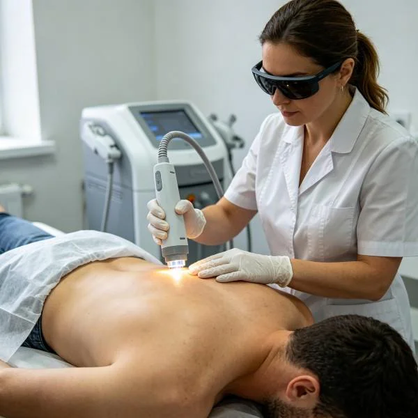
Deploying a multi-wavelength Class IV laser machine for pain treatment bypasses these physical limitations. Delivering dynamic power up to 30W creates a wide therapeutic window capable of resolving both acute neurogenic pain and chronic fibrotic changes.
Photobiomodulation actions operate across two clinical pathways:
- Immediate Neuro-Analgesic Conduction Block: High-density photon flux delivered over inflamed peripheral nerves inhibits A-delta and C pain-fiber transmission. Saturating local sensory receptors alters mitochondrial membrane potentials, slowing down axonal transport mechanisms and producing an immediate decrease in perceived pain intensity.
- Sustained Anti-Inflammatory and Regenerative Signaling: Photons hitting deep tissue stimulate vascular endothelial growth factor (VEGF) synthesis and accelerate fibroblast proliferation. Dense, disorganized Type III collagen fibers transition systematically into structured Type I collagen bundles. This clears calcific tendinopathies, resolves plantar fasciitis, and restores mechanical flexibility to thick muscle-tendon junctions without relying on long-term non-steroidal anti-inflammatory medications or invasive corticosteroid injections.
Standardized Clinical Practice and Detailed Treatment Record
The following treatment log outlines an integrated clinical protocol addressing complex anorectal pathology alongside chronic degenerative musculoskeletal inflammation.
Clinical Registry Record: FMX-2026-NE8841
| Parameter Group | Specification Detail | Clinical Target Value |
| Case Identification | Clinical Case ID | FMX-2026-NE8841 |
| Patient Demographics | Age / Biological Sex | 52 Years / Male |
| Primary Pathologies | Anorectal Diagnosis | Grade III Circumferential Prolapsing Internal Hemorrhoids |
| Secondary Pathologies | Musculoskeletal Diagnosis | Recalcitrant Left Knee Medial Joint Osteoarthritis (Kellgren-Lawrence Grade III) |
| Device Configuration | Operational Classification | Dual-Platform High-Intensity Class IV Medical Laser |
| Delivery Mechanics (Proctology) | Optical Delivery Fiber | 600-micron Radial Emission Fiber, 360-degree profile |
| Delivery Mechanics (Pain) | Transcutaneous Handpiece | Deep-Tissue Collimated Focus Handpiece with Dynamic Sensor |
| Wavelength Metrics (Proctology) | Dual-Spectrum Balance | 1470nm (70%) + 980nm (30%) |
| Wavelength Metrics (Pain) | Deep-Tissue Ratio | 810nm (40%) + 980nm (40%) + 1064nm (20%) |
| Operational Power (Proctology) | Wattage Setting | 8.5W to 10.0W Intermittent Pulse |
| Operational Power (Pain) | Wattage Setting | 15.0W Peak Duty-Cycle Pulsed Mode |
| Pulse Frequency (Proctology) | Repetition Rate | 3.0 Hz |
| Pulse Frequency (Pain) | Repetition Rate | 20.0 Hz modulated down to 5.0 Hz |
| Energy Dose Per Cushion (Hemorrhoid) | Total Delivered Joules | 240J to 280J per vascular node (Total: 820J) |
| Energy Dose Per Joint Session (Knee) | Total Delivered Joules | 3,600J per treatment session across four quadrants |
| Anesthetic Management (Proctology) | Anesthetic Type | Local Perianal Tumescent Infiltration (0.5% Lidocaine + Saline) |
| Topical Cooling (Pain Management) | Cutaneous Preservation | Integrated Contact Thermoelectric Chill Plate (4°C) |
Treatment Progression and Objective Clinical Metrics
| Treatment Stage | Anorectal Symptom Score (Goligher Criteria) | Hemorrhoid Node Volume Reduction (%) | Knee Pain Score (VAS 0-10 Scale) | Active Knee Flexion Range of Motion (Degrees) | Clinical Observations and Tissue Morphology |
| Baseline (Pre-Treatment) | Grade III Prolapse with active bleeding | 0% (Baseline Engorgement) | 8.5 / 10 | 85° | Submucosal cushions hyperemic, severe medial joint line tenderness |
| Day 3 (Post-Proctology) | Grade I (Non-prolapsing, minor swelling) | 45% Volume Reduction | 8.0 / 10 (Pre-Laser Pain Protocol) | 85° | Hemorrhoidal tissue retracted, no mucosal ulceration, minimal anal discomfort |
| Week 2 (Pain Session 4) | Grade I (Zero bleeding, complete stability) | 70% Volume Reduction | 4.0 / 10 | 105° | Proctological sites completely healed; medial knee joint crepitus significantly diminished |
| Week 4 (Pain Session 8) | Asymptomatic (Complete anatomical recovery) | 85% Shrinkage / Stable Fibrosis | 2.0 / 10 | 120° | Patient walking unaided for 5 km; vascular cushions retracted fully into anal canal |
| Week 8 (Final Evaluation) | Normal Anatomy, zero prolapse/bleeding | 90% Fibrotic Remodeling | 1.0 / 10 | 128° | Full functional mobility restored; zero analgesic dependence; sphincter tonus preserved |
Scientific Citations and Biophysical Frameworks
The clinical efficacy of dual-wavelength laser proctology rests on foundational work published by Dr. K. N. Poskus and colleagues in the journal International Journal of Colorectal Disease. Their randomized controlled trials evaluated submucosal diode laser coagulation against conventional excisional hemorrhoidectomy, demonstrating a statistically significant reduction in postoperative pain scores alongside preserved resting and squeeze anal sphincter pressures.
Thermal mitigation through controlled energy dispersion builds upon early biophysical models formulated by Dr. R. Rox Anderson and Dr. John A. Parrish in their landmark study on Selective Photothermolysis, published in Science. Their work demonstrated that confining thermal damage to target micro-structures depends on matching the laser pulse duration directly to the thermal relaxation time of targeted chromophores.
In musculoskeletal rehabilitation, high-irradiance photobiomodulation mechanisms align with research led by Dr. Tiina Karu, published in the Journal of Photochemistry and Photobiology B: Biology. Her research established that near-infrared laser irradiation stimulates terminal electron transfer within cytochrome c oxidase, boosting adenosine triphosphate (ATP) synthesis and triggering downstream anti-apoptotic gene expression.
Furthermore, clinical evaluations conducted by Dr. Ernesto Leal-Junior and associates in Lasers in Medical Science verified that high-intensity Class IV laser therapy accelerates musculoskeletal repair, attenuates muscular fatigue biomarkers, and downregulates pro-inflammatory cytokines significantly faster than low-intensity interventions.
Clinical Realities: High Intensity Laser vs Conventional Approaches
Moving from traditional excision and conservative therapy to advanced high-power laser therapy fundamentally changes patient recovery profiles.
Conventional surgical excision in sensitive anatomical zones relies on mechanical cutting, electrosurgical dissection, or suture ligation. In perianal procedures, scalpel excision creates open, irregular wounds in areas exposed to continuous bacterial loading. Electrocautery tips often generate temperatures exceeding 300 degrees Celsius, producing deep lateral thermal zones that denature adjacent smooth muscle bundles and damage sensitive nerve endings. Patients face weeks of painful dressing changes, narcotic analgesic reliance, wound drainage, and fear of defecation, with a recurring risk of anal stricture formation.
Deploying dual-wavelength therapeutic laser technology eliminates open, granulating surgical beds. The targeted 1470nm and 980nm light fields travel through micro-incisions via precision optical fibers, ablating the target vascular plexus or sinus tract from within. The process seals lymphatic vessels instantly, preventing local postoperative edema, accelerating tissue healing, and allowing patients to return home the same day without severe pain or narcotic prescriptions.
In chronic musculoskeletal pain management, standard practice often defaults to oral pharmaceuticals, intra-articular steroid injections, or arthroscopic debridement. These approaches carry well-documented drawbacks: non-steroidal anti-inflammatory drugs risk gastrointestinal and renal stress, repeated corticosteroid injections degrade joint cartilage and weaken tendon fibrils, and arthroscopic procedures introduce surgical infection risks and require prolonged recovery.
A high-intensity Class IV laser machine for pain treatment bypasses these hazards. Non-invasive, deep-reaching near-infrared energy penetrates directly through joint capsules and deep fascial layers. It relieves pain by calming irritated peripheral nerves and speeds up tissue healing by stimulating cellular mitochondria.
Clinical facilities adopt these advanced laser platforms to elevate their standard of care. By addressing the root causes of surgical recovery pain and chronic inflammation, they deliver predictable, long-term functional recovery across outpatient proctology and pain clinics.