 FotonMedix



Overcoming Chronic Patellar Tendinopathy in Competitive Sprinters
Synchronized multi-wavelength arrays bypass dense collagen scattering, microsecond duty cycle modulation eliminates cutaneous thermal buildup, and high peak photon density accelerates tenocyte metabolic repair within deep avascular tendon matrices.
Athletic training facilities face a brutal biological bottleneck every competition season. A collegiate sprinter pulls up lame during block starts, clutching the inferior pole of the patella where the tendon anchors into the tibial tuberosity. The diagnostic ultrasound confirms focal fiber discontinuity and localized hypoechogenicity, but the athlete cannot wait through an eight-month conservative recovery timeline. Traditional rest, nonsteroidal anti-inflammatory drugs, and eccentric decline board squats have stalled; the tendon core remains hypovascular, thick, and exquisitely tender to palpation. Standard physical therapy modalities hum away on the surface, yet their low-intensity energy scatters harmlessly across the skin and subcutaneous prepatellar bursa without altering the degenerate microenvironment inside the tendon core. Returning high-performance competitors to the track requires breaking through superficial soft tissue barriers and depositing therapeutic photon flux directly into the hypovascular tendon matrix.
Deep Optical Barriers and Photonic Attenuation in Dense Tendinous Tissue
The patellar tendon represents one of the most mechanically demanding and optically resistant structures in human anatomy. The patellar ligament comprises dense, parallel arrays of type I collagen fibers packed tightly together to withstand tensile forces exceeding ten times body weight during explosive deceleration and sprint propulsion. These tight, crystalline collagen arrays create extreme forward Mie scattering. Photons directed from a treatment handpiece do not simply pass through; they collide with dense protein fibrils, redirecting off-axis and spreading energy across adjacent soft tissues rather than delivering concentrated depth penetration.
When evaluating an indication of laser therapy for chronic overuse injuries, sports medicine clinicians must account for tissue-specific extinction coefficients. Photons must penetrate the epidermis, deep cutaneous vascular networks, prepatellar adipose layers, and the paratenon sheath before reaching the degenerated core of the tendon, which frequently lies 20 to 35 millimeters beneath the skin surface. Low-powered physical therapy systems operating at fractions of a Watt experience complete extinction within the first 4 to 6 millimeters of superficial tissue. High-power Class 4 systems solve this challenge by generating incident irradiance levels high enough to saturate superficial absorption bands and maintain a coherent forward-propagating photon flux into the deep collagen architecture.
Biological Extinction Layers in Patellar Tendon Irradiation:
[Cutaneous Layer]         --> Melanin absorption ceiling (managed by high beam velocity)
[Prepatellar Fat Pad]     --> Forward Mie scattering along lipid-water interfaces
[Paratenon Sheath]        --> Dense type I collagen scattering (requires high incident irradiance)
[Degenerate Core Matrix]  --> Target chromophore absorption (Cytochrome c oxidase & microvessels)
Strategic clinical execution relies on balancing key absorption bands within the near-infrared optical window. Water, hemoglobin, and melanin serve as the primary biological absorbing agents:
- 1470nm Selective Water Band: Extracellular fluid accumulation and microcystic mucin deposits characterize chronic tendinopathy. The 1470nm wavelength interacts directly with free and bound interstitial water molecules. Because its absorption coefficient in water is substantially higher than that of shorter infrared wavelengths, 1470nm energy mobilizes inflammatory fluid pockets, reduces localized swelling inside the rigid paratenon, and decreases hydrostatic pressure on compressed tenocytes.
- 980nm Hemoglobin Band: Degenerate tendon cores are notorious for poor vascularity, surrounded by aberrant, disorganized capillary neovascularization that drives nociception. The 980nm wavelength matches the optical absorption curve of deoxygenated and oxygenated hemoglobin. Radiant energy absorbed by blood vessels triggers localized photothermal vasodilation, clearing metabolic byproducts and promoting the influx of systemic growth factors.
- 810nm Cytochrome Window: Operating near the peak absorption peak of oxidized Cytochrome c oxidase, the 810nm wavelength maximizes photon transfer to mitochondrial respiratory complexes, accelerating intracellular energy synthesis.
High energy delivery into dense fibrous tissue creates risks of surface overheating. Because type I collagen and dermal melanin absorb radiant energy, continuous-wave emission at high wattages can quickly push surface temperatures beyond the 42 degrees Celsius safety threshold.
Temporal Gating across High-Peak Emission Cycles:
Emission Window (Pulse ON)  : |=========== High Peak Power Irradiance ===========|
                              [-------- Pulse Width Duration (t_on) --------]
Cooling Window (Pulse OFF)  : [ . . . . . Thermal Relaxation Phase (t_off) . . . . . ]
                              Net Result: High deep-tissue penetration without skin burns
Pulsed duty cycle control eliminates this operational obstacle. By pulsing emissions from 100Hz up to 20,000Hz, system controllers deliver high peak wattage while inserting precise off-duty cooling intervals. These off intervals match the thermal relaxation time of the skin, allowing vascular blood flow to carry away excess surface heat while photons penetrate deep into the dense fibrous tendon.
Tendinopathy Bioenergetics and Collateral Mechanical Remodeling
Deploying advanced laser therapy for athletes targets the cellular pathology of tendinopathy rather than merely deadening sensory nerve transmission. Chronic jumper’s knee does not represent an active inflammatory condition, as demonstrated by the histology models published by Leadbetter and the mechanical tendinopathy framework introduced by Cook and Purdam. Instead, it reflects a failed healing response characterized by tenocyte rounding, ground substance proliferation, microvascular ingrowth, and disruption of collagen fiber parallel alignment.
Under chronic tensile overload, tenocytes experience severe metabolic exhaustion. Mitochondrial membrane potential collapses, and intracellular adenosine triphosphate levels drop. Starved of energetic substrates, tenocytes lose their ability to synthesize structural proteins, instead producing weak, disorganized type III collagen that fails under explosive athletic loading.
Near-infrared photons directly target the binuclear copper and heme centers of mitochondrial Cytochrome c oxidase (Complex IV). Nitric oxide, which binds to these enzymatic sites under hypoxic stress and arrests cell respiration, dissociates via targeted photodissociation. This clears the pathway for oxygen binding and restores the proton pumping gradient across the inner mitochondrial membrane.
Intracellular Tenocyte Remodeling Cascade:
[Photonic Absorption]   --> Cytochrome c Oxidase Activation & NO Photolysis
         |
[Mitochondrial ATP]     --> Fuels Cellular Procollagen Type I Gene Transcription
         |
[Fibroblast Migration]  --> Reorganizes Extracellular Matrix from Type III to Type I Collagen
         |
[Neovascular Reset]     --> Downregulates Sensory C-Fiber Ingrowth and Substance P Release
This biological pathway drives several therapeutic responses:
- Upregulation of Type I Procollagen: Restoring mitochondrial ATP provides the energy required for tenocytes to transcribe collagen alpha chains, replacing weak type III collagen with high-tensile type I collagen fibrils.
- Transforming Growth Factor Beta Expression: High-intensity photon flux upregulates Transforming Growth Factor beta and basic Fibroblast Growth Factor, coordinating matrix metalloproteinase activity to break down disordered proteoglycans and realign collagen fibers along lines of functional stress.
- Eradication of Nociceptive Neovessels: The combination of targeted photothermal energy and restored tissue oxygenation shuts down the chaotic, non-functional capillary buds that accompany chronic tendinopathy, suppressing unmyelinated sensory C-fibers that transmit chronic pain.
- Reduction of Tenascin-C Accumulation: Downregulating mechanical stress proteins allows the tendon core to regain its normal fibrocartilaginous structure at the osteotendinous junction, restoring the tendon’s capacity to store and release elastic strain energy.
By rebuilding cellular metabolism and driving matrix reorganization, targeted high-intensity Class 4 laser therapy addresses the structural causes of chronic tendon failure.
Clinical Stratification and Laser Therapy Indications in Sports Medicine
Applying Class 4 systems in professional and collegiate athletics requires clear diagnostic stratification. Identifying a legitimate indication of laser therapy depends on distinguishing between mechanical structural tears, enthesopathies, and acute inflammatory bursitis:
- Acute Tendon Ruptures and High-Grade Tears: Full-thickness tears or partial tears exceeding 50 percent of the tendon cross-sectional area require surgical reconstruction or immobilization. High-intensity photonic therapy cannot structurally bridge completely severed mechanical gaps.
- Insertional Enthesopathy versus Mid-Substance Tendinopathy: Mid-substance tendinopathies respond rapidly to high-irradiance scanning because surrounding vascularity supports lymphatic drainage. Insertional enthesopathies—such as proximal patellar pole impingement or Achilles bursal spurs—require lower duty cycles, higher frequencies, and precise spot irradiation to avoid thermal buildup along periosteal margins.
- Peritendinous Edema and Bursal Distension: When chronic overuse triggers secondary deep infrapatellar bursitis, using 1470nm water-specific wavelengths accelerates fluid reabsorption, decompression of local tissue spaces, and restoration of joint mechanics.
- Myofascial Kinetic Chain Dysfunction: Treating isolated tendons without addressing kinetic contributors leads to high relapse rates. Protocols must address associated hypertonicity within the rectus femoris, vastus lateralis, and psoas major, relaxing the quadriceps extensor mechanism to lower resting tensile loads on the patellar tendon.
Calculating therapeutic dosage requires accounting for significant tissue scatter. Delivering 8 Joules per square centimeter to a degenerate tendon core positioned 25 millimeters below the skin surface requires a surface dose between 90 and 130 Joules per square centimeter to offset scattering and absorption across overlying tissues.
Clinical Case History: Division I Collegiate Track Facility
The following case record illustrates the diagnostic baseline, parameter selection, and clinical progression observed during high-power multi-wavelength laser therapy.
Patient Profile and Baseline Diagnostics
- Department Case ID: FTM-SPT-2026-3041
- Patient Age: 21 years
- Biological Sex: Male
- Sport / Event: Track and Field, 100m and 200m Sprint Competitor
- Presenting Condition: Severe chronic right proximal patellar tendinopathy (Jumper’s Knee) localized to the inferior patellar pole, exacerbated during acceleration and deceleration phases.
- Duration of Symptoms: 28 weeks of persistent functional limitation; prior interventions including platelet-rich plasma injection, dry needling, and six weeks of eccentric decline training failed to restore sprinting tolerance.
- Functional Baseline:
- Visual Analog Scale during Single-Leg Decline Squat: 8.5 / 10
- Visual Analog Scale during Normal Walking: 2.0 / 10
- Victorian Institute of Sport Assessment – Patella Score: 38 / 100 (severe impairment)
- Knee Range of Motion: Active knee flexion restricted to 118 degrees due to infrapatellar tightness; passive extension normal.
- Palpatory Examination: Extreme localized tenderness over the deep, posterior aspect of the inferior patellar pole; marked thickening of the proximal tendon border.
- Diagnostic Musculoskeletal Ultrasound:
- Marked proximal patellar tendon thickening measuring 8.4mm in anterior-posterior diameter (contralateral normal tendon measured 4.2mm).
- Prominent hypoechoic defect measuring 6.2mm x 4.1mm at the bone-tendon junction, consistent with localized mucoid degeneration and collagen disruption.
- Power Doppler revealed robust neovascularization within the hypoechoic core. No complete fiber tear.
Standardized Treatment Protocol and Kinetic Energy Matrix
The athlete completed 10 clinical sessions over a 3.5-week period (three sessions weekly during weeks 1 through 3, concluding with one session in week 4). Treatments were delivered using a multi-wavelength Class 4 laser platform utilizing synchronized 810nm, 980nm, and 1470nm emissions, administered with an optical quartz massage handpiece with real-time epidermal thermal tracking.
| Week | Session | Wavelength Ratio | Power (W) | Frequency (Hz) | Duty Cycle | Energy (J) | Area (cm2) |
| Wk 1 | S-01 | 810nm (60%) 980 (40%) | 10.0 W CW | Continuous | 100% | 4,200 J | 60 cm2 |
| Wk 1 | S-02 | 810/980/1470 (5:3:2) | 14.0 W P | 200 Hz | 60% | 5,880 J | 60 cm2 |
| Wk 1 | S-03 | 810/980/1470 (4:3:3) | 16.0 W P | 500 Hz | 50% | 6,720 J | 80 cm2 |
| Wk 2 | S-04 | 810/980/1470 (4:3:3) | 18.0 W P | 1,000 Hz | 50% | 7,560 J | 80 cm2 |
| Wk 2 | S-05 | 810/980/1470 (4:3:3) | 20.0 W P | 2,500 Hz | 40% | 7,680 J | 80 cm2 |
| Wk 2 | S-06 | 810/980/1470 (3:3:4) | 22.0 W P | 5,000 Hz | 40% | 8,448 J | 100 cm2 |
| Wk 3 | S-07 | 810/980/1470 (3:3:4) | 24.0 W P | 8,000 Hz | 35% | 8,064 J | 100 cm2 |
| Wk 3 | S-08 | 810/980/1470 (3:3:4) | 24.0 W P | 8,000 Hz | 35% | 8,064 J | 100 cm2 |
| Wk 3 | S-09 | 810/980/1470 (4:3:3) | 20.0 W P | 3,000 Hz | 50% | 8,400 J | 100 cm2 |
| Wk 4 | S-10 | 810/980 (50% : 50%) | 12.0 W CW | Continuous | 100% | 5,400 J | 60 cm2 |
Cumulative Clinical Progression
| Evaluation Stage | Decline Squat VAS | Walking VAS | VISA-P Score | Ultrasound AP Diameter | Neovascular Doppler Activity |
| Pre-Treatment | 8.5 / 10 | 2.0 / 10 | 38 / 100 | 8.4 mm | Grade 3+ (Intense flow) |
| Post-S-03 | 6.0 / 10 | 1.0 / 10 | 51 / 100 | 8.1 mm | Grade 3+ (Persistent) |
| Post-S-06 | 3.5 / 10 | 0.0 / 10 | 72 / 100 | 6.8 mm | Grade 1+ (Focal flow) |
| Post-S-08 | 1.5 / 10 | 0.0 / 10 | 86 / 100 | 5.4 mm | Absent / Normalized |
| Post-S-10 | 0.5 / 10 | 0.0 / 10 | 94 / 100 | 4.6 mm | Absent / Normalized |
| 60-Day Re-Check | 0.0 / 10 | 0.0 / 10 | 98 / 100 | 4.3 mm | Absent / Normalized |
Clinical Commentary on Treatment Milestones
During Sessions 1 through 3, treatment concentrated on reducing quadriceps hypertonicity and introducing the 1470nm wavelength to mobilize peritendinous fluid pockets around the deep infrapatellar bursa. Applying 10 Watts in continuous wave relaxed hypertonic rectus femoris fibers, reducing mechanical tension along the superior patellar border. By Session 3, single-leg decline squat pain had decreased from 8.5 to 6.0, allowing the athlete to perform basic rehabilitation drills without guarding.
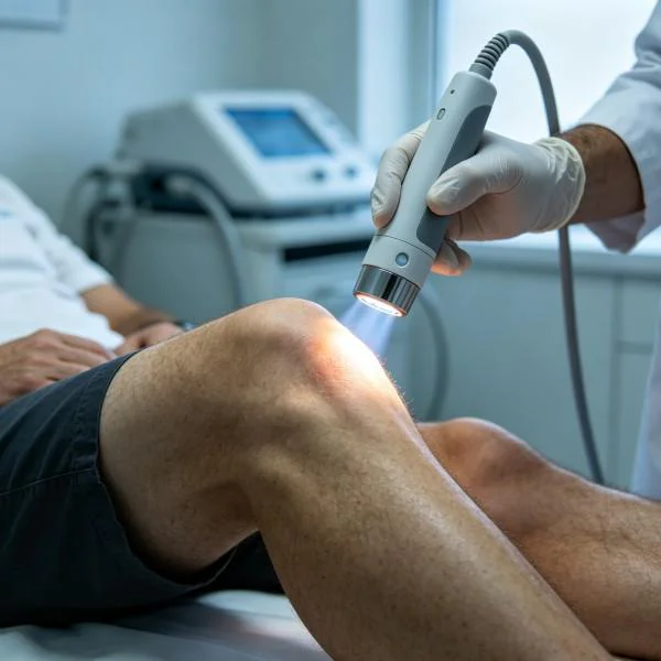
Sessions 4 through 8 transitioned to higher peak outputs, reaching 24.0 Watts. Operating across frequencies of 1,000Hz to 8,000Hz with 35 to 40 percent duty cycles delivered high photon flux to the osteotendinous junction without triggering epidermal thermal discomfort. By Session 6, follow-up ultrasound showed the anterior-posterior tendon thickness had reduced from 8.4mm to 6.8mm, while neovascular Doppler flow decreased from Grade 3+ to minor focal signals. The athlete began resisted sled pulls and light sprinting drills without post-exercise pain spikes.
Sessions 9 and 10 focused on consolidating collagen remodeling. By Session 10, the Victorian Institute of Sport Assessment score reached 94 out of 100, and pain during explosive single-leg decline squats dropped to 0.5 out of 10. Follow-up imaging confirmed the hypoechoic core had resolved into parallel collagen fibrils. The athlete returned to competitive sprint training, hitting sub-maximal competition velocities during week 5. At the 60-day follow-up, the tendon measured a normal 4.3mm on ultrasound, and the athlete registered personal-best split times without symptom recurrence.
Handpiece Scanning Velocity and Surface Thermal Control
Administering high-power Class 4 laser therapy over superficial tendons requires strict delivery discipline. Leaving the handpiece stationary over thin tissues like the patellar or Achilles tendon can elevate superficial skin temperatures past safe limits within seconds, causing pain and risk of burns.
Dynamic Movement Patterns Over the Extensor Mechanism:
[Zone 1: Suprapatellar Quadriceps]  <---> Transverse Sweeping Passes (5 cm/s, Continuous Wave)
[Zone 2: Superior Patellar Base]    <---> S-Curve Transverse Transitions across the Retinaculum
[Zone 3: Inferior Pole & Core]      <---> Overlapping Linear Strokes with Quartz Compression (3 cm/s)
[Zone 4: Tibial Tuberosity Foot]    <---> Cross-Hatch Standoff Pattern around the Enthesis
Key technical parameters govern clinical application:
- Kinetic Scanning Velocity: The delivery handpiece must maintain continuous motion across the treatment zone at speeds between 3 and 5 centimeters per second. This maintains forward photon penetration to target tissues while preventing local heat concentration on the skin.
- Mechanical Quartz Compression: Over the inferior patellar pole, the clinician uses a convex quartz contact head to apply 1.5 to 2.0 kilograms of manual pressure. This displaces superficial capillary blood, decreasing competitive light absorption by hemoglobin and increasing direct photon penetration into the avascular tendon core.
- Integrated Surface Pyrometry: Handpieces equipped with integrated thermal sensors monitor skin temperature in real time, maintaining cutaneous heat within the therapeutic window of 39.5 to 41.5 degrees Celsius. If the skin approaches 42.0 degrees Celsius, the operator increases scanning velocity or lowers the pulse duty cycle, preserving total energy delivery while protecting superficial tissues.
Advanced Photonic Modalities versus Conventional Sports Medicine Interventions
Managing chronic athletic overuse injuries often involves navigating trade-offs between speed of recovery, structural safety, and long-term tissue health. High-intensity Class 4 laser therapy provides a targeted non-invasive alternative across several common sports medicine approaches:
| Modality / Intervention | Penetration Depth | Underlying Mechanism | Clinical Limitations |
| Low-Level Light Therapy | 2 mm to 6 mm | Superficial Photochemistry | Underpowered for deep tendon cores |
| Platelet-Rich Plasma | Local Infiltration | Autologous Growth Factor Release | High cost, post-injection downtime |
| Corticosteroid Injections | Local Infiltration | Strong Anti-Inflammatory Action | High risk of tendon tear and atrophy |
| Extracorporeal Shockwave | Variable (Acoustic) | Mechanical Cavitation & Trauma | Significant procedural pain |
| Nonsteroidal Anti-Inflammatories | Systemic Distribution | COX-1 / COX-2 Enzyme Inhibition | Impairs long-term collagen remodeling |
| High-Power Class 4 Laser | 25 mm to 45 mm | Photobiomodulation & Perfusion | Requires clinical-grade equipment |
Low-Level Light Therapy operates in the milliwatt range. While useful for superficial conditions like digital tenosynovitis, it cannot produce the photon density needed to penetrate the dense fibrous structure of major weight-bearing tendons. High-power systems generate the irradiance required to drive therapeutic energy through thick connective tissue layers directly into the degenerate tendon core.
Autologous platelet-rich plasma injections supply concentrated growth factors to the injury site, but the procedure involves significant post-injection soreness, variable leukocyte concentrations, and several weeks of mandatory athletic rest. Corticosteroid injections offer rapid short-term pain relief, but repeated injections inhibit tenocyte protein synthesis, weaken collagen architecture, and raise the risk of catastrophic tendon rupture during high-impact sprint training.
Radial and focused shockwave therapies promote revascularization through mechanical cavitation, but treatment can be painful, sometimes causing local bruising that interrupts training schedules. Systemic anti-inflammatory medications suppress pain symptoms temporarily, but studies indicate they can inhibit collagen synthesis and blunt the body’s natural tendon repair pathways.
High-power multi-wavelength Class 4 laser therapy provides an effective alternative for sports performance teams. By combining deep tissue penetration, targeted wavelength absorption, and real-time thermal monitoring, this modality accelerates tenocyte cellular repair, normalizes neovascularization, and supports lasting structural remodeling without the systemic risks or prolonged downtime of invasive procedures.