 FotonMedix











Overcoming Dermal Burn Risks in Chronic Lumbar Radiculopathy
Deliver high forward photon flux directly into compressed spinal nerve roots. Target microvascular congestion and swollen perineural water matrices with dual-wavelength precision. Eliminate epidermal thermal saturation through sub-millisecond gated duty cycles.
A spine physical therapy clinic frequently encounters this difficult scenario: a 54-year-old interstate long-haul truck driver sits leaning hard to his right side on the consultation chair, clutching his left lower back, unable to rise without sharp, shooting electric pain radiating down his posterior thigh and into his calf. His MRI reports confirmed an L5-S1 disc protrusion with secondary foraminal stenosis and severe radicular impingement. He spent ten months cycling through high-dose oral gabapentin that left him chronically drowsy behind the wheel, three epidural steroid injections that only provided two weeks of transient relief, and dozens of visits to low-intensity therapy clinics where weak emitters did nothing to halt his nerve pain. He grew cynical after discovering that the steep class 4 laser therapy price at those facilities simply funded repeated, ineffective thirty-minute sessions of non-contact infrared wraps. Under the physical therapist’s thumbs, the left lumbar paraspinal gutter feels like a dense, fibrous knot, while the compressed nerve root 7 centimeters beneath remains hypoxic, inflamed, and unresponsive. When past operators dialed up continuous power on basic light therapy devices to force energy deep into the lumbar spine, the patient winced from sharp, burning sensations across his skin long before light reached the impinged nerve sheath.
Low-power modalities cannot overcome the steep optical scattering of thick lumbar erector spinae muscles and dense subcutaneous fat. When uncalibrated continuous systems run at higher wattages, epidermal heat builds up in seconds. The patient complains of unbearable skin burning, forcing the operator to stop the session well before the deep radicular target receives an adequate therapeutic dose.
Overcoming this anatomical impasse requires projecting high-irradiance optical energy through dense paraspinal musculature directly to the nerve root while shielding the surface skin from thermal saturation.
Optical Penetration Across Thick Lumbar Musculature and Fascia
Reaching a compressed spinal nerve root presents an intense biophysical challenge. Photons must penetrate the stratum corneum, traverse thick lumbar adipose tissue, navigate dense posterior thoracolumbar aponeuroses, and pass through deep multifidus and longissimus muscles before reaching the foraminal zone. Under the Kubelka-Munk optical scattering model, light scatters across dense fibrous interfaces, creating an exponential drop-off in forward energy.
Inside human biological tissue, three primary endogenous absorbers control photon distribution: melanin, microvascular hemoglobin, and interstitial water.
In chronic L5-S1 radiculopathy, the primary pathology involves mechanical compression and severe microvascular congestion of the spinal nerve root, located 6.0 to 8.5 centimeters below the posterior lumbar skin. Class III low-level therapy systems fail to penetrate this depth; more than 97% of their light scatters into shallow dermal and muscular layers. The nerve root remains starved of photons, locked in an ongoing cycle of endoneurial hypoxia and neuropathic firing.
Overcoming this steep decay curve requires a high-intensity class iv therapeutic laser capable of delivering high peak power without exceeding surface thermal thresholds.
Stratum Corneum & Lumbar Adipose (Forward Scattering Trap)
         │
         ▼
Thoracolumbar Aponeurosis & Multifidus (Dense Muscular Barrier)
         │ (1470nm Interstitial Water Absorption & Decongestion)
         ▼
Radicular Perineural Capillary Beds (Ischemic Capillary Congestion)
         │ (980nm Hemoglobin Hyperemia & NO Offloading)
         ▼
Compressed L5-S1 Nerve Root & DRG (Mitochondrial ATP Restoration)
Targeting hemoglobin and water chromophores simultaneously transforms how compressed neural tissue responds:
At 980nm, photon capture aligns with absorption peaks for oxygenated and deoxygenated hemoglobin. In chronic radiculopathy, mechanical compression of the nerve root chokes venous return, creating localized ischemia and trapping inflammatory cytokines like interleukin-6 and tumor necrosis factor-alpha. The 980nm wavelength triggers immediate localized vasodilation, prompting circulating red blood cells to offload bound nitric oxide, clearing sluggish microcirculation and restoring oxygen delivery to starving axons.
At 1470nm, photon absorption shifts toward bound water molecules within swollen perineural sheaths and thick fascial planes. Interstitial water absorbs 1470nm light at rates significantly higher than in the 800nm zone. This controlled absorption creates micro-thermal fields that break down rigid, polymerized proteoglycan matrices, clearing stagnant inflammatory edema around the compressed intervertebral foramen and reducing mechanical pressure on the nerve.
Delivering this combined optical power deep into the lumbar spine without scorching the skin requires precise pulse gating.
Duty Cycle Mechanics and Cutaneous Thermal Clearance
Continuous-wave light delivery encounters a hard biological limit over the lumbar spine: the adipose-rich subcutaneous layer absorbs surface heat quickly, but cannot dissipate it through microvascular clearance as fast as thinner skin. When continuous photons strike the lower back, surface temperatures spike within seconds, setting off intense burning sensations.
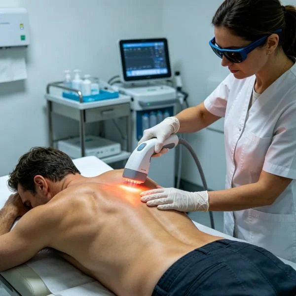
Precision microsecond pulse gating eliminates this surface thermal build-up. By slicing high-intensity light into microsecond bursts separated by controlled dark intervals, deep articular structures receive high peak irradiance while the sensitive skin cools down safely between pulses.
$$\text{Duty Cycle (\%)} = \left( \frac{\tau_{\text{pulse}}}{\tau_{\text{pulse}} + \tau_{\text{interval}}} \right) \times 100$$
Where $\tau_{\text{pulse}}$ represents active optical delivery and $\tau_{\text{interval}}$ represents the thermal relaxation pause.
Applying 25W of continuous laser energy over the lumbar paraspinal zone triggers uncomfortable skin heating within 4 seconds. In contrast, running that same power through a 25% duty cycle delivers powerful peak bursts that penetrate through dense lumbar muscles, while the 75% dark phase allows surface tissues to shed heat safely into the air and the moving handpiece.
Capillary blood flow carries away residual surface heat, while the hypoperfused, compressed nerve root absorbs high photon density, initiating cellular healing without epidermal irritation.
Multi-Wavelength Synergies in Spinal Neurovascular Rehab
Treating complex spinal nerve compression with a single wavelength leaves critical parts of the pathology unaddressed. Lumbar radiculopathy involves skin, adipose layers, thick fascial sheaths, deep paraspinal muscle bellies, and inflamed neural tissue. Each distinct anatomical layer features unique water content, vascular density, and optical scattering properties.
Combining coordinated wavelengths establishes an unbroken therapeutic gradient across all tissue depths:
[ Deep Spinal Tissue Interaction Matrix ]

810nm  ──► Mitochondrial Cytochrome c Oxidase (Surge in Axonal ATP Synthesis)
980nm  ──► Radicular Capillary Perfusion & NO Release (Flushing Inflammatory Fluid)
1064nm ──► Deep Lumbar Paraspinal Mass (Forward Directional Penetration)
1470nm ──► Perineural Water Matrices (Decongesting Foraminal Edema)
Pairing 980nm with 1470nm produces immediate biomechanical and circulatory improvements. The 980nm beam opens compressed microvascular beds, delivering fresh oxygen to ischemic nerve roots. At the same time, 1470nm targets bound water molecules inside the swollen nerve sheath, softening rigid collagen cross-links and draining localized fluid.
Clinicians use this dual-action delivery to loosen tight, guarded spinal structures, making subsequent manual therapy and lumbar decompression exercises far more productive.
Clinical Efficiency with Class IV Deep Tissue Laser Therapy
High-power platforms have transformed how specialized spine centers and physical therapy practices manage severe radiculopathy and discogenic pain.
Therapists managing chronic lumbar radiculopathy, piriformis syndrome, or post-laminectomy pain syndrome often spend extensive time on slow, painful manual interventions. Low-intensity modalities require 35 to 45 minutes of scanning to deliver minimal joules through deep lumbar musculature. Delivering class iv deep tissue laser therapy cuts treatment times to under ten minutes while driving meaningful, therapeutic energy directly to the injury site.
Rehabilitation practices evaluating high-output systems recognize clear clinical and operational advantages:
First, practitioners achieve meaningful therapeutic energy doses in 7 to 9 minutes, optimizing daily patient throughput and eliminating physical strain from aggressive manual friction.
Second, rapid pain relief breaks the patient’s antalgic guarding, allowing active spinal stabilization exercises earlier in their treatment plan.
Third, reliable deep-tissue cellular repair restores microvascular blood flow, clearing radicular shooting pain and supporting long-term disc preservation.
Clinical Case Documentation
The following case record details the clinical application of multi-wavelength high-intensity laser therapy for severe, treatment-resistant L5-S1 lumbar radiculopathy that had failed to respond to epidural steroid injections, oral anticonvulsants, and conventional modalities.
Case Record Details
Case Identification: SPINE-2026-R8204
Patient Demographics: 54-year-old male, long-haul freight operator
Primary Diagnosis: Subacute-on-Chronic L5-S1 Disc Protrusion with Left S1 Radiculopathy (Wiltse Classification: Left Paracentral Disc Herniation with Severe Lateral Recess and Foraminal Stenosis)
Chronicity: 10 months of worsening radicular pain; failed three fluoroscopically guided epidural steroid injections, failed high-dose gabapentin (2400mg/day) due to severe sedation
Clinical Assessment: Sharp, shooting electric shock pain traveling down the left posterior thigh, posterolateral calf, and lateral foot border. Straight Leg Raise (SLR) test positive on the left side at 28 degrees with severe radiating pain (Numeric Pain Rating Scale: 8/10 sitting or standing, 6/10 constant dull ache at rest). Absent left Achilles tendon reflex (0/4), along with weakness in left ankle plantarflexion (4-/5 manual muscle test).
| Session ID | Phase Protocol | Wavelength Configuration | Average Power (W) | Pulse Frequency & Duty Cycle | Scan Area (cm2) | Treatment Duration | Total Energy (Joules) | Clinical Response Metric |
| Session 01 | Radicular Desensitization | 980nm (75%) + 1470nm (25%) | 15.0W | 100 Hz @ 20% Duty Cycle | 200 | 06:40 | 6,000 J | Resting NPRS drops from 6/10 to 4/10; leg burning softens |
| Session 03 | Foraminal Decongestion | 980nm (45%) + 1470nm (55%) | 18.0W | 250 Hz @ 30% Duty Cycle | 160 | 08:20 | 9,000 J | SLR increases from 28° to 44°; paraspinal guarding eases |
| Session 06 | Axonal Biostimulation | 810nm (30%) + 980nm (45%) + 1470nm (25%) | 22.0W | 800 Hz @ 40% Duty Cycle | 220 | 08:20 | 11,000 J | Radiating pain retreats above the knee; NPRS down to 3/10 |
| Session 09 | Deep Nerve Root Remodeling | 980nm (40%) + 1470nm (60%) | 20.0W | 20 Hz @ 25% Duty Cycle (Deep Impact Mode) | 140 | 07:25 | 8,900 J | Plantarflexion strength restored to 5/5; SLR hits 72° |
| Session 12 | Kinetic Consolidation | 980nm (50%) + 1470nm (50%) | 16.0W | Continuous Wave (Dynamic Scan) | 240 | 08:00 | 7,680 J | Zero resting or sitting pain; return to driving duties |
Case Progression Notes
Sessions 01 through 03 focused on relieving perineural inflammation, reducing lateral recess venous congestion, and dampening unmyelinated C-fiber excitability. The combination of 980nm and 1470nm cleared inflammatory edema around the S1 nerve root, reducing nocturnal throbbing and allowing the patient to sit comfortably for twenty minutes by the end of the first week.
Sessions 04 through 08 targeted the deep multifidus muscle contracture and ischemic foraminal sheath. High water absorption at 1470nm softened the rigid fascial bands, while 980nm and 810nm stimulated capillary blood flow to flush out metabolic waste and recruit reparative cells. Straight Leg Raise testing improved from 28° to 60° without shooting electric sensations.
By Session 12, the patient showed complete centralization of radicular symptoms, with pain confined to a faint, occasional ache in the left gluteal region. Motor strength in left ankle plantarflexion fully normalized, the Achilles tendon reflex returned to a normal 2/4 baseline, and the patient completed an eight-hour continuous driving assessment without leg numbness or antalgic posturing.
Biological Mechanisms of Spinal Nerve Decompression
High-intensity deep tissue laser therapy stimulates healing through integrated cellular and extracellular mechanisms:
Photonic Absorption (Mitochondria & Perineural Water Matrix)
         │
         ├──► Dissociation of Inhibitory Nitric Oxide ──► Accelerated Axonal ATP Synthesis
         │
         ├──► Interstitial Hydration Cleaving ────────► Clearance of Foraminal Fluid Stasis
         │
         └──► Endothelial Cell Activation ─────────────► Vasa Nervorum Reperfusion
When photons hit cytochrome c oxidase inside stressed axons and Schwann cells, they displace inhibitory nitric oxide molecules. This allows oxygen to bind efficiently, speeding up the mitochondrial electron transport chain and driving a surge in adenosine triphosphate (ATP) production. Chronically fatigued cells regain the metabolic energy needed to maintain axonal membrane potentials.
Simultaneously, 1470nm photons excite water molecules bound within the dense proteoglycan gel surrounding the compressed nerve root. The resulting micro-thermal fields break down rigid collagen cross-links, converting viscous ground substance into a fluid state and opening pathways for venous drainage.
Downstream gene signaling shifts rapidly. Inflammatory cytokines like interleukin-1-beta, tumor necrosis factor-alpha, and cyclooxygenase-2 decrease, while nerve growth factor and vascular endothelial growth factor rise.
These biochemical changes prompt Schwann cells to remyelinate damaged nerve segments and restore normal nerve conduction velocities, eliminating neuropathic firing.
Moving Beyond Injections and Spinal Fusion
Conventional management for severe lumbar radiculopathy relies heavily on oral opioids, repeated epidural cortisone injections, and invasive spinal surgery like microdiscectomy or fusion.
Systemic medications dull central pain perception while doing nothing to resolve the mechanical ischemia or axonal degeneration of the nerve root. Repeated cortisone injections provide short-term symptom relief, but they suppress local collagen synthesis, elevate blood glucose, and can weaken adjacent vertebral bone over time. Spinal surgery decompresses the nerve mechanically, but introduces surgical risks like dural tears, epidural fibrosis, adjacent segment disease, and long postoperative recovery times.
High-power Class IV laser therapy provides a focused, non-invasive alternative. It avoids systemic drug toxicity, preserves spinal stability, and eliminates surgical trauma. Clinicians can deliver thousands of therapeutic joules deep into compressed intervertebral foramina and ischemic paraspinal muscles within minutes.
Applying high-intensity multi-wavelength phototherapy targets radicular nerve pain at its biological and vascular source. Foraminal edema drains, vasa nervorum circulation rebounds, and damaged nerve fibers heal cleanly. It turns disabling spinal nerve compression into a predictable, lasting return to active, pain-free living.