 FotonMedix



Overcoming Flexor Tendon Sheath Adhesions in Performance Thoroughbreds
Synchronized multi-wavelength photonic arrays penetrate dense fibrous tenosynovial margins, selective 1470nm and 980nm chromophore absorption disperses organized synovial fluid protein conglomerates, and microsecond duty cycle modulation protects sensitive palmar neurovascular bundles from heat accumulation.
Racehorse rehabilitation barns encounter a debilitating orthopedic barrier during conditioning resumes. A three-year-old Thoroughbred colt finishes a morning gallop favoring the left forelimb, showing localized filling and effusion tracking along the digital flexor tendon sheath proximal to the fetlock joint. Palpation elicits pain during passive fetlock extension, while diagnostic ultrasonography reveals severe chronic tenosynovitis accompanied by dense, non-functional fibrous adhesions binding the superficial digital flexor tendon to the inner tenosynovial lining. Standard management pathways carry severe performance compromises. Intermittent cold water hydrotherapy, intrasynovial corticosteroid injections, and stall confinement yield only transient fluid reduction; once speed training resumes, repetitive friction inside the scarred sheath reignites tenosynovial effusion and chronic lameness. When trainers inquire about equine cold laser therapy, they encounter underpowered low-level devices unable to penetrate the thick fibrous wall of the inflamed digital sheath. When assessing whether invasive tenoscopic adhesiolysis justifies the operational downtime and equine laser surgery risk profile, syndicates must calculate both procedural impact and equine laser therapy cost over an extended season. Restoring friction-free gliding mechanics to the equine digital flexor tendon sheath requires delivering high peak irradiance directly into the scarred tenosynovial interface to dissolve dense adhesions and reactivate cellular matrix remodeling.
Anatomical Scattering and Near-Infrared Attenuation in the Distal Equine Limb
The digital flexor tendon sheath represents an intricate, highly resistant optical boundary in equine sports medicine. Enclosing the superficial and deep digital flexor tendons as they pass the fetlock joint and pastern, the sheath is shielded by dense equine skin, thin subcutaneous fascia, the palmar annular ligament, and thick fibrous tenosynovium. In competitive racehorses, therapeutic photons directed from an external handpiece must penetrate through 25 to 45 millimeters of dense fibrous and synovial tissues to reach deep annular adhesions and the interior gliding surfaces of the digital flexor tendons.
As light traverses these dense anatomical strata, Rayleigh and Mie scattering rapidly disperse forward-propagating photons. Equine tenosynovial tissues exhibit high optical attenuation due to organized collagenous lamellae in the palmar annular ligament and dense proteinaceous exudates within the synovial cavity. In low-power modalities operating in the milliwatt range, forward photon coherence dissolves within the first few millimeters of superficial dermis, leaving the scarred sheath completely untreated. High-intensity Class 4 systems overcome this limitation by generating sufficient incident irradiance to saturate superficial scattering centers, establishing a coherent forward-propagating photon flux that penetrates the deep tenosynovial compartment.
Equine Distal Metacarpophalangeal Stratification and Extinction Curve:
[Equine Cutaneous Coat]       ===> Incident High-Density Flux (100% Surface Irradiance)
              |
[Dense Palmar Annular Band]   ---> Severe Mie Scattering across Transverse Collagen (38% Loss)
              |
[Fibrotic Tenosynovial Wall]  ---> Synovial Protein Scatter & Capillary Attenuation (42% Loss)
              |
[Intrasynovial Gliding Space] ---> Residual Forward Photonic Field (Target Fluence: 7-11 J/cm2)
Clinical efficacy relies on matching laser emissions to primary tissue chromophore absorption characteristics:
- 1470nm Hydro-Targeted Synovial Clearance: Chronic tenosynovitis causes excessive production of high-viscosity inflammatory effusion, high-molecular-weight hyaluronic acid fragments, and stagnant exudate within the unyielding synovial cavity. The 1470nm wavelength interacts directly with free water molecules. Its elevated absorption profile heats and mobilizes stagnant synovial effusion, accelerating lymphatic clearance through the fibrous sheath wall, reducing intracavitary pressure, and easing friction against the palmar annular ligament.
- 980nm Hemoglobin Absorption and Perfusion: The gliding surfaces of flexor tendons possess limited direct vascularity, relying on synovial fluid diffusion and tiny vincula tendinum microvessels. The 980nm wavelength matches the optical absorption curve of oxygenated and deoxygenated hemoglobin. Absorption of 980nm photons triggers localized photothermal vasodilation, stimulating endothelial nitric oxide synthesis and reopening closed microcapillary loops within the synovial villi to clear inflammatory metabolites.
- 810nm Mitochondrial Cytochrome Activation: Matching the peak absorption band of Cytochrome c oxidase, this wavelength stimulates the respiratory electron transport chain, driving the cellular adenosine triphosphate synthesis required for synoviocyte repair and extracellular matrix remodeling.
Delivering high-energy Class 4 laser power to the distal equine limb introduces critical thermal management concerns. The fetlock and pastern regions possess virtually no subcutaneous adipose buffering, placing nerves, blood vessels, and dense ligaments directly beneath the skin. Continuous-wave energy absorbed by epidermal melanin and dermal water can accumulate rapidly, and if skin temperatures exceed 42 degrees Celsius, thermal nociceptors trigger limb retraction and risk cutaneous blistering.
Pulsed Temporal Emission Dynamics across Equine Distal Sheaths:
Emission Phase (Pulse ON)  : |=========== High Peak Power Irradiance ===========|
                             [-------- Controlled Pulse Duration (t_on) --------]
Thermal Clearance (Pulse OFF): [ . . . . Cutaneous Thermal Relaxation (t_off) . . . . ]
                             Net Outcome: Deep tenosynovial saturation without skin burns
Microsecond duty cycle modulation provides the necessary clinical protection. Operating across frequencies from 100Hz to 15,000Hz, system processors deliver high peak wattage to penetrate dense annular ligaments while inserting brief off-duty rest intervals. These intervals match the thermal relaxation time of the skin, allowing cutaneous capillary blood flow to dissipate heat while forward-moving photons penetrate deep into the scarred tenosynovial compartment.
Synoviocyte Bioenergetics and Adhesion Dissolution Pathways
Targeting flexor tendon sheath adhesions addresses the underlying failed cellular healing response rather than merely suppressing local pain. Histological models documented in equine orthopedic studies by Smith and Wright demonstrate that chronic tenosynovitis is marked by synovial lining hyperplasia, sub-synovial fibrosis, tenocyte rounding, and organized fibrin bands that cross-link the tendon epitenon to the sheath wall.
Under repetitive frictional trauma, tenocytes and synoviocytes suffer metabolic exhaustion. Mitochondrial membrane potentials drop, leading to decreased intracellular adenosine triphosphate synthesis. Lacking energy, synoviocytes produce abnormal, low-molecular-weight proteoglycans and inflammatory cytokines that stimulate fibroblast proliferation, resulting in dense, unorganized type III collagen adhesions that restrict longitudinal tendon gliding during galloping.
Near-infrared photons directly target the primary mitochondrial photoacceptor, Cytochrome c oxidase (Complex IV). Under hypoxic and ischemic stress caused by microvascular compression, nitric oxide binds to the enzyme’s catalytic center, halting mitochondrial respiration. Irradiating the injured tissue dislodges nitric oxide through targeted photolysis, allowing oxygen to bind and restoring the proton pumping gradient across the inner mitochondrial membrane.
Biological Progression of Tenosynovial Photobiomodulation:
[Photonic Absorption]   --> Complex IV Photolysis of Inhibitory Nitric Oxide
         |
[Mitochondrial ATP]     --> Fuels Type B Synoviocyte Matrix Synthesis
         |
[MMP Regulation]        --> Balances MMP-1/MMP-13 to Dissolve Dense Fibrinous Adhesions
         |
[Hyaluronan Synthesis]  --> Restores High-Molecular-Weight Synovial Fluid Gliding Layer
This biological pathway drives several therapeutic responses:
- Matrix Metalloproteinase Activation: Photonic stimulation balances the expression of matrix metalloproteinases (MMP-1 and MMP-13) and tissue inhibitors of metalloproteinases, dissolving disorganized fibrinous adhesions between the tendon epitenon and synovial wall without damaging healthy underlying tendon fibers.
- Restoration of High-Molecular-Weight Hyaluronan: Elevated intracellular ATP fuels Type B synoviocytes to synthesize high-viscosity, native-weight hyaluronic acid, re-establishing low-friction boundary lubrication between the flexor tendons and the annular ligament.
- Depletion of Local Pain Mediators: High-intensity photon flux downregulates substance P and calcitonin gene-related peptide expression within palmar digital sensory nerve terminals, reducing mechanical pain during extreme fetlock extension.
- Resolution of Synovial Effusion: Selective photothermal absorption by water accelerates lymphatic drainage through the synovial membrane, decompressing the surrounding digital sheath and easing pressure on the palmar annular ligament.
By stimulating synoviocyte cellular metabolism and promoting adhesion remodeling, targeted high-intensity Class 4 laser therapy addresses the structural causes of chronic tenosynovial restriction.
Clinical Stratification and Differential Diagnosis in Equine Distal Limb Pathology
Accurate clinical and sonographic assessment ensures energy is applied to correct tissue targets. Establishing appropriate treatment protocols requires differentiating between tenosynovial adhesions, structural tendon core lesions, and osseous impingements:
- Complete Annular Ligament Desmotomy Candidates: Severe, circumferential constriction of the palmar annular ligament with deep digital flexor tendon notching and loss of vascular flow requires surgical transection. Photobiomodulation cannot mechanically sever thick, fibrotic constriction rings that strangle underlying tendons.
- Chronic Non-Constrictive Tenosynovitis with Fibrous Bands: The primary indication for deep multi-wavelength intervention. Horses display persistent Grade 2 to 3 lameness, synovial distension proximal to the fetlock, pain on passive distal flexion, and sonographic confirmation of hyperechoic adhesion bands tethering the tendon without structural transection.
- Secondary Digital Flexor Tendon Longitudinal Tears: Marginal tears of the deep digital flexor tendon frequently shed fibrinous debris into the sheath, perpetuating chronic tenosynovitis. High-intensity multi-wavelength laser therapy mobilizes inflammatory fluid, softens surrounding adhesions, and stimulates cellular repair along the disrupted tendon margins.
- Proximal Sesamoidian Enthesopathy: When chronic tenosynovial inflammation extends to the proximal sesamoidean bones or distal sesamoidean ligaments, protocols must combine higher total energy with precise handpiece scanning to treat both soft tissue and bone-ligament interfaces.
Calculating the correct therapeutic dose requires accounting for tissue scatter and absorption. Because overlying annular ligaments and fibrous sheath walls attenuate up to 94 percent of surface photons, delivering an effective target dose of 7 to 11 Joules per square centimeter to a gliding interface 30 millimeters deep requires surface-applied doses ranging from 110 to 160 Joules per square centimeter across the palmar fetlock region.
Comprehensive Clinical Case History: Thoroughbred Performance Center
The following case record details the clinical baseline, parameter matrix, and objective recovery trajectory observed during high-power multi-wavelength laser intervention.
Patient Profile and Baseline Diagnostics
- Department Case ID: FTM-EQ-2026-8812
- Patient Subject: 3-year-old Thoroughbred Colt
- Discipline: Flat Racing (Under training for stakes-grade 1,600m events)
- Presenting Condition: Severe chronic tenosynovitis of the left forelimb digital flexor tendon sheath, accompanied by thick synovial effusion, reduced range of motion, and performance decline.
- Duration of Symptoms: 16 weeks of recurrent lameness; unresponsive to stall rest, two intra-articular triamcinolone acetonide injections, and four weeks of systemic phenylbutazone administration.
- Functional Baseline:
- AAEP Lameness Scale: Grade 3 / 5 (consistently visible at a trot on a straight line, severe on a left-hand circle).
- Distal Limb Flexion Test: Markedly positive (exacerbation of lameness to Grade 4/5 for 20 strides).
- Digital Sheath Palpation: Tense, fibrous swelling proximal to the palmar annular ligament with painful resistance during extreme fetlock passive dorsiflexion.
- Diagnostic Anesthesia:
- Palmar digital nerve block provided no significant improvement.
- Low four-point nerve block eliminated 90% of lameness, isolating pathology to the fetlock and distal sheath compartment.
- Diagnostic High-Frequency Musculoskeletal Ultrasound:
- Markedly distended digital flexor tendon sheath with synovial wall thickening measuring 3.4mm (contralateral normal sheath measured 1.1mm).
- Prominent hyperechoic adhesion band measuring 14mm in length bridging the palmar border of the superficial digital flexor tendon to the parietal layer of the sheath.
- Heterogeneous, proteinaceous synovial effusion with no complete structural tendon tears. Palmar annular ligament thickened to 2.8mm (normal: less than 1.5mm).
Standardized Treatment Protocol and Kinetic Energy Matrix
The horse underwent a course of 12 clinical sessions across 4 consecutive weeks (three sessions per week for four weeks). Treatment was delivered using a multi-wavelength Class 4 clinical platform featuring synchronized 810nm, 980nm, and 1470nm emissions, administered with an optical quartz contact roller handpiece with integrated surface pyrometry.
| Week | Session | Wavelength Ratio | Power (W) | Frequency (Hz) | Duty Cycle | Energy (J) | Area (cm2) |
| Wk 1 | S-01 | 810nm (60%) 980 (40%) | 12.0 W CW | Continuous | 100% | 5,400 J | 90 cm2 |
| Wk 1 | S-02 | 810/980/1470 (5:3:2) | 15.0 W P | 250 Hz | 60% | 7,200 J | 90 cm2 |
| Wk 1 | S-03 | 810/980/1470 (4:3:3) | 18.0 W P | 500 Hz | 50% | 8,640 J | 100 cm2 |
| Wk 2 | S-04 | 810/980/1470 (4:3:3) | 20.0 W P | 1,000 Hz | 50% | 9,600 J | 100 cm2 |
| Wk 2 | S-05 | 810/980/1470 (4:3:3) | 22.0 W P | 2,500 Hz | 45% | 9,900 J | 110 cm2 |
| Wk 2 | S-06 | 810/980/1470 (3:3:4) | 24.0 W P | 4,000 Hz | 40% | 9,600 J | 110 cm2 |
| Wk 3 | S-07 | 810/980/1470 (3:3:4) | 26.0 W P | 6,000 Hz | 40% | 10,400 J | 120 cm2 |
| Wk 3 | S-08 | 810/980/1470 (3:3:4) | 26.0 W P | 6,000 Hz | 40% | 10,400 J | 120 cm2 |
| Wk 3 | S-09 | 810/980/1470 (4:3:3) | 22.0 W P | 3,000 Hz | 50% | 10,560 J | 120 cm2 |
| Wk 4 | S-10 | 810/980/1470 (4:3:3) | 20.0 W P | 1,500 Hz | 50% | 9,600 J | 100 cm2 |
| Wk 4 | S-11 | 810/980/1470 (4:3:3) | 16.0 W P | 500 Hz | 50% | 7,680 J | 90 cm2 |
| Wk 4 | S-12 | 810/980 (50% : 50%) | 12.0 W CW | Continuous | 100% | 5,400 J | 90 cm2 |
Cumulative Clinical Progression
| Checkpoint | AAEP Lameness Grade | Flexion Response | Synovial Effusion | Sheath Wall Thickness | Adhesion Band Status |
| Pre-Treatment | Grade 3 / 5 | Grade 4 / 5 | Severe (Tense) | 3.4 mm | Dense (14 mm linear) |
| Post-S-03 | Grade 2 / 5 | Grade 3 / 5 | Moderate (Pliable) | 2.9 mm | Unchanged (14 mm) |
| Post-S-06 | Grade 1 / 5 | Grade 2 / 5 | Mild Distension | 2.2 mm | Thinning (8 mm partial) |
| Post-S-08 | Grade 0.5 / 5 | Grade 1 / 5 | Trace Effusion | 1.8 mm | Fragmented (Focal) |
| Post-S-10 | Grade 0 / 5 | Grade 0.5 / 5 | Resolved | 1.4 mm | Absent / Dissolved |
| Post-S-12 | Grade 0 / 5 | Grade 0 / 5 | Resolved | 1.2 mm | Free Gliding Restored |
| 90-Day Follow | Grade 0 / 5 | Grade 0 / 5 | Resolved | 1.1 mm | Normal Gliding Mechanics |
Clinical Commentary on Treatment Milestones
During Sessions 1 through 3, treatment concentrated on relaxing the distal flexor apparatus and introducing the 1470nm wavelength to mobilize dense synovial effusion around the palmar annular ligament. Continuous-wave delivery at 12.0 Watts across the distal metacarpus promoted vascular inflow and softened peritendinous tension. By Session 3, lameness improved from Grade 3 to Grade 2, and digital sheath fluid tension shifted from rigid to pliable.
Sessions 4 through 8 transitioned to higher peak outputs, running from 20.0 to 26.0 Watts. Operating at frequencies between 1,000Hz and 6,000Hz with 40 to 45 percent duty cycles delivered high photon flux to the interior of the sheath while keeping skin temperatures below 40.8 degrees Celsius. By Session 6, follow-up ultrasound showed the synovial wall thickness had decreased from 3.4mm to 2.2mm, with the fibrous adhesion band thinning and fragmenting. The horse began controlled walking and light trotting on a straight line without lameness exacerbation.
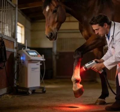
Sessions 9 through 12 focused on consolidating synoviocyte repair and restoring low-friction gliding mechanics. Operating at higher peak pulses and transitioning to continuous-wave emissions stimulated long-term tenocyte protein synthesis and synovial membrane normalization. By Session 12, baseline lameness resolved completely, flexion tests were negative, and ultrasound confirmed total dissolution of the adhesion band with free longitudinal tendon excursion. The colt returned to regular track conditioning, completing timed breezes within 10 weeks. At the 90-day follow-up, the sheath wall measured a normal 1.1mm on ultrasound, with zero recurrence of effusion or lameness under race-pace training.
Handpiece Scanning Velocity and Surface Thermal Control
Administering high-power Class 4 laser therapy over the distal equine limb requires strict operational discipline. Because the skin overlying the palmar annular ligament and sesamoid bones is thin and contains minimal adipose padding, leaving the handpiece static will rapidly elevate skin temperatures beyond safe thresholds.
Handpiece Trajectory along the Equine Digital Sheath Complex:
[Zone 1: Supracarpal Inflow Corridor] <---> Longitudinal Sweeping Strokes (5 cm/s, CW Delivery)
[Zone 2: Palmar Annular Ligament]     <---> Overlapping Concentric Spirals with Quartz Pressure (3 cm/s)
[Zone 3: Intersesamoidean Space]      <---> Transverse Cross-Hatch Scanning across Synovial Pouches
[Zone 4: Distal Digital Pastern]      <---> Linear Contouring along the Flexor Manica
Key technical parameters govern clinical application:
- Kinetic Scanning Velocity: The treatment optic must maintain continuous motion across the target area at speeds between 3 and 5 centimeters per second. This maintains forward photon penetration to deep target structures while preventing localized heat accumulation on the skin surface.
- Mechanical Quartz Compression: Over the palmar annular ligament and digital sheath pouch, the clinician applies firm manual pressure (1.5 to 2.0 kg) using a convex quartz roller handpiece. This displaces superficial venous and capillary blood, decreasing competitive light absorption by hemoglobin and increasing direct photon penetration into the deep avascular tenosynovial gliding space.
- Real-Time Thermal Monitoring: The handpiece incorporates an integrated infrared pyrometer that measures real-time skin surface temperature. The clinical objective is to maintain skin temperature within the therapeutic window of 38.5 to 41.0 degrees Celsius. If surface temperatures approach 41.5 degrees Celsius, the operator accelerates scanning speed or reduces the pulse duty cycle, preserving total energy delivery while protecting superficial tissues.
High Intensity Laser Integration in Equine Distal Limb Care
Managing chronic digital flexor tenosynovitis highlights the trade-offs inherent in conventional veterinary modalities. High-intensity Class 4 laser therapy provides a targeted, non-invasive alternative across several common equine treatment approaches:
| Treatment Modality | Target Depth | Primary Mechanism | Clinical Limitations |
| Low-Level Light Therapy | 2 mm to 6 mm | Superficial Photochemistry | Cannot penetrate dense annular ligaments |
| Intrasynovial Corticosteroids | Direct Synovial Cavity | Receptor-Mediated Anti-Inflammatory | Weakens collagen fibrils; laminitis risk |
| Tenoscopic Adhesiolysis | Direct Intracavitary | Mechanical Blade / Laser Debridement | Surgical risks, portal scarring, high cost |
| Hyaluronic Acid Infiltration | Direct Synovial Cavity | Viscosupplementation | Transient effect; leaves adhesions intact |
| Systemic NSAID Phenylbutazone | Systemic Circulation | Systemic COX Inhibition | Gastrointestinal ulceration; hides injury |
| Class 4 Multi-Wave Laser | 25 mm to 45 mm | Photobiomodulation & Fluid Removal | Requires clinical-grade equipment |
Low-Level Light Therapy operates in the milliwatt range. While it can aid superficial skin lacerations or coronary band abrasions, its low energy cannot penetrate the thick palmar annular ligament to reach the scarred digital sheath. High-power systems deliver the incident irradiance needed to maintain an effective forward-propagating photon field that reaches the deep tenosynovial interface.
Intrasynovial corticosteroid injections provide rapid temporary relief by suppressing inflammation, but repeated injections inhibit tenocyte protein synthesis, weaken collagen architecture, and carry risks of steroid-induced laminitis and joint infection. Tenoscopic surgery allows direct mechanical release of adhesions, but surgical portals can form secondary scar tissue, and the procedure requires general anesthesia, systemic antibiotics, and prolonged stall rest.
Intrasynovial hyaluronic acid injections provide temporary lubrication, but the injected fluid degrades quickly in the presence of active inflammatory enzymes, leaving fibrous adhesions mechanically intact. Systemic nonsteroidal anti-inflammatory drugs like phenylbutazone suppress pain symptoms temporarily, but long-term administration carries documented risks of right dorsal colitis and gastric ulcers while failing to stimulate structural matrix repair.
When evaluating equine laser therapy cost alongside clinical outcomes, high-power multi-wavelength Class 4 laser therapy bridges the gap between conservative rest protocols and invasive surgical interventions. By combining deep tissue penetration, targeted wavelength absorption, and real-time surface thermal monitoring, this modality accelerates synoviocyte cellular repair, mobilizes deep tenosynovial effusion, and dissolves fibrotic adhesions without the structural risks or prolonged downtime of invasive procedures.