 FotonMedix



Overcoming High-Grade Hamstring Junction Strains in Track Athletes
Synchronized multi-wavelength photonic arrays bypass thick gluteal-bicipital fascia, sub-millisecond duty cycle modulation suppresses cutaneous thermal buildup, and high peak irradiance delivers regenerative photon density directly into deep avascular musculotendinous junctions.
Sports medicine departments face an urgent recovery timeline whenever an elite sprinter pulls up abruptly mid-stride, grabbing the posterior thigh during high-speed upright acceleration. The field evaluation points straight to a classic proximal biceps femoris musculotendinous junction strain. Magnetic resonance imaging confirms substantial intermuscular fluid accumulation, myofiber architectural disruption, and intramuscular hematoma tracking along the long head of the biceps femoris. The competitive schedule offers no room for passive six-month rest schedules or conservative stagnation. Traditional cryotherapy, passive stretching protocols, and basic isometric contractions provide minimal restorative stimulus; the deep fascial junction remains congested, fibrotic, and prone to re-injury upon maximum sprint velocity. Standard electrotherapy and low-powered light devices merely graze the superficial dermal margins, losing their energy in the thick subcutaneous adipose and muscular mass of the posterior thigh. Returning power athletes safely to maximum velocity requires piercing deep fibrous barriers to deposit targeted photon density directly into the damaged musculotendinous architecture.
Photonic Extinction Curves and Deep Musculoskeletal Scattering Dynamics
Irradiating the proximal hamstring complex presents an exceptionally challenging optical task in physical rehabilitation. The target anatomy—the proximal myotendinous junction of the biceps femoris long head near its ischial tuberosity origin—sits beneath dense tissue strata. In high-performance sprinters, photons directed from a clinical delivery handpiece must penetrate cutaneous tissue, thick superficial adipose layers, dense gluteal and femoral fascia lata, and the proximal belly of the semitendinosus before encountering the damaged myotendinous interface at depths from 40 to 65 millimeters.
Each biological interface produces marked photonic scatter. In high-density musculoskeletal tissue, forward beam propagation decays exponentially via Mie scattering against closely packed collagen bundles, myofibril filaments, and intermuscular fascial sheets. Underpowered modalities delivering mere milliwatts or low continuous wattages suffer total optical extinction within the superficial 5 to 10 millimeters, failing to alter cellular repair cascades within the deep injury bed. Advanced Class 4 platforms resolve this mechanical threshold by providing high incident irradiance that overcomes superficial scattering, creating an effective forward-propagating photon field that penetrates into deep musculotendinous tears.
Sub-Gluteal Photonic Attenuation and Energy Penetration Gradient:
[Cutaneous & Adipose Layers]  ===> Incident High-Density Flux (100% Incident Irradiance)
             |
[Dense Deep Fascia Lata]      ---> Severe Mie Scattering & Lateral Beam Spreading (35% Attenuation)
             |
[Proximal Muscle Mass]        ---> Capillary Bed & Myoglobin Absorption Filter (50% Attenuation)
             |
[Deep Musculotendinous Tear]  ---> Forward Effective Photonic Field (Target Fluence: 6-10 J/cm2)
Clinical efficacy relies on matching laser emissions to primary tissue chromophore absorption characteristics. Water, deoxygenated hemoglobin, oxygenated hemoglobin, and intracellular myoglobin represent the principal near-infrared absorbing agents:
- 1470nm Hydro-Targeted Resonance: Muscle tears trigger massive local capillary disruption, resulting in interstitial edema, intramuscular hematoma, and inflammatory exudates. The 1470nm wavelength interacts selectively with free water molecules. Its high water absorption profile allows it to heat and mobilize trapped interstitial seroma and inflammatory fluid, decompressing congested myofascial compartments and alleviating mechanical pressure on local sensory nerve terminals.
- 980nm Hemoglobin and Myoglobin Stimulation: Acute and subacute muscle strains generate localized tissue ischemia within disrupted fiber margins. The 980nm wavelength matches the peak absorption spectrum of hemoglobin and intramuscular myoglobin. Targeted photon absorption stimulates localized photothermal vasodilation, promoting microvascular perfusion to wash out necrotic debris, cellular breakdown products, and lactic accumulations.
- 810nm Cytochrome Window: Aligning with the absorption peak of mitochondrial Cytochrome c oxidase in its oxidized state, the 810nm spectrum maximizes electron transport chain activity to stimulate cellular adenosine triphosphate synthesis.
Delivering high photon flux through muscular tissue requires rigorous thermal management. Continuous-wave energy absorbed by water and hemoglobin converts directly into heat. If cutaneous temperatures exceed 42 degrees Celsius, thermal nociceptive fibers fire, causing patient guarding and increasing the risk of superficial thermal injury.
Pulse-Width Timing across High-Irradiance Delivery Cycles:
Laser Emission Window (ON)  : |=========== High Peak Power Pulse Window ===========|
                              [-------- Controlled Pulse Duration (t_on) --------]
Thermal Clearance (OFF)     : [ . . . . . Cutaneous Thermal Relaxation (t_off) . . . . . ]
                              Target Result: Maximum deep tissue saturation without epidermal burn risk
Microsecond duty cycle modulation provides the necessary safeguard. By pulsing emissions from 100Hz to 15,000Hz, system processors deliver high peak wattage to drive photons deep into the muscle belly while inserting off-duty intervals that match cutaneous thermal relaxation times. Dermal microcirculation clears accumulated heat during the off cycle, protecting the skin surface while photons penetrate to the injured myotendinous junction.
Cellular Bioenergetics and Myofibrillar Structural Remodeling
Deploying advanced laser therapy for athletes targets muscle strain pathophysiology at the cellular level. Musculotendinous junction strains involve ruptured sarcomere units, sheared basement membranes, and disrupted extracellular matrix anchors. If cellular repair pathways stall, damaged muscle tissue heals via dense fibrotic scar deposition driven by excessive type III collagen and myofibroblast overactivation. This unorganized scar tissue lacks normal muscle elasticity, forming a permanent weak link prone to recurrent tearing during maximal eccentric contraction.
Near-infrared photons directly target the primary mitochondrial photoacceptor, Cytochrome c oxidase (Complex IV), inside injured satellite cells, myoblasts, and capillary endothelial cells. Under hypoxic and ischemic stress caused by vascular disruption, nitric oxide binds to the enzyme’s catalytic center, halting mitochondrial respiration. Irradiating the injured tissue dislodges nitric oxide through targeted photolysis, restoring cellular oxygen utilization and reactivating mitochondrial adenosine triphosphate generation.
Biological Progression of Photobiomodulation in Muscle Repair:
[Photonic Absorption]   --> Photolysis of Nitric Oxide from Complex IV
         |
[Mitochondrial ATP]     --> Activates Satellite Cell Proliferation and Nuclear Division
         |
[Myoblast Realignment]  --> Upregulates MyoD & Myogenin for Functional Myotube Assembly
         |
[Matrix Remodeling]     --> Balances MMP-1/MMP-2 to Prevent Disorganized Fibrotic Scars
Downstream restorative cascades follow this metabolic reactivation:
- Satellite Cell Activation and Differentiation: Elevated intracellular adenosine triphosphate fuels the proliferation of quiescent satellite cells located beneath the basal lamina. Upstream expression of muscle-specific regulatory transcription factors (MyoD and Myogenin) accelerates the differentiation of these cells into myoblasts, which fuse into multinucleated myotubes to bridge damaged muscle gaps.
- Regulation of Transforming Growth Factor Beta 1: High-intensity photon flux tempers excessive Transforming Growth Factor beta 1 expression, reducing aberrant fibroblast proliferation and preventing dense, unorganized collagen scar formation in favor of flexible, parallel functional connective tissue.
- Angiogenic Factor Upregulation: Photonic energy upregulates Vascular Endothelial Growth Factor and basic Fibroblast Growth Factor, stimulating capillary sprouting across the damaged zone to restore microvascular blood flow and nutrient delivery to regenerating myofibers.
- Myofibrillar Realignment and Tensile Resilience: The combination of cellular energy restoration and controlled physical reconditioning guides collagen fiber deposition along natural mechanical traction lines, restoring muscle tensile strength during explosive sprinting strides.
By stimulating satellite cell activity and regulating extracellular matrix repair, targeted Class 4 laser protocols restore structural integrity while minimizing the risk of chronic fibrotic re-tearing.
Diagnostic Staging and Laser Therapy Indications in Sports Performance
Accurate clinical and radiographic staging ensures targeted energy delivery to injured tissues. Determining a valid indication of laser therapy requires differentiating between acute structural disruption, secondary fascial restrictions, and neurogenic referred pain:
- Complete Grade III Muscle Avulsions: Complete proximal hamstring avulsions from the ischial tuberosity with structural retraction require orthopedic surgical refixation. Photobiomodulation cannot bridge retracted, detached tendons.
- Grade I and Grade II Myotendinous Strains: The optimal indication for deep multi-wavelength intervention. Patients present with focal pain during passive straight leg raises, sharp inhibition during resisted prone knee flexion, and palpable defects or localized swelling along the posterior thigh. MRI or high-resolution ultrasound confirms structural fiber disruption without complete mechanical detachment.
- Secondary Sciatic Neurofascial Tethering: Chronic or recurrent hamstring injuries frequently generate dense perineural scar adhesions that entrap the adjacent sciatic nerve, producing pseudoradicular symptoms down the posterior limb. High-power multi-wavelength laser therapy softens fibrotic entrapment zones, restoring normal neural excursion.
- Kinetic Chain Compensation Patterns: Treating isolated muscle bellies without addressing mechanical contributors leads to high relapse rates. Clinicians must address compensatory hypertonicity throughout the ipsilateral gluteus medius, piriformis, and gastrocnemius muscles to re-establish balanced biomechanical loading across the posterior hip and knee joints.
Effective treatment requires calculating total volumetric energy deposition based on target tissue depth. Because skin, subcutaneous fat, and muscle scatter or absorb up to 95 percent of surface photons, delivering an effective therapeutic dose of 6 to 9 Joules per square centimeter to a muscle tear located 5 centimeters deep requires surface-applied doses ranging from 120 to 180 Joules per square centimeter across the posterior thigh.
Detailed Clinical Case History: Elite Athletic Performance Facility
The following case record illustrates the diagnostic baseline, energy delivery matrix, and rehabilitation timeline seen during high-power multi-wavelength laser intervention.
Patient Profile and Baseline Diagnostics
- Department Case ID: FTM-SPT-2026-4412
- Patient Age: 24 years
- Biological Sex: Female
- Sport / Event: Professional Track and Field, 100m Hurdles and 4x100m Relay Competitor
- Presenting Condition: Acute, high-grade Grade II tear of the right proximal biceps femoris musculotendinous junction sustained during block clearance training.
- Duration of Symptoms: 4 days post-injury; the athlete was non-weight-bearing without crutches, displaying antalgic gait, intense localized spasm, and extensive subcutaneous ecchymosis tracking down the posterior thigh.
- Functional Baseline:
- Visual Analog Scale during Active Prone Knee Flexion: 9.0 / 10
- Visual Analog Scale during Normal Ambulation (with crutches): 4.5 / 10
- Active Knee Flexion Range of Motion: Restricted to 40 degrees (contralateral: 135 degrees) due to pain and muscular guarding.
- Passive Straight Leg Raise: Limited to 32 degrees (contralateral: 85 degrees) by sharp posterior thigh pain.
- Isometric Hamstring Strength (Handheld Dynamometer at 30 degrees knee flexion): 8.2 kg force (contralateral: 34.6 kg force).
- Diagnostic Musculoskeletal Ultrasound and 3.0T MRI:
- High-grade Grade II structural tear at the proximal biceps femoris myotendinous junction, situated 6.5cm distal to the ischial tuberosity origin.
- Fluid collection and intramuscular hematoma measuring 4.8cm in longitudinal dimension, with 30% cross-sectional myofibril disruption.
- Marked perilesional edema with preserved bone-tendon anchor integrity at the ischium.
Standardized Treatment Protocol and Kinetic Energy Matrix
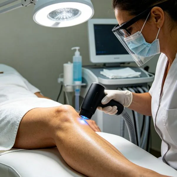
The athlete completed 11 clinical sessions across 3.5 weeks (four sessions during week 1, three sessions weekly in weeks 2 and 3, concluding with one session in week 4). Treatments were delivered using a multi-wavelength Class 4 clinical laser platform utilizing synchronized 810nm, 980nm, and 1470nm emissions, administered with an optical quartz roller handpiece with real-time epidermal thermal tracking.
| Week | Session | Wavelength Ratio | Power (W) | Frequency (Hz) | Duty Cycle | Energy (J) | Area (cm2) |
| Wk 1 | S-01 | 810/980/1470 (3:2:5) | 12.0 W P | 500 Hz | 40% | 5,760 J | 150 cm2 |
| Wk 1 | S-02 | 810/980/1470 (3:2:5) | 14.0 W P | 1,000 Hz | 40% | 6,720 J | 150 cm2 |
| Wk 1 | S-03 | 810/980/1470 (4:3:3) | 16.0 W P | 2,000 Hz | 50% | 7,680 J | 180 cm2 |
| Wk 1 | S-04 | 810/980/1470 (4:3:3) | 18.0 W P | 2,500 Hz | 50% | 8,640 J | 180 cm2 |
| Wk 2 | S-05 | 810/980/1470 (4:3:3) | 20.0 W P | 4,000 Hz | 45% | 8,640 J | 200 cm2 |
| Wk 2 | S-06 | 810/980/1470 (4:3:3) | 22.0 W P | 5,000 Hz | 40% | 8,448 J | 200 cm2 |
| Wk 2 | S-07 | 810/980/1470 (4:3:3) | 24.0 W P | 6,000 Hz | 40% | 9,216 J | 200 cm2 |
| Wk 3 | S-08 | 810/980/1470 (4:4:2) | 24.0 W P | 8,000 Hz | 35% | 8,064 J | 200 cm2 |
| Wk 3 | S-09 | 810/980/1470 (4:4:2) | 22.0 W P | 10,000 Hz | 40% | 8,448 J | 180 cm2 |
| Wk 3 | S-10 | 810/980 (50% : 50%) | 18.0 W CW | Continuous | 100% | 8,640 J | 150 cm2 |
| Wk 4 | S-11 | 810/980 (50% : 50%) | 15.0 W CW | Continuous | 100% | 7,200 J | 150 cm2 |
Cumulative Clinical Progression
| Evaluation Stage | Active Flexion VAS | Walking VAS | Active Flexion ROM | Straight Leg Raise | Dynamometer Force | Hematoma Volume (US) |
| Pre-Treatment | 9.0 / 10 | 4.5 / 10 | 40 degrees | 32 degrees | 8.2 kg | 4.8 cm longitudinal |
| Post-S-02 | 6.5 / 10 | 2.5 / 10 | 65 degrees | 46 degrees | 12.8 kg | 3.4 cm longitudinal |
| Post-S-04 | 4.0 / 10 | 1.0 / 10 | 92 degrees | 62 degrees | 19.5 kg | 1.8 cm longitudinal |
| Post-S-07 | 2.0 / 10 | 0.0 / 10 | 118 degrees | 76 degrees | 27.4 kg | Resolved / Trace fluid |
| Post-S-10 | 0.5 / 10 | 0.0 / 10 | 132 degrees | 84 degrees | 33.1 kg | Organized myotubes |
| 45-Day Follow | 0.0 / 10 | 0.0 / 10 | 135 degrees | 85 degrees | 35.2 kg | Full structural repair |
Clinical Commentary on Recovery Phases
Sessions 1 and 2 prioritized rapid fluid clearance and pain reduction without overheating fragile healing margins. Delivering 12 to 14 Watts with high 1470nm weighting targeted interstitial hematoma and edema around the biceps femoris sheath. By Session 2, ultrasound imaging showed the longitudinal hematoma dimension had decreased from 4.8cm to 3.4cm, and the athlete ceased using crutches for normal ambulation.
Sessions 3 through 7 transitioned to progressive cellular biostimulation and tissue repair. Utilizing peak powers up to 24 Watts at frequencies between 2,000Hz and 6,000Hz (40 to 50 percent duty cycles) delivered high photon density to the deep muscle junction while maintaining epidermal temperatures below 41.2 degrees Celsius. By Session 7, active knee flexion expanded to 118 degrees, handheld dynamometer strength improved to 27.4 kg, and ultrasound confirmed total resolution of the fluid pocket, revealing linear fibroblastic bridging across the myotendinous junction. The athlete began supervised pool workouts and bodyweight eccentric loading.
Sessions 8 through 11 focused on tissue remodeling and strengthening. Running frequencies from 8,000Hz up to continuous wave emissions promoted functional collagen realignment and vascularization. By Session 10, isometric hamstring strength matched 95 percent of the uninjured limb (33.1 kg versus 34.6 kg), and pain-free passive straight leg raise reached 84 degrees. Ultrasound imaging showed parallel myofiber continuity without dense fibrotic scarring. The athlete progressed through track acceleration workouts, cleared full-speed hurdle drills during week 5, and resumed competition without reinjury. At the 45-day follow-up, hamstring output exceeded baseline values, with symmetrical flexibility and zero localized tenderness.
Handpiece Scanning Velocity and Surface Thermal Control
Applying high-intensity Class 4 laser therapy over the posterior thigh requires strict operational technique. Static placement of a high-power beam over skin will quickly cause surface heat accumulation, triggering patient pain and thermal injury.
Handpiece Trajectory along the Posterior Kinetic Chain:
[Zone 1: Ischial Origin Footprint]   <---> Overlapping Concentric Rings with Quartz Compression (3 cm/s)
[Zone 2: Musculotendinous Core]      <---> Longitudinal Stripping Passes along Myofiber Axes (4 cm/s)
[Zone 3: Distal Muscle Bellies]      <---> Transverse Broad Sweeps to Clear Lymphatic Channels (5 cm/s)
The clinical application uses a dynamic scanning approach:
- Kinetic Scanning Rate: The treatment head must maintain continuous movement across the tissue at speeds between 3 and 5 centimeters per second. This maintains forward photon penetration to deep tissue layers while preventing heat accumulation on the skin.
- Mechanical Quartz Compression: Over thick hamstring musculature, applying 2.0 to 2.5 kilograms of manual pressure with a quartz roller or contact head blanches cutaneous and superficial capillary blood. Removing superficial hemoglobin absorbers reduces optical attenuation, allowing photons to penetrate into deeper muscle layers.
- Real-Time Thermal Monitoring: Handpieces with integrated infrared sensors monitor surface skin temperatures in real time, keeping the skin within the safe therapeutic window of 39.5 to 41.5 degrees Celsius. If surface temperatures near 42.0 degrees Celsius, the operator accelerates scanning speed or lowers the duty cycle, maintaining total energy delivery while protecting the skin.
High Intensity Laser Integration in Sports Performance
Managing acute and chronic muscle tears in high-performance athletes requires weighing treatment efficacy against rehabilitation timelines and long-term tissue resilience. High-intensity Class 4 laser therapy offers a non-invasive approach across common sports medicine interventions:
| Modality / Approach | Effective Depth | Primary Biological Mechanism | Practical Limitations |
| Low-Level Light Therapy | 2 mm to 8 mm | Superficial Photochemistry | Ineffective for deep muscle bellies |
| Cryotherapy / Ice Packs | 5 mm to 12 mm | Vasoconstriction & Conduction | Slows cellular repair & protein synthesis |
| Therapeutic Ultrasound | 15 mm to 30 mm | Acoustic Wave Cavitation & Heat | Risk of periosteal pain & localized hot spots |
| Platelet-Rich Plasma | Local Infiltration | Autologous Growth Factor Release | High cost; prolonged post-injection downtime |
| Intramuscular Injections | Local Infiltration | Anesthetic / Corticosteroid Block | Softens connective tissue; risk of re-rupture |
| High-Power Class 4 Laser | 35 mm to 65 mm | Photobiomodulation & Fluid Removal | Requires clinical-grade equipment |
Low-Level Light Therapy operates at milliwatt levels, producing insufficient energy to penetrate past the superficial fascia into deep hamstring muscle bellies. High-power systems deliver the incident irradiance needed to maintain an effective forward-propagating photon field that reaches the myotendinous junction.
Cryotherapy produces superficial vasoconstriction that relieves acute pain, but prolonged icing impairs lymphatic drainage, slows cellular repair, and delays the synthesis of structural proteins essential for muscle recovery. Therapeutic ultrasound generates deep heat through acoustic cavitation, but energy reflection off underlying femoral bone surfaces can cause periosteal irritation and uneven heating in dense muscle tissue.
Platelet-rich plasma therapy delivers autologous growth factors directly into torn tissues, but post-injection inflammation causes significant pain and requires several weeks of mandatory athletic rest before intensive loading can resume. Corticosteroid injections relieve pain rapidly, but repeated exposure weakens connective tissue, inhibits protein synthesis in regenerating myoblasts, and increases the risk of catastrophic re-rupture during explosive sprint acceleration.
High-power multi-wavelength Class 4 laser therapy bridges the gap between acute injury management and high-performance return-to-sport protocols. By combining deep tissue penetration, targeted wavelength absorption, and real-time surface thermal monitoring, this modality accelerates satellite cell differentiation, mobilizes localized hematomas, and drives organized myofibrillar remodeling without the structural risks or prolonged downtime of invasive interventions.