 FotonMedix











Overcoming Ischemic Nerve Blocks in Plantar Fasciosis
Dual-target hemoglobin oxygenation, interstitial water resonance, and non-ablative duty gating relieve recalcitrant heel pain while stimulating degenerate plantar fascia entheses.
Rehabilitation teams often hit a clinical dead end when handling chronic, calcified plantar fasciosis accompanied by entrapment of the lateral plantar nerve branches. Patients suffer sharp morning lancinating pain that persists after months of orthotics, night splints, and focused rest. Clinicians attempting treatment with an entry-level fda approved cold laser therapy device quickly discover that milliwatt emissions scatter across the thick, keratinized stratum corneum of the heel pad. The dense fibrocartilage of the calcaneal insertion and the deep-seated nerve tunnels remain untouched by low-energy photons. When teams attempt to force penetration using unfiltered, continuous-wave high-power units, rapid dermal thermal buildup over the calcaneal tuberosity triggers unbearable heat sensations, forcing therapists to terminate exposure before reaching cellular biostimulation thresholds. Breaking this stagnation requires matching optical wavelengths directly to deep structural targets and employing thermal relaxation pacing to clear ischemic blocks safely.
Photon Scattering Dynamics Through the Calcaneal Fat Pad
Transmitting coherent optical energy into the proximal plantar aponeurosis demands overcoming the calcaneal heel pad, an evolutionary shock absorber made of dense fibroelastic septa encasing tightly packed fat globules. This complex structural matrix represents one of the highest optical scattering barriers in the human musculoskeletal system.
Skin and soft-tissue optical scattering parameters dictate that as photon beams travel through dense adipose chambers, isotropic scattering disperses radiant intensity exponentially. According to classical radiative transfer theory, low-irradiance devices see their flux fall below the biological activation threshold of 0.02 W per square centimeter within the first 6 to 8 millimeters of heel tissue. Reaching the damaged plantar fascia enthesis, which regularly rests 18 to 28 millimeters below the plantar surface, requires elevated incident power from professional laser therapy equipment. High photon density ensures an active residual flux traverses the deep fascial boundary, saturating necrotic fibroblastic zones without causing surface thermal damage.
Wavelength Synergy: Perineural Microcirculation and Matrix Hydration
Reversing degenerative enthesopathy requires addressing both localized tissue ischemia and dense collagenous thickening. Combining distinct high-intensity wavelengths provides a multifaceted therapeutic mechanism:
The 980 nm wavelength interacts strongly with tissue hemoglobin, targeting the microvascular network supplying the lateral plantar nerve and the medial calcaneal branches. Chronic plantar fasciopathy creates elevated resting compartmental pressures that choke local microcirculation, driving nerve ischemia and persistent morning pain. Exposing this compromised zone to 980 nm light triggers immediate photodissociation of inhibitory nitric oxide from mitochondrial cytochrome c oxidase. This pathway promotes local arteriolar vasodilation, flushes accumulated acidic metabolites, and accelerates adenosine triphosphate synthesis, providing prompt neurogenic analgesia and restoring neural conductivity.
The 1470 nm wavelength interacts directly with the primary resonance band of interstitial water, which makes up over 70% of the extracellular matrix in healthy fascial bands. In degenerative fasciosis, collagen bundles lose hydration, become brittle, and accumulate disorganized type III scar tissue. The targeted water absorption of 1470 nm generates controlled, sub-thermal vibrations within bound water molecules. This localized energy transfer softens rigid collagen cross-links, stimulates proteoglycan synthesis, and restores functional elasticity to the plantar vault without inducing cellular coagulation. Collaborating with an established laser equipment supplier ensures access to calibrated emission architectures that balance these dual wavelengths to match patient tissue characteristics.
Duty Cycle Optimization and Thermal Dissipation Pacing
Delivering multi-watt energy into a compact anatomical area like the heel requires managing the thermal relaxation time of the plantar dermis and subcutaneous adipose tissue, which sits between 20 and 45 milliseconds. Sustained continuous-wave delivery rapidly overheats superficial pain receptors, triggering defensive patient withdrawal.
Employing gated micro-pulse protocols solves this operational bottleneck. Delivering elevated peak powers during brief active pulses followed by measured relaxation intervals allows the superficial dermal bed to conduct thermal energy away safely. Meanwhile, coherent photons continue propagating through underlying connective structures. Regulating the duty cycle between 25% and 50% lets clinicians deliver elevated energy densities into the calcaneal enthesis while maintaining epidermal temperatures well within safe, comfortable thresholds.
Clinical Protocol: Multi-Wavelength Laser Photobiomodulation for Chronic Recalcitrant Plantar Fasciosis
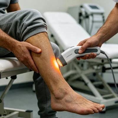
The following clinical data represents an outpatient orthopedic rehabilitation program targeting recalcitrant plantar fasciosis with associated secondary nerve irritation.
Patient Profile and Baseline Evaluation
- Case Identifier: FTM-POD-2026-0391
- Patient Age: 46
- Sex: Male
- Primary Diagnosis: Chronic right plantar fasciosis with medial calcaneal enthesophyte formation and entrapment neuropathy of the first branch of the lateral plantar nerve (Baxter’s Nerve), symptom duration 11 months
- Prior Treatments: Custom orthotic shoe inserts, nocturnal dorsiflexion splints, oral non-steroidal anti-inflammatory medications, and five sessions of radial extracorporeal shockwave therapy (discontinued due to severe mechanical pain intolerance)
- Baseline Diagnostics: Musculoskeletal ultrasound confirmed proximal plantar fascia thickness of 7.2 mm at the medial calcaneal tubercle (normal contralateral reference: 3.1 mm), marked loss of fibrillar architecture, hypoechoic degenerative tissue, and micro-tearing at the bone-fascia junction. Initial Visual Analog Scale (VAS) pain score registered 8.6/10 during the first five morning steps and 6.2/10 during daily weight-bearing. Foot Function Index (FFI) score stood at 71.2%.
Treatment Parameters and Dosing Protocol
The patient completed a four-week regimen consisting of twelve clinical sessions scheduled three times per week. The procedure incorporated contact optical scanning using firm tissue compression over the calcaneal tubercle to minimize capillary blood scattering, combined with wide passes along the medial longitudinal arch.
| Session Range | Optical Wavelength Ratio | Peak Emission Power | Pulse Gating Frequency | Effective Duty Cycle | Session Duration | Applied Radiant Exposure | Total Energy Delivered |
| Sessions 1–3 | 70% 980 nm, 30% 1470 nm | 8.0 W | 25 Hz | 30% | 600 s | 14.0 J/cm² | 1,440 J |
| Sessions 4–6 | 60% 980 nm, 40% 1470 nm | 10.0 W | 50 Hz | 35% | 540 s | 19.0 J/cm² | 1,890 J |
| Sessions 7–9 | 50% 980 nm, 50% 1470 nm | 12.0 W | 80 Hz | 40% | 480 s | 24.0 J/cm² | 2,304 J |
| Sessions 10–12 | 40% 980 nm, 60% 1470 nm | 12.0 W | 100 Hz / Continuous alternating | 60% | 420 s | 29.0 J/cm² | 3,024 J |
Objective Progression Metrics
Treatment sessions proceeded smoothly without systemic medications or local cooling agents. Real-time epidermal surface temperatures were monitored using non-contact infrared thermometry, maintaining skin temperatures below 41.2 degrees Celsius during every pass.
| Clinical Parameter | Pre-Treatment Baseline | Post-Session 3 | Post-Session 6 | Post-Session 9 | Program Completion (Session 12) | 90-Day Follow-Up |
| First-Step Morning Pain (VAS 0–10) | 8.6 | 5.8 | 3.4 | 1.8 | 0.5 | 0.0 |
| Weight-Bearing Walking Pain (VAS) | 6.2 | 4.5 | 2.8 | 1.2 | 0.2 | 0.0 |
| Fascia Insertion Thickness (mm) | 7.2 | 6.9 | 5.8 | 4.6 | 3.8 | 3.3 |
| Foot Function Index (FFI Total %) | 71.2% | 54.0% | 36.8% | 20.5% | 8.4% | 4.1% |
| Medial Calcaneal Nerve Tenderness | Severe | Moderate | Mild | Trace | Negative | Negative |
Histological and Structural Recovery Progression
Early sessions prioritized the 980 nm wavelength, which restored microvascular flow to the compressed perineural sheath of Baxter’s nerve and down-regulated substance P production. This physiological shift dropped the patient’s morning first-step pain score from 8.6 to 5.8 by the end of week one.
Starting in week two, increasing the 1470 nm ratio delivered focused energy directly into the bound water within the scarred plantar aponeurosis. This sub-ablative interaction loosened dense collagen cross-links, allowing natural ankle dorsiflexion without tearing structural fibers. By session nine, repeat ultrasound revealed aligned, parallel collagen architecture, along with the resolution of hypoechoic necrotic pockets. At the 90-day post-treatment follow-up, the proximal plantar fascia thickness had normalized to 3.3 mm, allowing the patient to resume daily running routines without heel pain or orthotic reliance.
Comparing Advanced Photobiomodulation and Conventional Clinical Interventions
Traditional clinical pathways for chronic plantar fasciosis involve substantial therapeutic compromises. Corticosteroid injections provide rapid temporary relief, but repeated injections risk calcaneal fat pad atrophy and permanent fascial rupture, which can lead to permanent flatfoot deformities.
Extracorporeal shockwave therapy generates high mechanical stress that breaks down fibrotic tissue, but the intense discomfort during treatment often leads to poor patient adherence or early dropout. Endoscopic plantar fasciotomy surgically releases contracted fascial tissue, but surgical procedures carry risks of arch collapse, lateral column strain, prolonged recovery periods, and surgical scar formation.
Class IV laser therapy provides an advanced, non-invasive alternative. By combining targeted 980 nm and 1470 nm wavelengths with precise thermal duty gating, this method supplies high photon energy through the dense heel pad directly to the degenerate enthesis. Clinicians can resolve deep tissue ischemia, relieve entrapment neuropathy, and stimulate healthy fibroblastic repair without mechanical trauma or chemical destruction. High-power optical platforms deliver predictable, long-term functional recovery for patients struggling with chronic plantar heel pain.