 FotonMedix



Overcoming Peroneal Microvascular Stasis in Tarsal Tunnel Entrapment
Synchronized multi-wavelength arrays bypass the fibrotic flexor retinaculum, selective dual-band chromophore resonance relieves epineurial capillary stasis, and microsecond duty cycle modulation prevents epidermal heat accumulation over denervated distal tissues.
The late afternoon schedule in an outpatient foot and ankle surgical center regularly encounters an agonizing clinical scenario. A middle-aged retail manager sits with their foot elevated, describing a persistent, searing ache radiating from behind the medial malleolus into the medial longitudinal arch and plantar digits. Sensation over the heel and plantar vault feels blunt and deadened, punctuated by sudden bouts of lancinating paresthesias that worsen after prolonged standing. Clinical palpation directly over the tarsal tunnel triggers a sharp distal Tinel sign shooting along the medial and lateral plantar nerves. Conservative attempts have hit a dead end: rigid medial arch orthotics fail to arrest the compression, repeated blind cortisone injections within the fibro-osseous canal risk plantar fascial rupture without restoring nerve mobility, and oral gabapentinoids produce chronic daytime somnolence without improving local tissue blood flow. Standard clinical therapy units fail because their diffuse, low-energy output scatters across the thickened retinaculum and edematous subcutaneous tissues, failing to alter the underlying neural ischemia. Re-establishing normal sensory conduction requires delivering high-power photonic flux through dense fibrous retinacular boundaries to restore microvascular perfusion within the compressed nerve corridor.
Optical Penetration Gradients and Structural Extinction in the Medial Ankle
Targeting the posterior tibial neurovascular bundle beneath the flexor retinaculum represents an intricate anatomical and optical challenge. The target anatomy—the posterior tibial nerve, its bifurcation into the medial and lateral plantar nerves, and the accompanying venae comitantes—lies enclosed within the rigid fibro-osseous tarsal tunnel beneath 18 to 30 millimeters of dense connective tissue. Photons directed from a cutaneous handpiece must pass through the stratum corneum, dermal capillary beds, dense subretinacular fat, and the fibrous flexor retinaculum (laciniate ligament) before reaching the ischemic epineurium.
As photons traverse this dense fibrous landscape, structural collagen bundles and surrounding fascial planes create intense Rayleigh and Mie scattering. In chronic compressive entrapments, mechanical friction and chronic low-grade inflammation cause reactive thickening of the flexor retinaculum, forming a dense barrier that deflects incident light into lateral scatter paths. Low-power modalities emitting continuous fractions of a Watt experience rapid spatial extinction within the first few millimeters of superficial skin, producing no measurable physiological impact on the compressed nerve below. High-intensity Class 4 systems generate the incident irradiance required to overcome this scattering barrier, establishing an effective forward-propagating photon flux that penetrates the deep fibro-osseous canal.
Medial Retinacular Photonic Attenuation and Tissue Penetration Gradient:
[Cutaneous & Dermal Layer]    ===> Incident High-Density Flux (100% Incident Irradiance)
              |
[Dense Flexor Retinaculum]   ---> Severe Mie Scattering along Packed Collagen Bundles (36% Loss)
              |
[Subretinacular Adipose Bed] ---> Forward Photonic Dispersion across Lipid Interfaces (40% Loss)
              |
[Posterior Tibial Epineurium]--> Residual Forward Photonic Field (Target Dose: 6-10 J/cm2 Fluence)
Within the rapidly developing podiatry lasers market, equipment selection depends on matching emissions precisely to target biological chromophores. Peripheral nerve repair requires coordinated interaction across three primary physiological absorbers: water, hemoglobin, and mitochondrial photoacceptors:
- 1470nm Hydro-Selective Interstitial Clearance: Chronic nerve entrapment generates venous congestion and stagnant interstitial fluid within the epineurial space, creating high hydrostatic fluid pressure that strangulates delicate endoneurial capillaries. The 1470nm wavelength interacts directly with free water molecules. Its high water absorption profile generates localized photothermal fluid mobilization, accelerating lymphatic outflow from the tight tarsal tunnel, decreasing intraneural pressure, and relieving mechanical strain on compressed axonal membranes.
- 980nm Hemoglobin Absorption and Perfusion: The microvessels supplying the nerve trunk—the vasa nervorum—undergo chronic vasoconstriction and endothelial hypoxia under mechanical entrapment. The 980nm band matches the optical absorption curves of oxygenated and deoxygenated hemoglobin. Absorption of 980nm photons triggers localized photothermal vasodilation, stimulating endothelial nitric oxide synthesis and reopening closed microcapillaries within the vasa nervorum to re-establish oxygen delivery to starving axons.
- 810nm Cytochrome Activation: Aligning with the absorption peak of mitochondrial Cytochrome c oxidase, this wavelength stimulates the respiratory electron transport chain, driving the cellular adenosine triphosphate synthesis required for axonal repair and maintenance of resting membrane potentials.
Delivering high-energy Class 4 laser power to neuropathic feet introduces critical thermal management concerns. Because diabetic patients suffer from blunted or absent nociception, they cannot provide normal sensory feedback if surface temperatures rise dangerously. Continuous-wave energy absorbed by epidermal melanin and dermal water can accumulate rapidly, and if skin temperatures exceed 42 degrees Celsius, tissue burns can develop unnoticed on denervated skin.
Gated Pulse Mechanics for Denervated Retinacular Tissue:
Emission Phase (Pulse ON)  : |=========== High Peak Power Irradiance ===========|
                             [-------- Controlled Pulse Duration (t_on) --------]
Thermal Clearance (Pulse OFF): [ . . . . Cutaneous Thermal Relaxation (t_off) . . . . ]
                             Net Outcome: Deep neurovascular perfusion without skin burns
Microsecond duty cycle gating provides the necessary clinical protection. Operating across frequencies from 100Hz to 12,000Hz, system processors deliver high peak wattage to penetrate thickened retinacular fibers while inserting brief off-duty rest intervals. These intervals match the thermal relaxation time of the skin, allowing cutaneous capillary blood flow to dissipate heat while forward-moving photons penetrate deep into the ischemic vasa nervorum.
Microvascular Restoration and Axonal Bioenergetics
Deploying high-power professional laser vascular treatment devices targets the physiological drivers of nerve entrapment rather than temporarily masking sensory nerve transmission. Peripheral neuropathic degeneration begins with microangiopathy of the vasa nervorum. Chronic mechanical compression damages capillary endothelial cells, leading to basement membrane thickening and reduced endoneurial capillary blood flow. The resulting hypoxia starves the axonal cytoskeleton of nutrients, disabling active axonal transport, reducing intra-epidermal nerve fiber density, and leading to spontaneous, erratic firing in unmyelinated C-fibers alongside conduction failure in myelinated A-beta fibers.
Irradiating the ischemic lower limb with a specialized neuro laser restores mitochondrial respiration within both the vascular endothelium and the starving axons. Cytochrome c oxidase (Complex IV) functions as the key mitochondrial photoacceptor. Under ischemic conditions, excess nitric oxide binds to the enzyme’s catalytic copper and heme centers, shutting down the electron transport chain. Near-infrared photons trigger targeted photolysis of this bound nitric oxide, restoring oxygen binding and re-establishing the proton gradient across the inner mitochondrial membrane.
Biological Progression of Neurovascular Photobiomodulation:
[Photonic Absorption]     --> Complex IV Cytochrome c Oxidase Activation & NO Release
           |
[Endothelial Nitric Oxide] --> Vasa Nervorum Capillary Vasodilation & Micro-Revascularization
           |
[Mitochondrial ATP Surge] --> Restores Axonal Na+/K+ ATPase Pump & Fast Axonal Transport
           |
[Functional Regeneration]  --> Upregulates Nerve Growth Factor & Restores Intra-Epidermal Fibers
This metabolic reactivation drives four distinct regenerative stages:
- Vasa Nervorum Revascularization: Increased endothelial nitric oxide and upregulated Vascular Endothelial Growth Factor stimulate neo-angiogenesis within the epineurial and perineurial sheaths, restoring functional capillary blood flow to chronically ischemic nerve trunks.
- Restoration of the Axonal Sodium-Potassium Pump: Elevated cellular adenosine triphosphate levels repolarize the axonal membrane by fueling the sodium-potassium ATPase pump, stabilizing irritable nerve membranes and suppressing the spontaneous ectopic firing that causes severe burning dysesthesia.
- Upregulation of Neurotrophic Factors: High-intensity photon flux upregulates local transcription of Nerve Growth Factor, Brain-Derived Neurotrophic Factor, and Neurotrophin-3, accelerating Schwann cell proliferation, promoting remyelination of thinned nerve sheaths, and restoring intra-epidermal sensory nerve endings.
- Reduction of Oxidative Stress and Advanced Glycation Byproducts: Normalizing mitochondrial electron flow prevents electron leakage, reducing reactive oxygen species generation, suppressing NF-kappa-B activation, and clearing chronic inflammatory markers from surrounding connective tissue.
Through these combined pathways, targeted Class 4 laser therapy addresses the underlying vascular ischemia and metabolic deficits that drive peripheral nerve entrapment.
Clinical Stratification and Diagnostic Staging in Lower Extremity Neuropathy
Selecting appropriate candidates requires systematic clinical and diagnostic staging. Identifying suitable clinical presentations depends on differentiating between functional ischemic neuropathy, advanced mechanical deformities, and critical limb ischemia:
- Critical Limb Ischemia with Gangrenous Tissue Breakdown: Patients presenting with resting ischemic pain, absent pedal pulses, non-healing necrotic plantar ulcers, and transcutaneous oxygen pressure below 30 mmHg require urgent vascular surgery for arterial revascularization. Photobiomodulation cannot substitute for open surgical bypass or endovascular intervention in limbs facing acute gangrene.
- Diabetic Peripheral Sensorimotor Neuropathy with Vasa Nervorum Ischemia: The primary indication for deep multi-wavelength intervention. Patients display symmetrical stocking-distribution sensory loss, diminished vibratory perception, loss of protective 10g monofilament sensation, burning nocturnal dysesthesia, and reduced microvascular Doppler flow without complete large-vessel occlusion.
- Secondary Tarsal Tunnel Entrapment Syndrome: Chronic nerve swelling often leads to mechanical compression of the posterior tibial nerve beneath the flexor retinaculum at the medial malleolus (double crush phenomenon). High-intensity multi-wavelength laser therapy mobilizes inflammatory fluid, reduces endoneurial swelling, and eases mechanical pressure within the fibro-osseous tunnel.
- Charcot Neuroarthropathy (Active Acute Phase): Active Charcot foot with acute joint temperature elevation, bone fragmentation, and joint subluxation requires immediate non-weight-bearing immobilization. Applying thermal or photobiomodulatory modalities to an actively collapsing acute Charcot joint is contraindicated until the osteoclastic stage stabilizes.
Determining treatment dosage requires accounting for significant optical attenuation through dense retinacular and adipose layers. Because fibrous tissue and overlying fat scatter or absorb up to 95 percent of surface photons, delivering an effective therapeutic dose of 6 to 9 Joules per square centimeter to the tibial nerve branches at a depth of 25 millimeters requires surface doses between 110 and 150 Joules per square centimeter across the retromalleolar sulcus and tarsal canal.
Comprehensive Clinical Case History: Outpatient Podiatric Specialty Center
The following case record details the clinical baseline, parameter matrix, and objective recovery trajectory observed during high-power multi-wavelength laser intervention.
Patient Profile and Baseline Diagnostics
- Department Case ID: FTM-POD-2026-9142
- Patient Age: 56 years
- Biological Sex: Female
- Clinical History: Longstanding pronated foot mechanics complicated by mild metabolic syndrome; 18 months of standing occupational stress as an educational administrator.
- Presenting Condition: Severe right-sided tarsal tunnel entrapment neuropathy accompanied by persistent plantar dysesthesia, distal burning sensation across the first through third toes, and sharp nocturnal throbbing.
- Duration of Symptoms: 32 weeks of progressive functional decline; unresponsive to rigid medical-grade orthotics, a local methylprednisolone injection within the retromalleolar canal, and oral duloxetine (60mg daily, discontinued due to severe gastrointestinal nausea).
- Functional Baseline:
- Visual Analog Scale for Plantar Burning and Throbbing: 8.0 / 10
- Visual Analog Scale for Weight-Bearing Ambulation: 6.5 / 10
- Manchester Foot Pain and Disability Index: 42 / 60 (severe functional impairment)
- Semmes-Weinstein Monofilament Examination: Complete absence of 10g monofilament perception across the medial plantar nerve distribution (great toe and first two metatarsal heads).
- Provocative Testing: Strongly positive Tinel sign elicited over the right retromalleolar sulcus with distal radiation; positive Valleix sign tracking proximally into the deep calf.
- Diagnostic Musculoskeletal Ultrasound and High-Resolution MRI:
- Cross-sectional area of the right posterior tibial nerve at the retromalleolar sulcus enlarged to 18.4 square millimeters (contralateral asymptomatic nerve measured 8.6 square millimeters).
- Marked subretinacular epineurial fluid cuffing and thickening of the flexor retinaculum measuring 2.8mm (normal: less than 1.2mm).
- Power Doppler ultrasound revealed engorged, sluggish microvascular flow within the surrounding vasa comitantes without discrete space-occupying ganglion cysts or schwannomas.
Standardized Treatment Protocol and Kinetic Energy Matrix
The patient underwent a course of 12 clinical sessions across 4 consecutive weeks (three sessions per week for four weeks). Treatment was delivered using a multi-wavelength Class 4 clinical platform featuring synchronized 810nm, 980nm, and 1470nm emissions, administered with an optical quartz contact handpiece with integrated surface pyrometry.
| Week | Session | Wavelength Ratio | Power (W) | Frequency (Hz) | Duty Cycle | Energy (J) | Area (cm2) |
| Wk 1 | S-01 | 810nm (60%) 980 (40%) | 10.0 W CW | Continuous | 100% | 4,500 J | 90 cm2 |
| Wk 1 | S-02 | 810/980/1470 (5:3:2) | 12.0 W P | 200 Hz | 60% | 5,760 J | 90 cm2 |
| Wk 1 | S-03 | 810/980/1470 (4:3:3) | 14.0 W P | 400 Hz | 50% | 6,720 J | 100 cm2 |
| Wk 2 | S-04 | 810/980/1470 (4:3:3) | 16.0 W P | 800 Hz | 50% | 7,680 J | 100 cm2 |
| Wk 2 | S-05 | 810/980/1470 (4:3:3) | 18.0 W P | 1,500 Hz | 45% | 7,290 J | 110 cm2 |
| Wk 2 | S-06 | 810/980/1470 (3:3:4) | 20.0 W P | 3,000 Hz | 40% | 7,680 J | 110 cm2 |
| Wk 3 | S-07 | 810/980/1470 (3:3:4) | 22.0 W P | 5,000 Hz | 40% | 8,448 J | 120 cm2 |
| Wk 3 | S-08 | 810/980/1470 (3:3:4) | 22.0 W P | 5,000 Hz | 40% | 8,448 J | 120 cm2 |
| Wk 3 | S-09 | 810/980/1470 (4:3:3) | 20.0 W P | 2,500 Hz | 50% | 8,400 J | 120 cm2 |
| Wk 4 | S-10 | 810/980/1470 (4:3:3) | 18.0 W P | 1,000 Hz | 50% | 7,560 J | 110 cm2 |
| Wk 4 | S-11 | 810/980/1470 (4:3:3) | 16.0 W P | 500 Hz | 50% | 6,720 J | 100 cm2 |
| Wk 4 | S-12 | 810/980 (50% : 50%) | 12.0 W CW | Continuous | 100% | 5,400 J | 90 cm2 |
Cumulative Clinical Progression
| Checkpoint | Resting VAS | Walking VAS | MFPDI Score | Tibial Nerve CSA | Monofilament Sites | Tinel Sign Status |
| Pre-Treatment | 8.0 / 10 | 6.5 / 10 | 42 / 60 | 18.4 mm2 | 0 / 5 intact | Positive (Instant) |
| Post-S-03 | 5.5 / 10 | 4.5 / 10 | 33 / 60 | 16.1 mm2 | 1 / 5 intact | Positive (Delayed) |
| Post-S-06 | 3.0 / 10 | 2.5 / 10 | 21 / 60 | 12.8 mm2 | 3 / 5 intact | Faint Tingling |
| Post-S-09 | 1.5 / 10 | 1.0 / 10 | 10 / 60 | 10.4 mm2 | 5 / 5 intact | Absent / Negative |
| Post-S-12 | 0.0 / 10 | 0.0 / 10 | 3 / 60 | 9.0 mm2 | 5 / 5 intact | Absent / Negative |
| 90-Day Follow | 0.0 / 10 | 0.0 / 10 | 2 / 60 | 8.8 mm2 | 5 / 5 intact | Absent / Negative |
Clinical Commentary on Progress Phases
During Sessions 1 through 3, treatment concentrated on preparing the lower limb vascular tree and introducing the 1470nm wavelength to mobilize interstitial edema around the tarsal tunnel. Continuous-wave delivery at 10.0 Watts across the posterior calf musculature promoted arterial inflow toward the tibial bifurcations. By Session 3, ultrasound imaging showed the cross-sectional area of the swollen tibial nerve decreased from 18.4 to 16.1 square millimeters, while resting burning pain dropped from 8.0 to 5.5, allowing the patient to sleep without waking from painful paresthesias.
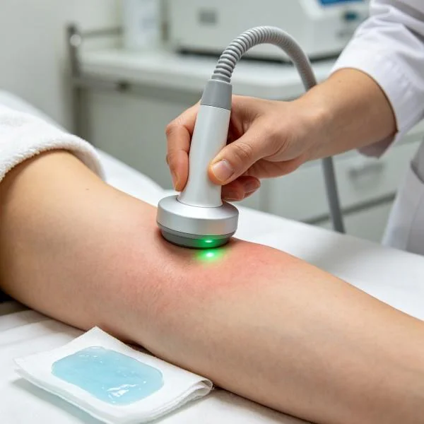
Sessions 4 through 8 increased peak output settings up to 22.0 Watts, using frequencies between 800Hz and 5,000Hz with 40 to 50 percent duty cycles. This configuration delivered high photon flux to the plantar digital branches and the deep medial calcaneal nerve without raising epidermal temperatures above 40.5 degrees Celsius. By Session 6, protective 10g monofilament sensation returned to 3 of 5 plantar sites, the tibial nerve cross-sectional area contracted to 12.8 square millimeters, and daytime walking pain dropped to 2.5. The patient reported walking through normal store corridors without wearing oversized protective clogs.
Sessions 9 through 12 focused on consolidating neural recovery and supporting microvascular remodeling. Operating at higher peak pulses and transitioning to continuous-wave emissions stimulated long-term capillary angiogenesis and axonal transport. By Session 12, protective sensation was restored across all 5 plantar test sites, the Manchester Foot Pain and Disability score dropped from 42 to 3, and the posterior tibial nerve normalized to 9.0 square millimeters on ultrasound. At the 90-day follow-up examination, the clinical gains remained stable, with full preservation of protective sensation, normal walking endurance, and zero recurrence of nocturnal burning pain.
Handpiece Scanning Velocity and Surface Thermal Control
Administering high-power Class 4 laser therapy over neuropathic extremities requires strict procedural discipline. Because diabetic patients suffer from impaired temperature sensation, leaving the handpiece stationary over calloused skin or prominent metatarsal heads risks localized heat accumulation and painless thermal injury.
Handpiece Trajectory along the Lower Extremity Neurovascular Chain:
[Zone 1: Popliteal & Calf Trunks]   <---> Broad Longitudinal Passes (5 cm/s, CW Delivery)
[Zone 2: Medial Tarsal Tunnel]      <---> Overlapping Spirals with Quartz Compression (3 cm/s)
[Zone 3: Central Plantar Vault]     <---> S-Curve Transverse Sweeps along the Neurovascular Bundle
[Zone 4: Metatarsal Digital Heads]  <---> Grid Standoff Scanning across Terminal Capillary Loops
Key technical parameters govern clinical application:
- Kinetic Scanning Velocity: The treatment optic must maintain continuous motion across the target field at speeds between 3 and 5 centimeters per second. This maintains forward photon penetration to deep nerve trunks while preventing heat accumulation in the superficial keratin layers.
- Mechanical Quartz Compression: Over the medial tarsal tunnel, the clinician applies firm manual pressure (1.5 to 2.0 kg) using a flat quartz contact head. This displaces superficial venous and capillary blood, decreasing competitive light absorption by hemoglobin and increasing direct photon penetration into the deep posterior tibial nerve.
- Real-Time Thermal Monitoring: The handpiece uses an integrated infrared pyrometer to measure skin surface temperature in real time, keeping tissue within the safe therapeutic window of 38.5 to 40.5 degrees Celsius. If surface temperatures approach 41.0 degrees Celsius, the operator accelerates scanning speed or reduces the pulse duty cycle, preserving total energy delivery while protecting denervated skin.
High Intensity Laser Integration in Lower Extremity Neuropathy Care
Managing diabetic peripheral neuropathy highlights the trade-offs inherent in conventional pharmacological and physical interventions. High-intensity Class 4 laser therapy provides a targeted, non-invasive alternative across several common clinical pathways:
| Modality / Approach | Penetration Depth | Underlying Mechanism | Clinical Limitations |
| Low-Level Light Therapy | 2 mm to 6 mm | Superficial Photochemistry | Cannot penetrate thick plantar calluses |
| Gabapentinoid Medications | Systemic Distribution | Voltage-Gated Calcium Block | Dizziness, lethargy; leaves ischemia untreated |
| Tricyclic Antidepressants | Systemic Distribution | Serotonin-Norepinephrine Block | Anticholinergic toxicity, cardiac risks |
| Surgical Nerve Decompression | Direct Canal Margin | Mechanical Retinaculum Release | Invasive; infection risk in diabetic feet |
| Transcutaneous Neuro-Stim | 5 mm to 10 mm | Gate Control Pain Suppression | Transient effect; does not restore tissue blood flow |
| Class 4 Multi-Wave Laser | 20 mm to 45 mm | Angiogenesis & Axonal Perfusion | Requires clinical-grade equipment |
Low-Level Light Therapy operates in the milliwatt range. While it can aid superficial digital ulcer healing, its low energy cannot overcome the optical scattering barriers of thick plantar calluses and fibrous fat pads to reach deep nerve trunks. High-power systems deliver the incident irradiance needed to maintain an effective forward-propagating photon field that reaches the ischemic vasa nervorum.
Systemic medications such as gabapentin, pregabalin, and duloxetine act centrally to dampen pain perception, but they cause documented side effects like dizziness, sedation, and mental clouding while leaving the underlying microvascular ischemia and axonal starvation completely untreated. Tricyclic antidepressants carry similar anticholinergic risks and potential cardiac side effects, particularly in older patients with cardiovascular comorbidities.
Surgical tarsal tunnel decompression mechanically releases the retinaculum to relieve nerve compression, but invasive open surgery carries elevated risks of wound dehiscence and deep foot infections in diabetic patients with impaired healing capacity. Transcutaneous electrical nerve stimulation temporarily alters sensory transmission via gate control mechanisms, but its effects fade once the unit is switched off, and it does not improve endoneurial blood flow or reverse metabolic axonal deficits.
High-power multi-wavelength Class 4 laser therapy bridges the gap between purely symptomatic pharmaceuticals and invasive surgical procedures. By combining deep tissue penetration, targeted wavelength absorption, and real-time surface thermal monitoring, this modality accelerates microvascular reperfusion of the vasa nervorum, stimulates mitochondrial energy production in damaged axons, and restores protective sensation without systemic side effects or invasive procedural risks.