 FotonMedix











Resolving Chronic Tenocyte Exhaustion in Deep Plantar Tears
High-power multi-spectral photobiomodulation stimulates deep fibrocartilaginous repair, activates stagnant tenocyte metabolism via mitochondrial photo-dissociation, and prevents structural tissue degradation through microsecond duty cycle regulation.
Few musculoskeletal conditions challenge physical therapists and orthopedic surgeons like chronic, degenerative plantar fasciopathy and deep flexor digitorum longus insertional tears. A marathon runner or active worker presents with debilitating medial heel pain that has dragged on for over nine months. First steps in the morning cause intense, lancinating pain that makes weight-bearing nearly impossible. Biomechanical offloading, night splints, custom orthotics, and platelet-rich plasma injections have yielded only transient, partial relief. The anatomical reality explains this therapeutic wall: the plantar aponeurosis and deep pedal tendons are subject to relentless cyclical tensile shear forces, yet they possess a sparse vascular network and an extremely low baseline metabolic turnover rate. At the enthesis, where fibrocartilage anchors into the calcaneus, mechanical strain creates an avascular zone of chronic hypoxia. Tenocytes within this niche undergo cellular exhaustion, downregulating normal collagen expression and producing mucinous, mucoid degeneration marked by unorganized extracellular matrix, micro-calcifications, and high interstitial fluid pressure that chokes remaining micro-capillaries.
Reversing this degenerative spiral demands an intervention capable of depositing substantial photonic energy directly into dense, deep collagenous matrices. Low-output therapeutic modalities fail here because the plantar fat pad, thickened skin, and deep fascial layers scatter and attenuate superficial photons before they reach the enthesis. When utilizing laser therapy for soft tissue injuries, the clinician must achieve a balance of high photonic flux, target chromophore affinity, and dynamic beam delivery to saturate the ischemic footprint at depths of three to four centimeters while safeguarding the thick, sensitive epidermal layer against heat accumulation.
Spectral Balancing and Hemodynamic Recruitment
Restoring structural competence to degenerate plantar fibers requires resolving two simultaneous barriers: mechanical edema compressing micro-vessels and cellular hypoxia stalling matrix synthesis. Multi-wavelength Class IV medical systems achieve this via simultaneous spectral targeting.
The 980 nm wavelength acts directly on intravascular chromophores. At 980 nm, light absorption by deoxygenated and oxygenated hemoglobin produces precise, controlled thermal micro-disturbances within capillary loops and terminal arterioles. This localized thermal energy induces rapid relaxation of vascular smooth muscle, expanding vascular lumen diameter and significantly accelerating regional micro-circulation. This sudden influx of arterial blood clears accumulated inflammatory catabolites and elevates local tissue partial pressure of oxygen, providing the biological foundation required for fibroblast and tenocyte survival.
In contrast, the 1470 nm wavelength addresses the dense, fluid-logged extracellular matrix. Water molecules within the interstitial fluid and edematous tissue beds display an absorption coefficient for 1470 nm that is orders of magnitude higher than near-infrared wavelengths below 900 nm. By interacting strongly with interstitial water, 1470 nm light alters the local viscosity of congested exudate, enhances hydraulic conductivity within the ground substance, and accelerates superficial and deep lymphatic drainage. Reducing this localized interstitial compartment pressure relieves mechanical compression on adjacent micro-vessels and desensitizes hyperactive unmyelinated nociceptive fibers. Operating these wavelengths together produces an optimal environment: 1470 nm decompresses swollen, painful tissue layers, enabling the hemodynamic flush driven by 980 nm to oxygenate the deepest collagen bundles.
Mitochondrial Photobiomodulation and Structural Collagen Deposition
At the cellular level, coherent near-infrared photons directly target the enzymatic machinery driving connective tissue regeneration. The central engine of this response is Cytochrome c Oxidase, situated within the inner mitochondrial membrane.
In chronic, degenerate tendinopathy, persistent mechanical and oxidative stress leads to excessive accumulation of endogenous nitric oxide. Nitric oxide binds with high affinity to the heme and copper centers of Cytochrome c Oxidase, displacing oxygen and paralyzing cellular respiration. This halts the proton-pumping function across the inner mitochondrial membrane, drastically dropping adenosine triphosphate (ATP) production and arresting active protein synthesis.
When exposed to coherent infrared radiation:
- Target photons are absorbed by the chromophores of Cytochrome c Oxidase, facilitating the photo-dissociation of bound nitric oxide from the catalytic sites.
- Free oxygen immediately binds to the unlocked enzyme, restoring electron transport down the respiratory chain and generating an immediate surge in ATP synthesis.
- Dissociated nitric oxide diffuses into local vascular endothelium, activating soluble guanylyl cyclase to maintain long-lasting micro-vascular perfusion.
- The sudden availability of cellular ATP fuels the metabolic activity of dormant tenocytes, driving transcription of genes responsible for producing procollagen alpha chains.
- Fibroblasts and tenocytes resume production of dense, high-tensile type I collagen while downregulating the deposition of mechanically weak, chaotic type III reticular fibers.
This bioenergetic jumpstart is fundamental when applying laser therapy for tendon injuries, turning a chronically degenerated, avascular enthesis into an active site of structural remodeling.
Thermal Kinetics and Duty Cycle Precision
Delivering therapeutic photon densities into tough, dense plantar structures necessitates high power output to overcome physical scatter and absorption losses. However, unchecked continuous-wave delivery risks excessive heat build-up at the dermal-adipose boundary, which can cause surface discomfort or localized thermal tissue trauma.
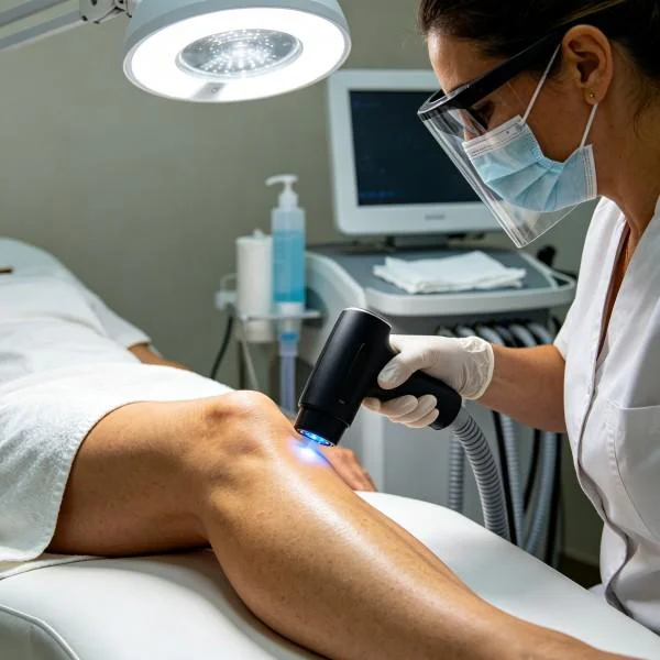
To maximize deep photon penetration without thermal degradation, clinicians employ gated pulse modes and manipulate the duty cycle:
- The duty cycle governs the ratio between active photon emission and the thermal relaxation period.
- Utilizing lower duty cycles (30% to 50%) allows clinicians to emit high peak power spikes. These short bursts propel high photon counts deep through the plantar fat pad, satisfying the threshold fluence required for cellular photobiomodulation in the deep aponeurosis.
- During the off-cycle, the target and overlying tissues dissipate thermal energy via the micro-circulatory bed, preventing tissue temperatures from reaching the 42 to 45 degree Celsius threshold where pain receptors fire and structural proteins risk thermal denaturation.
- Modulating pulse repetition rates between low frequencies (such as 20 to 100 Hz for acute neural hyperalgesia) and high frequencies (up to 5,000 Hz for chronic, dense structural fibrosis) alters mechanical matrix tension and encourages functional fiber reorientation.
Clinical Protocol: Degenerative Plantar Fasciopathy and Deep Tendon Tear
The following case illustrates the clinical deployment of high-power Class IV laser therapy for chronic, recalcitrant plantar soft tissue degeneration.
Clinical Profile and Baseline Status
- Case Identifier: REHAB-POD-2026-1104
- Patient Profile: 46-year-old female, school administrator and recreational long-distance walker
- Primary Diagnosis: Chronic Degenerative Plantar Fasciopathy with partial thickness interstitial tearing at the calcaneal insertion (Left Foot)
- Chronicity: 11 months of continuous symptoms, refractory to custom orthotics, night splints, corticosteroid iontophoresis, and deep friction massage
- Baseline Diagnostic Ultrasound: Marked fusiform thickening of the medial band of the plantar fascia measuring 6.9 mm at the calcaneal enthesis (asymptomatic right foot: 3.2 mm). Intrasubstance focal hypoechoic defect measuring 3.5 mm in width, indicating an interstitial tear. Prominent cortical irregularity and traction osteophyte at the calcaneal origin, accompanied by loss of parallel fibrillar architecture.
- Baseline Functional Metrics: Morning first-step Visual Analog Scale (VAS) pain score: 8/10; post-weightbearing walking VAS score: 6/10. Foot Function Index (FFI): 64%. Physical examination demonstrates acute point tenderness over the medial calcaneal tubercle and a positive Windlass test eliciting sharp insertional pain.
Therapeutic Parameters and Protocol Progression
The patient received a structured course of 10 therapeutic sessions over five weeks. A Class IV multi-wavelength laser system was utilized, delivering energy via a wide-beam optical handpiece using slow, overlapping cross-hatch scanning over the heel pad, followed by focused linear passes along the medial longitudinal arch.
| Treatment Metric | Phase 1: Sessions 1–3 (Edema Clearance & Nociceptive Gating) | Phase 2: Sessions 4–7 (Metabolic Stimulation & Tenogenesis) | Phase 3: Sessions 8–10 (Collagen Realignment & Consolidation) |
| Wavelength Ratio | 65% 1470 nm, 35% 980 nm | 45% 1470 nm, 55% 980 nm | 25% 1470 nm, 75% 980 nm |
| Emission Mode | Pulsed | Pulsed | Continuous Wave & Superpulsed Hybrid |
| Peak Output Power | 16 W | 22 W | 14 W Average |
| Pulse Frequency (Hz) | 40 Hz | 400 Hz | 3,000 Hz |
| Duty Cycle (%) | 35% | 50% | 70% |
| Target Surface Area | 35 cm² (Heel pad and medial arch) | 25 cm² (Direct calcaneal enthesis) | 20 cm² (Focused insertion site) |
| Target Fluence | 80 J/cm² | 150 J/cm² | 175 J/cm² |
| Total Delivered Joules | 2,800 J | 3,750 J | 3,500 J |
| Treatment Frequency | 3 sessions / week | 2 sessions / week | 1 session / week |
Structured delivery of laser therapy for ligament injuries and dense aponeurotic tears follows this exact technical staging. In early sessions, higher 1470 nm energy decompresses tense heel pads and stabilizes hyperactive nerve endings. As symptoms shift from an acute inflammatory flare to mechanical healing, the protocol transitions toward deep 980 nm emission to maximize tenocyte ATP output and drive collagen remodeling.
Clinical Progress and Structural Recovery Metrics
Detailed clinical tracking documented objective, progressive tissue repair throughout the intervention:
Session 3 (Conclusion of Week 1)
Morning first-step VAS pain dropped from 8/10 to 4/10. The patient reported a noticeable reduction in that severe “stone bruise” sensation when stepping out of bed. Peritendinous swelling around the medial calcaneal process decreased, and the Windlass test produced mild ache rather than sharp pain.
Session 7 (Conclusion of Week 3)
Diagnostic ultrasound revealed that the plantar fascia thickness decreased from 6.9 mm to 4.8 mm. The 3.5 mm interstitial hypoechoic tear site showed significant echogenic filling, indicating active granular matrix deposition. Foot Function Index (FFI) score improved from 64% to 28%. The patient was able to complete 45 minutes of continuous flat-ground walking without triggering an inflammatory flare.
Session 10 (Conclusion of Week 5)
Follow-up high-resolution sonography confirmed structural consolidation: the plantar fascia stabilized at a healthy 3.6 mm thickness, with clear, continuous parallel hyperechoic lines extending across the calcaneal boundary. The focal tear had fully resolved with no residual hypoechoic gaps. Morning VAS pain registered at 0/10, with walking VAS at 0/10. The Foot Function Index improved to 6%, indicating complete functional recovery and a successful return to low-impact recreational exercise without relapse.
Structural Photobiomodulation Compared with Conventional Therapy
The clinical management of deep fibrous injuries has long relied on passive or palliative treatments that often fail to alter the underlying degenerative pathology. Nonsteroidal anti-inflammatory drugs provide temporary pain relief, yet they suppress normal tenocyte proliferation and impair long-term collagen tensile strength. In chronic insertional tendinopathy, inflammation is rarely the dominant feature; instead, the issue is structural necrosis and cellular exhaustion, rendering long-term pharmacological suppression counterproductive.
Corticosteroid injections, while capable of reducing short-term pain, carry well-documented structural risks. Corticosteroids arrest cellular protein synthesis, decrease tendon elasticity, and induce permanent atrophy of the plantar fat pad, dramatically increasing the lifetime incidence of catastrophic aponeurotic rupture. Extracorporeal shockwave therapy offers a non-invasive mechanical alternative, but it relies on delivering repeated acoustic microtrauma to stimulate healing. This process is often intensely painful for the patient, requires high compliance over multiple agonizing sessions, and can cause significant bruising and localized tissue disruption.
Class IV laser therapy offers a distinct, biologically supportive therapeutic mechanism. Rather than chemically suppressing tissue responses or inducing mechanical trauma, it works at the bioenergetic level:
- Completely Non-Invasive Delivery: Photons glide through skin, adipose tissue, and plantar aponeurotic layers to reach deep entheses without causing micro-structural trauma or patient discomfort.
- Dual Action on Edema and Blood Flow: Tailored wavelength combinations clear chronic interstitial swelling via 1470 nm water absorption while opening micro-capillary pathways through 980 nm hemoglobin targeting.
- Metabolic Matrix Regeneration: Instead of degrading extracellular proteins, Class IV photobiomodulation activates Cytochrome c Oxidase, boosting cellular ATP to drive the rapid deposition of resilient type I collagen.
- Controlled Thermodynamic Safety: Precise duty cycle modulation allows operators to deliver high total energy doses to deep tissue layers while preventing thermal accumulation on sensitive skin surfaces.
Deploying high-power Class IV laser therapy directly addresses the cellular dormancy and hypovascular limits that stall soft tissue recovery, giving clinicians an effective, evidence-backed tool to permanently resolve challenging tendon and ligament pathologies.