 FotonMedix



Resolving Neurovascular Entrapment in Cervical Spondylosis
High-peak irradiance penetrates deep paraspinal connective tissues, synchronized dual-spectrum targeting resolves neural canal microvascular congestion, and pulse frequency gating controls energy delivery to decompress impinged nerve roots without thermal damage.
A familiar sequence of events unfolds every day in specialized spinal outpatient clinics. A patient sits stiffly on the examination chair, bracing their chin against their chest because any slight neck extension sends an electric shock down the medial scapular border and into the forearm. Conservative pathways have run their course. Multiple rounds of chiropractic manipulation yielded only fleeting relief followed by muscular guarding. Transcutaneous electrical nerve stimulation units vibrate superficial sensory fibers without altering the compressive pathology around the exiting cervical root. Oral methylprednisolone tapers provided brief respite, but symptoms rebounded sharply once systemic medication ceased. The clinical impasse remains unchanged: superficial modalities cannot penetrate the dense fibrous and muscular barriers of the posterior neck, leaving deep mechanical and biochemical entrapment unresolved.
Deep Spatial Tissue Barriers and Near-Infrared Photonic Dissipation
Delivering therapeutic photon flux into the neural exit pathways of the cervical spine presents a complex optical problem. The target anatomy—the hypertrophied ligamentum flavum, uncovertebral arthritic osteophytes, and the compressed dural sleeve of the C7 root—lies beneath 40 to 60 millimeters of dense tissue. Photons must pass through the stratum corneum, the dermis, thick fibrofatty subcutaneous tissue, the trapezius, the splenius cervicis, and the deep rotatores and multifidi before reaching the target site.
Biological tissues act as a turbid scattering medium. As light travels inward, incident parallel light beams rapidly convert into diffuse, isotropic photon clouds driven by Mie scattering against cell membranes and collagen fibril bundles. In underpowered modalities, forward-propagating photon coherence dissolves within millimeters, leaving the deeper tissues untreated.
Sub-Axial Cervical Cross-Section and Photonic Scatter Gradients:
[Cutaneous Surface]      ===> Incident Direct Photon Beam (100% Irradiance)
          |
[Fibro-Adipose Layer]    ---> High Mie Scattering & Angular Diversion (30% Loss)
          |
[Splenius & Multifidus]  ---> Dense Collagen Fibril Scatter & Capillary Absorption (55% Loss)
          |
[Foraminal Corridor]     ---> Forward Residual Effective Photon Cloud (Effective 5-8 J/cm2 Target)
High-intensity Class 4 systems generate the photon density required to overcome these scattering barriers. Achieving clinical success with a modern therapeutic laser machine requires understanding how specific wavelengths interact with primary tissue chromophores:
- 1470nm Water Resonance: Water molecules in extracellular and intracellular matrices show a pronounced absorption peak at 1470nm. In cervical spondylotic radiculopathy, persistent mechanical irritation causes severe intraneural edema, perineural venous stasis, and localized tissue swelling within the rigid bony neural foramen. The 1470nm wavelength interacts directly with this localized fluid collection, promoting fluid mobilization and reducing intraneural pressure.
- 980nm Hemoglobin Resonance: The 980nm band matches the optical absorption curve of oxygenated and deoxygenated hemoglobin. As energy transfers to microvascular structures, local endothelial nitric oxide releases, inducing sustained capillary vasodilation. This process accelerates venous drainage, clears acidic inflammatory metabolites, and restores capillary perfusion to ischemic neural tissues.
High energy delivery into deep tissue requires careful thermal management. As high-intensity continuous-wave energy enters tissue, heat accumulates rapidly within the superficial vascular plexuses and epidermal melanin layers. If temperature climbs past 42 degrees Celsius, thermal nociceptors activate and patient tolerance drops.
Duty Cycle Dynamic During High-Power Pulsing:
Phase A (Pulse ON)  : |=========== High Irradiance Emission ===========| (Photonic Infiltration)
                      [------- Active Pulse Width (t_on) --------]
Phase B (Pulse OFF) : [ . . . . Thermal Dissipation Window (t_off) . . . . ] (Convective Heat Sink)
                      Net Outcome: Deep photon accumulation with zero epidermal thermal distress
Pulsed duty cycle gating solves this technical problem. Operating across adjustable frequencies from 50Hz to 15,000Hz, system processors deliver high peak energy while interspersing rest intervals that match the thermal relaxation time of cutaneous tissue. During the pulse-off window, dermal microcirculation dissipates heat, preventing superficial thermal accumulation while maintaining consistent forward photon transfer into the deeper cervical architecture.
Mitochondrial Energetics and Radicular Pain De-Escalation
The biological rationale for laser treatment for cervical spondylosis rests on photobiomodulation within chronically compressed nerve fibers and surrounding connective tissues. When an intervertebral disc protrudes or uncinate spurs encroach on the neuroforaminal space, local microcirculation diminishes, starving axons of oxygen and glucose. Under chronic hypoxia, intracellular adenosine triphosphate levels drop precipitously, disabling the sodium-potassium ATPase pump and leaving nerve fibers in a persistent state of hyperexcitability.
Targeted near-infrared photons penetrate the neural sheath to interact with mitochondrial Cytochrome c oxidase (Complex IV). Nitric oxide molecules that bind to and inhibit the enzyme’s active catalytic pockets are displaced via targeted photodissociation. This clears the pathway for molecular oxygen to bind once again, restoring the mitochondrial electron transport chain.
Biological Response Pathway within Irradiated Neural Sheaths:
[Photonic Absorption]     --> Complex IV Cytochrome c Oxidase Activation
         |
[Nitric Oxide Release]    --> Mitochondrial Proton Gradient Restored
         |
[ATP Synthesis Cascade]   --> Restores Na+/K+ ATPase Pump Function in Axons
         |
[Membrane Stabilization]  --> Suppresses Spontaneous Ectopic Neuroma Discharge
This restoration triggers several beneficial cellular cascades:
- Restoration of Axonal Polarization: Re-establishing cellular energy reserves reactivates the sodium-potassium pump, returning the resting membrane potential of the irritated dorsal root ganglion toward its normal negative baseline (-70mV) and curbing ectopic neuropathic signaling.
- Downregulation of Inflammatory Mediators: Photonic stimulation dampens transcription factor NF-kappa-B, decreasing downstream production of prostaglandin E2, interleukin-6, and matrix metalloproteinases, which otherwise drive discogenic inflammation and ligamentous thickening.
- Calcitonin Gene-Related Peptide Depletion: High-intensity photon flux downregulates substance P and calcitonin gene-related peptide expression within sensory dorsal horn synapses, dampening central pain amplification.
- Schwann Cell Proliferation: Upregulation of cyclic AMP promotes local glial regeneration, accelerating the repair of damaged myelin sheaths and reducing mechanical hypersensitivity along the affected nerve.
Through these combined pathways, targeted therapeutic laser therapy halts the self-sustaining cycle of inflammation and tissue ischemia, addressing the biochemical environment that perpetuates chronic radicular neck pain.
Clinical Examination, Foraminal Mapping, and Patient Selection
Appropriate patient selection requires careful clinical examination and precise foraminal mapping. Spondylotic cervical spine conditions typically present in several distinct forms:
- Mechanical Facet Syndrome: Characterized by localized paraspinal pain, muscular hypertonicity, and pain aggravated by extension and ipsilateral rotation. Spurling’s maneuver may trigger local discomfort without radiating down the arm. The primary target for photon delivery is the zygapophyseal joint capsule, situated approximately 25 to 35 millimeters beneath the skin surface.
- Cervical Spondylotic Radiculopathy: The optimal indication for deep multi-wavelength treatment. Patients present with sharp shooting pain, motor weakness, diminished deep tendon reflexes, and dermatomal dysesthesia. When MRI confirms neuroforaminal narrowing without unstable spondylolisthesis or structural disc extrusion, high-intensity laser protocols provide a targeted, non-invasive therapeutic pathway.
- Progressive Cervical Myelopathy: Involving direct central spinal cord compression marked by upper motor neuron signs, such as lower extremity spasticity, clonus, gait instability, and loss of hand fine motor control. These patients require urgent orthopedic or neurosurgical spine evaluation. Photobiomodulation is not indicated as a primary therapy for structural cord compression with cord signal changes.
Targeting deep spinal structures requires delivering an adequate volumetric dose. Calculating surface dosage alone is inadequate; superficial tissues absorb and scatter up to 96 percent of incident photons. To ensure an effective therapeutic dose of 6 to 9 Joules per square centimeter reaches the inflamed nerve interface at a depth of 4 centimeters, surface protocols must deliver between 110 and 160 Joules per square centimeter across the paraspinal field.
Detailed Clinical Case History: Outpatient Spine Center
The following case demonstrates the clinical evaluation, energy delivery matrix, and recovery trajectory seen during structured high-intensity laser intervention.
Patient Profile and Baseline Diagnostics
- Department Case ID: FTM-RAD-2026-1102
- Patient Age: 48 years
- Biological Sex: Female
- Occupation: Executive Financial Auditor (sustained neck flexion and document analysis: 9 to 11 hours daily)
- Presenting Condition: Intense left cervicobrachial pain radiating along the posterior shoulder, triceps, and extending through the dorsal forearm into the middle finger.
- Duration of Symptoms: 20 weeks of persistent pain; previous physical therapy, cervical collar immobilization, and oral celecoxib (200mg daily) failed to provide lasting relief.
- Functional Baseline:
- Visual Analog Scale for Axial Neck Pain: 5.5 / 10
- Visual Analog Scale for Left Arm Radicular Pain: 8.0 / 10
- Neck Disability Index: 52% (severe functional impairment)
- Active Cervical Spine Range of Motion: Left lateral rotation restricted to 18 degrees (normal: 45); extension restricted to 10 degrees with severe arm pain reproduction.
- Neurological Examination:
- Sensory: Hypoesthesia to pinprick and light touch along the left C7 dermatome.
- Motor: Left triceps strength graded at 4-/5 on the Medical Research Council scale; left wrist flexor strength graded at 4/5.
- Deep Tendon Reflexes: Left triceps reflex markedly reduced (0 to trace) compared to contralateral side (2+).
- Provocative Testing: Positive left Spurling’s maneuver reproducing lancinating brachialgia; positive Upper Limb Tension Test (median nerve bias).
- Magnetic Resonance Imaging (Cervical Spine, 3.0T):
- Multilevel degenerative changes most pronounced at C6-C7.
- Left posterolateral uncinate osteophyte combined with a 3.4mm broad-based subligamentous disc bulge causing marked neural exit foraminal stenosis and direct impingement of the left exiting C7 nerve root.
- Localized soft tissue edema within the adjacent facet capsule. Central spinal canal dimension preserved.
Standardized Treatment Protocol and Kinetic Energy Matrix
The patient completed a course of 12 clinical sessions across 4 consecutive weeks, attending three times weekly for three weeks, followed by two sessions in the final week. Treatment was administered using a multi-wavelength Class 4 clinical laser platform utilizing synchronized 810nm, 980nm, and 1470nm emissions, fitted with an optical collimation handpiece with integrated surface thermal feedback.
+-------------------------------------------------------------------------------------------------------+
| Week | Session | Wavelength Ratio   | Power (W) | Frequency (Hz) | Duty Cycle | Energy (J) | Area (cm2) |
+-------------------------------------------------------------------------------------------------------+
| Wk 1 | S-01    | 810nm (70%) 980 (30)| 8.0 W CW  | Continuous     | 100%       | 4,000 J    | 80 cm2     |
| Wk 1 | S-02    | 810/980/1470 (5:3:2)| 12.0 W P  | 150 Hz         | 60%        | 5,400 J    | 80 cm2     |
| Wk 1 | S-03    | 810/980/1470 (4:3:3)| 15.0 W P  | 300 Hz         | 50%        | 6,300 J    | 100 cm2    |
| Wk 2 | S-04    | 810/980/1470 (4:3:3)| 16.0 W P  | 600 Hz         | 50%        | 6,720 J    | 100 cm2    |
| Wk 2 | S-05    | 810/980/1470 (4:3:3)| 18.0 W P  | 1,200 Hz       | 50%        | 7,560 J    | 100 cm2    |
| Wk 2 | S-06    | 810/980/1470 (3:3:4)| 20.0 W P  | 2,000 Hz       | 40%        | 7,680 J    | 120 cm2    |
| Wk 3 | S-07    | 810/980/1470 (3:3:4)| 22.0 W P  | 4,000 Hz       | 40%        | 8,448 J    | 120 cm2    |
| Wk 3 | S-08    | 810/980/1470 (3:3:4)| 22.0 W P  | 6,000 Hz       | 40%        | 8,448 J    | 120 cm2    |
| Wk 3 | S-09    | 810/980/1470 (3:2:5)| 24.0 W P  | 8,000 Hz       | 35%        | 8,064 J    | 120 cm2    |
| Wk 4 | S-10    | 810/980/1470 (4:3:3)| 20.0 W P  | 2,000 Hz       | 50%        | 8,400 J    | 120 cm2    |
| Wk 4 | S-11    | 810/980/1470 (4:3:3)| 16.0 W P  | 1,000 Hz       | 50%        | 6,720 J    | 100 cm2    |
| Wk 4 | S-12    | 810/980 (60% : 40%) | 10.0 W CW | Continuous     | 100%       | 4,800 J    | 80 cm2     |
+-------------------------------------------------------------------------------------------------------+
Cumulative Clinical Progression
+----------------------------------------------------------------------------------------------------+
| Checkpoint    | Neck VAS | Arm VAS | NDI (%) | C7 Motor Grade | Triceps Reflex | Spurling's Sign   |
+----------------------------------------------------------------------------------------------------+
| Pre-Treatment | 5.5 / 10 | 8.0 / 10| 52 %    | 4- / 5         | Trace (0/1+)   | Positive (Instant)|
| Post-S-03     | 4.0 / 10 | 5.5 / 10| 40 %    | 4 / 5          | Trace (0/1+)   | Positive (Delayed)|
| Post-S-06     | 2.5 / 10 | 3.0 / 10| 24 %    | 4+ / 5         | 1+ (Emerging)  | Mild Discomfort   |
| Post-S-09     | 1.0 / 10 | 1.0 / 10| 12 %    | 5 / 5          | 2  (Normal)    | Negative          |
| Post-S-12     | 0.0 / 10 | 0.0 / 10| 4 %     | 5 / 5          | 2  (Normal)    | Negative          |
| 90-Day Follow | 0.0 / 10 | 0.0 / 10| 4 %     | 5 / 5          | 2  (Normal)    | Negative          |
+----------------------------------------------------------------------------------------------------+
Clinical Commentary on Progress Phases
During Sessions 1 through 3, treatment targeted superficial muscular guarding across the upper trapezius and levator scapulae while introducing the 1470nm wavelength to mobilize inflammatory fluid around the left C6-C7 foramen. Continuous delivery at 8.0 Watts reduced baseline muscle tone. By Session 3, adding pulsed multi-wavelength emissions decreased the arm pain Visual Analog Scale score from 8.0 to 5.5, with radiating symptoms retreating from the fingers back toward the elbow.
During Sessions 4 through 8, therapy advanced to higher peak power settings (16.0 to 22.0 Watts) utilizing higher frequencies (600Hz to 6,000Hz) with 40 to 50 percent duty cycles. This configuration delivered deep photon flux to the uncovertebral interface while keeping epidermal temperatures under 40.8 degrees Celsius. By Session 6, triceps motor strength improved to 4+/5, and sensation returned to the middle finger. The patient reported sleeping uninterrupted through the night without waking from radicular paresthesia.
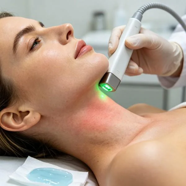
Sessions 9 through 12 focused on consolidating neural recovery and promoting tissue repair. High-frequency pulse trains at 8,000Hz stimulated axonal repair pathways while minimizing surface thermal accumulation. By Session 12, both neck and arm pain scores dropped to zero, the Neck Disability Index reached a normal 4 percent, and left cervical rotation expanded to 43 degrees without symptom reproduction. At the 90-day follow-up, the patient maintained full cervical range of motion and complete functional independence, working full workdays without symptom recurrence.
Handpiece Delivery Dynamics and Epidermal Thermal Management
Safely delivering high-power Class 4 laser therapy requires strict adherence to moving-beam application techniques. Because high photon intensities are used, leaving the handpiece static over pigmented skin or superficial bony landmarks risks localized heat spikes that cause discomfort or thermal injury.
Handpiece Motion Architecture:
[Zone 1: C2-C4 Paraspinal Baseline]   <---> Rapid Transverse Smoothing Sweep (4 cm/s)
[Zone 2: C5-C7 Foraminal Epicenter]   <---> Overlapping Figure-Eight Concentrated Passes (3 cm/s)
[Zone 3: Scapular Levator Insertion]  <---> Longitudinal Stripping Strokes toward Lymphatic Gates
The clinical application uses a dynamic scanning approach:
- Controlled Scanning Velocity: The treatment optic moves continuously across the target area at a uniform velocity of 3 to 5 centimeters per second. This rate deposits therapeutic energy into underlying structures while keeping the skin well within comfortable thermal limits.
- Tissue Compression Mechanics: Over dense muscular areas, clinicians apply gentle mechanical contact using a curved optical glass massage head. This direct contact blanches superficial dermal capillary beds; reducing superficial hemoglobin absorption allows photons to penetrate deeper into underlying fascia and facet structures. In areas over superficial bony prominences, such as the C7 spinous process, clinicians switch to a non-contact standoff distance of 20 to 30 millimeters, using quick, sweeping passes to disperse heat.
- Surface Thermal Monitoring: The handpiece incorporates an integrated infrared pyrometer that measures real-time skin surface temperature. The clinical objective is to maintain skin temperature within the therapeutic window of 39.0 to 41.5 degrees Celsius. If surface temperatures approach 42.0 degrees Celsius, the operator increases handpiece velocity or reduces the duty cycle, maintaining continuous photon delivery without exceeding epidermal thermal thresholds.
High Intensity Laser Integration in Spine Care
Treating chronic cervical spondylosis has long highlighted the trade-offs inherent in conventional treatment approaches. High-intensity Class 4 laser therapy provides a targeted, non-invasive alternative across several common clinical pathways:
+------------------------------------------------------------------------------------------------------+
| Treatment Modality      | Target Depth        | Primary Mechanism         | Clinical Bottlenecks     |
+------------------------------------------------------------------------------------------------------+
| Low-Level Light Therapy | 2 mm to 8 mm        | Superficial Photochemistry| Cannot penetrate to root |
| Mechanical Traction     | Axial Spine Segment | Foraminal Widening        | Transient decompression  |
| Oral NSAID Therapy      | Systemic Circulation| Systemic COX Inhibition   | Organ toxicity concerns  |
| Epidural Corticosteroid | Local Foraminal     | Receptor-Mediated Block   | Invasive; structural loss|
| Surgical Foraminotomy   | Direct Neural Canal | Surgical Bony Resection   | Structural trauma risk   |
| Class 4 Multi-Wave Laser| 35 mm to 60 mm      | Photobiomodulation & Edema| Requires high power unit |
+------------------------------------------------------------------------------------------------------+
Low-Level Light Therapy operates in the milliwatt range. While it helps superficial soft tissue strains, its low energy cannot overcome the scattering barriers of deep posterior neck musculature. High-power systems deliver the incident irradiance needed to maintain an effective forward-propagating photon field that reaches the compressed nerve root within the intervertebral foramen.
Mechanical traction temporarily separates vertebral bodies and widens the neural exit canal, but its mechanical benefits diminish once axial loading returns during upright posture. It does not resolve intraneural edema or stimulate mitochondrial energy production within ischemic axons. High-intensity laser therapy targets local tissue biochemistry directly, accelerating fluid resorption and downregulating persistent pro-inflammatory cascades.
Systemic anti-inflammatory medications carry well-documented gastrointestinal, renal, and cardiovascular risks when used long-term, and their systemic distribution provides little active drug concentration to poorly vascularized degenerative discs. Epidural corticosteroid injections introduce strong anti-inflammatory agents directly to the target nerve root, but repeated injections risk weakening surrounding supportive ligaments and carry hazards of dural puncture or infection. Surgical foraminotomy effectively removes structural impingements, but tissue resection can alter local biomechanics, occasionally leading to adjacent-segment degeneration and requiring lengthy postoperative rehabilitation.
High-power multi-wavelength Class 4 laser therapy bridges the gap between passive modalities and invasive procedures. By combining deep spatial penetration, tissue-specific wavelength targeting, and safe thermal management, this modality provides spine clinicians with a reproducible, non-surgical tool to relieve nerve root compression and address the structural drivers of degenerative cervical spine disease.