 FotonMedix




Overcoming Forefoot Ischemia and Refractory Calcaneal Entrapment
Targeted near-infrared photonic flux restores stalled endothelial capillary networks, downregulates sensory nociceptive pathways, and stimulates tenocyte matrix remodeling without collateral cutaneous burns.
Arterial insufficiency in the distal extremity combined with chronic biomechanical fascial contracture presents an intractable barrier in outpatient lower-limb clinics. When patients present with non-healing trans-metatarsal amputation margins, atonic neuro-ischemic ulcers, or severe calcified plantar fasciitis, standard conservative regimens routinely hit a physiological wall. Clinicians frequently encounter severe photon attenuation across calloused dermal layers and hypoperfused subcutaneous matrices, leaving deep microvascular networks and scarred ligamentous origins undertreated. Resolving these intertwined pathologies demands multi-wavelength photonic control capable of driving regenerative energy through dense tissue barriers while preserving friable wound margins.
Photon Attenuation Dynamics across Fibrotic Connective and Vascular Beds
Biological tissue in the human foot exhibits extreme optical heterogeneity characterized by high scattering coefficients and layered chromophore absorption. When photons target the plantar surface or the margins of a chronic distal stump, light must cross a dense, hyperkeratotic stratum corneum, fibrous subcutaneous fat pads, and thick fascial envelopes before reaching the deep plantar aponeurosis or dormant capillary loops situated several centimeters beneath the surface.
Transmittance through these dense lower-limb tissues follows an exponential decay profile dictated by scattering and selective molecular absorption. Melanin in the epidermal basal layer and water in edematous interstitial spaces absorb specific spectral bands, while cross-linked, disorganized collagen fibers scatter uncollimated beams in multiple directions. Low-power, single-spectrum modalities lose operational irradiance within the first few millimeters of tissue, generating mild surface warmth while failing to deliver therapeutic photon densities to ischemic wound beds or degenerated plantar fascia.
Deploying high-power, multi-wavelength configurations overcomes this penetration barrier by matching discrete spectral outputs to specific physiological targets. An 810nm output directly targets the oxidized copper centers of cytochrome c oxidase in mitochondrial membranes, accelerating electron transport, increasing adenosine triphosphate synthesis, and stimulating reparative cellular metabolism.
Pairing this with a 980nm wavelength leverages strong affinity for both oxyhemoglobin and water. This induces a brief, controlled micro-thermal response that triggers local capillary dilation, clearing out accumulated inflammatory chemical mediators like bradykinin, prostaglandin E2, and substance P.
Adding a 1064nm output extends light penetration even deeper into dense fibrous envelopes. Because 1064nm encounters minimal melanin absorption and low scatter, it penetrates through thick fibrous sheaths to treat damaged intra-articular capsules and deep muscle-tendon junctions.
Where tissue breakdown involves tortuous feeding vessels or localized venous reflux, deploying professional laser vascular treatment devices emitting at 1470nm seals incompetent micro-channels. Interstitial water within vascular endothelial walls absorbs 1470nm light roughly forty times more readily than 980nm light. This high affinity enables precise photocoagulation of abnormal micro-vessels within a tight 1 to 1.5 millimeter margin, preventing erratic lateral thermal spread.
This integrated multi-spectral profile establishes high-intensity laser therapy for slow-healing injuries as an essential clinical protocol to revive dormant granulating tissue and repair recalcitrant soft tissue attachments.
Thermal Clearance Balancing via Calibrated Pulse Duty Cycles
Continuous-wave energy delivery carries high clinical risks when treating neuropathy-compromised or poorly perfused extremities. Delivering unmodulated laser energy generates steady heat buildup within tissue layers. If continuous photon absorption outpaces passive capillary heat clearance, local temperatures climb toward the 43-degree Celsius threshold, risking protein denaturation, microvascular thrombosis, and secondary tissue breakdown.
Modern treatment protocols avoid this risk by matching energy delivery to the Thermal Relaxation Time of the targeted structures. Thermal relaxation time represents the duration needed for treated cellular structures or microvascular envelopes to disperse half of their absorbed heat through passive capillary conduction.
Applying calibrated pulse duty cycles breaks up energy delivery into microsecond bursts:
Duty Cycle (%) = [Pulse On Duration / (Pulse On Duration + Pulse Off Duration)] x 100
Restricting the active pulse duration within the tissue cooling window allows deep neurovascular junctions, damaged tenocytes, and stagnant wound margins to absorb high-intensity photon bursts without excessive heat retention. The alternating pulse-off intervals provide enough quiet time for surrounding viable capillaries to disperse residual warmth safely into adjacent tissues.
When treating chronic non-healing ulcers or dense plantar fascial insertions, selecting duty cycles between 20% and 40% permits peak power outputs up to 28W. These brief, high-energy pulses penetrate dense tissue boundaries, triggering deep photobiomodulation while keeping surface temperatures well within safe, comfortable levels.
Clinical Realities within the Evolving Podiatry Lasers Market
Outpatient foot and ankle clinics handle some of the most stubborn pathologies in modern medicine, including non-healing neuro-ischemic wounds, recalcitrant plantar fasciitis, and deep-seated fungal dystrophies.
The broader podiatry lasers market has long been dominated by low-power units that produce superficial warmth without sufficient photon density to stimulate deep cellular repair. Modern specialized practices are moving away from these underpowered systems, adopting multi-wavelength Class IV platforms that transition easily between fine microvascular coagulation, deep soft-tissue photobiomodulation, and non-contact wound decontamination.
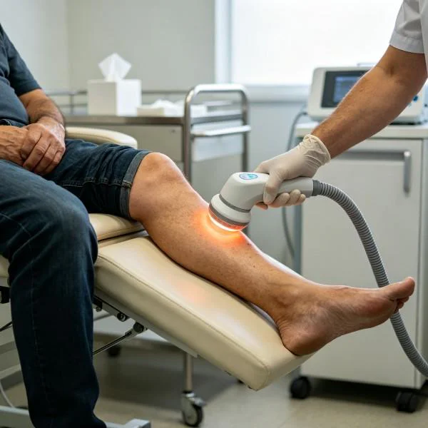
In recalcitrant neuropathic ulcers, repeated mechanical curettage frequently tears away newly formed, fragile epithelial buds. Non-contact laser irradiation decontaminates the ulcer bed without physical contact. Calibrated photon delivery disrupts resistant bacterial biofilms while simultaneously stimulating local fibroblasts, tenocytes, and capillary endothelial cells along the wound perimeter.
For chronic degenerative plantar fasciitis and recalcitrant heel pain, high photon densities pass straight through the thick plantar fascia and dense calcaneal fat pad. This restores mitochondrial energy production in strained connective tissues, breaks down chronic fascial adhesions, and relieves chronic heel pain without relying on corticosteroid injections that risk fascial rupture.
Restoring Microcirculatory Perfusion in Atonic Extremity Wounds
Chronic wound failure is driven by sustained microvascular stagnation. Vascular endothelial cells in long-standing neuro-ischemic ulcers or post-amputation wounds enter functional senescence, losing their capacity to form patent tubular capillaries. As a consequence, the wound core remains hypoxic, acidic, and trapped in an unresolving inflammatory cycle.
Deploying Class IV medical laser systems breaks this ischemic stalemate through three distinct biological steps:
- Capillary Sprouting and Endothelial Renewal: High photon irradiance triggers the release of vascular endothelial growth factor (VEGF) and basic fibroblast growth factor (bFGF) from perivascular pericytes. This initiates organized capillary sprouting from healthy peripheral margins into the ischemic ulcer core.
- Biofilm Decontamination: Targeted near-infrared wavelengths generate mild, localized photodynamic reactions within bacterial cell walls. This suppresses Pseudomonas aeruginosa and Staphylococcus aureus colonies without generating resistant bacterial strains or causing chemical trauma to fragile granulating margins.
- Extracellular Matrix Reorganization: Photon absorption accelerates procollagen mRNA transcription in local fibroblasts, helping the wound transition from disorganized provisional granulation to dense, well-aligned Type I collagen matrices that resist shear stress during ambulation.
Standardized Clinical Practice and Detailed Treatment Record
The following treatment log outlines an integrated clinical protocol addressing a chronic non-healing trans-metatarsal surgical stump wound paired with recalcitrant insertional plantar fasciitis in the contralateral limb.
Clinical Registry Record: FMX-2026-PU8190
| Parameter Group | Specification Detail | Clinical Target Value |
| 案例识别 | 临床病例编号 | FMX-2026-PU8190 |
| Patient Demographics | Age / Biological Sex | 61 Years / Male (Peripheral Artery Disease) |
| Primary Pathologies | Wound Diagnosis | Non-Healing Trans-Metatarsal Amputation Margin (Wagner Grade 2; 9-Month Stagnation) |
| Secondary Pathologies | Musculoskeletal Diagnosis | Recalcitrant Contralateral Insertional Plantar Fasciitis with Enthesophyte Formation |
| Device Configuration | Operational Classification | Multi-Wavelength High-Intensity Class IV Medical Laser |
| Delivery Mechanics (Wound Bed) | Non-Contact Handpiece | Defocused Collimated Handpiece with Thermal Distance Ring (22mm spot size) |
| Delivery Mechanics (Plantar Fascia) | Transcutaneous Handpiece | Deep-Tissue Collimated Handpiece with Real-Time Thermal Contact Ring |
| Wavelength Balance (Wound Bed) | Tri-Spectrum Ratio | 810nm (45%) + 980nm (35%) + 1064nm (20%) |
| Wavelength Balance (Plantar Fascia) | Quad-Spectrum Ratio | 810nm (40%) + 915nm (15%) + 980nm (25%) + 1064nm (20%) |
| Operational Power (Wound Bed) | Wattage Setting | 8.0W Average Power (Gated Pulsed Mode) |
| Operational Power (Plantar Fascia) | Wattage Setting | 18.0W Peak Duty-Cycle Pulsed Mode |
| Pulse Frequency (Wound Bed) | Repetition Rate | 10.0 Hz modulated down to 2.5 Hz |
| Pulse Frequency (Plantar Fascia) | Repetition Rate | 15.0 Hz modulated down to 4.0 Hz |
| Energy Dose Per Wound Session | 总输出焦耳数 | 1,350J across stump dehiscence line and periwound skin |
| Energy Dose Per Fascia Session | 总输出焦耳数 | 4,200J across medial calcaneal tubercle and plantar fascia band |
| Surface Preservation System | Cutaneous Safeguard | Continuous Infrared Non-Contact Thermal Monitoring (Target < 39°C) |
治疗进展与客观临床指标
| 治疗阶段 | Wound Surface Area (Square Centimeters) | Wound Granulation Quality (%) | Plantar First-Step Pain Score (VAS 0-10) | Plantar Fascia Thickness on Ultrasound (mm) | Functional Status and Tissue Observations |
| 基线(治疗前) | 4.6 cm² (Sluggish, ischemic margin) | 10% (Fibrotic, pale wound bed) | 8.5 / 10 (Severe morning pain) | 6.8 mm (Marked hypoechoic thickening) | Stump wound edges rolled and atonic; patient non-ambulatory due to bilateral foot pain |
| 第 7 天(第 3 節) | 3.8 cm² | 30% (Early pink capillary buds) | 6.0 / 10 | 5.9 mm | Wound exudate converted to serous; inflammatory erythema cleared along stump seam |
| Week 3 (Session 7) | 2.2 cm² | 75% (Healthy hyperemic bed) | 3.5 / 10 | 4.8 mm (Active matrix realignment) | Robust epidermal migration advancing from wound edges; patient tolerating partial weight-bearing |
| Week 6 (Session 12) | 0.6 cm² | 95% (Dense vascular coverage) | 1.0 / 10 | 3.9 mm | Stump wound contracting cleanly without hyperkeratosis; morning heel stiffness resolved |
| Week 10 (Final Evaluation) | 0.0 cm² (Complete structural closure) | 100% Fully Epithelialized | 0.0 / 10 (Pain-free ambulation) | 3.2 mm (Normal physiological thickness) | Full durable epidermal restoration over stump; patient walking independently in custom orthotics |
Scientific Citations and Biophysical Frameworks
The clinical efficacy of multi-wavelength photobiomodulation in treating non-healing lower-extremity wounds is supported by research led by Dr. Raymond J. Lanzafame and colleagues, published in Photomedicine and Laser Surgery. Their clinical evaluations demonstrated that near-infrared laser irradiation stimulates cellular mitochondrial output, accelerates wound closure rates, and upregulates microvascular perfusion in chronically hypoperfused tissue beds.
Targeted closure of incompetent microvascular channels builds on biophysical models established by Dr. R. Rox Anderson and Dr. John A. Parrish in their landmark study on Selective Photothermolysis, published in Science. Their work proved that matching laser pulse duration directly to the thermal relaxation time of targeted vascular structures produces selective endothelial photocoagulation without damaging surrounding parenchymal cells.
Further confirmation of cellular repair pathways comes from research by Dr. Tiina Karu, published in the Journal of Photochemistry and Photobiology B: Biology. Her research confirmed that near-infrared photon absorption activates cytochrome c oxidase within mitochondrial membranes, increasing adenosine triphosphate synthesis and initiating downstream gene expressions that drive cell proliferation and tissue repair.
In lower-extremity vascular medicine, clinical trials conducted by Dr. Thomas E. Serena and associates in the Journal of Wound Care showed that applying Class IV multi-wavelength laser therapy significantly suppresses local bacterial bioburden, accelerates wound edge migration, and shortens complete healing times in recalcitrant diabetic foot ulcers compared to standard moist wound therapy.
Clinical Realities: High Intensity Laser vs Conventional Approaches
Moving away from conventional wound care toward high-power Class IV laser therapy improves outcomes across both podiatric and outpatient vascular clinics.
Standard management of chronic lower-extremity wounds often relies on passive symptom care. Patients undergo repetitive mechanical debridement, receive rotating topical antimicrobial dressings, and face long courses of systemic antibiotics, carrying risks of drug-resistant infections, gastrointestinal upset, and persistent wound breakdown. When conservative measures fail, patients face aggressive surgical revascularization, extensive skin grafting, or progressive toe and partial-foot amputations that permanently impair mobility.
High-intensity Class IV laser therapy provides a tissue-preserving alternative that treats the underlying ischemia directly. By delivering dense photon flux through calloused wound perimeters, it restores microcirculatory blood flow, suppresses bacterial colonization, and stimulates dormant repair cells without mechanical trauma or chemical toxicity. Patients achieve steady, measurable wound closure and regain durable skin integrity while avoiding repetitive, painful debridements.
In managing chronic peripheral vascular lesions, standard options often default to chemical sclerotherapy or electrocautery. Chemical sclerotherapy can trigger localized tissue necrosis or allergic reactions, while electrocautery generates high temperatures exceeding 300 degrees Celsius that leave deep, carbonized wounds prone to secondary infection.
Utilizing dedicated high-power vascular laser systems provides a clean, precise approach. Matching wavelength absorption directly to hemoglobin and water enables instantaneous microvascular closure without open skin breakdown, post-procedural pain, or extended downtime.
Modern podiatry and vascular centers adopt these advanced laser systems to elevate their standard of care. By resolving the true physiological causes of poor wound healing and microvascular stasis, they deliver reliable, accelerated functional recovery that restores patient mobility and prevents limb loss.