 FotonMedix




克服椎间盘突出复发和全身性缺氧
靶向水峰值光气化可在不引起环状炭化的情况下缩小椎间盘内体积,微血管内照射可增强红细胞变形性,而动态脉冲占空比门控可防止热性继发损伤。
一名患者一瘸一拐地走进脊柱介入治疗室,一边捂着下背部,一边因坐骨神经痛导致左小腿直至脚外侧麻木无力。此前,患者已尝试过硬膜外类固醇注射、高剂量加巴喷丁及物理治疗,但均未获得持久疗效。 骨科医生建议进行开放式微创椎间盘切除术,但患者因需六周的康复期以及可能出现永久性硬膜外纤维化的风险而犹豫不决。与此同时,诊所管理者也面临着关于手术预算的质疑,因为潜在患者不断询问:与医院进行的开放式脊柱手术相比,激光椎间盘消融术的费用是多少。 手术室内的临床障碍纯粹是生物物理性的:无血管的髓核被困在坚硬的纤维环内,维持着过高的椎间盘内压力,从而压迫神经根,而周围的棘旁微血管则处于受压和缺氧状态。 若要在不进行手术切除的情况下缓解神经根痛,必须快速对髓核核心进行减压,并配合全身循环支持以消除神经源性炎症。
经皮椎间盘减压术的生物物理机制
经皮激光椎间盘减压术(PLDD)的原理是使髓核内的微量水分汽化。由于椎间盘是一个被坚韧的胶原纤维囊包裹的封闭液压空间,因此即使仅去除极少量的椎间盘内物质,也能立即使压力下降。
发色团亲和力与光学吸收动力学
要针对椎间盘组织进行治疗,必须使光能与髓核的主要结构成分——水——精确匹配。在退变性椎间盘中,虽然水分含量下降,但与周围致密的纤维环相比,髓核的核心部分仍主要由水构成。
采用双发射IV类高强度系统,将精确的光热切割与组织回收相结合:
- 1470nm波长的定向水汽化:1470nm波长发出的光与细胞内水的吸收峰相吻合。 该波长的吸收系数是标准近红外光谱的四十倍,这意味着辐射能量会在超薄边界层内瞬间转化为热量。该光束可蒸发核内水分,且不会向外散射,从而避免穿透后纵韧带或导致相邻脊神经根过热。
- 980nm波长对血红蛋白的刺激及凝血作用:980nm波长可靶向作用于相邻肉芽组织区内的脱氧血红蛋白和氧合血红蛋白池。这会触发瞬时的微血管内凝血,从而阻断环形撕裂裂隙处的炎症组织向内生长,且不会引起侧向炭化。
通过动态占空比门控实现散热
通过直径为400微米或600微米的光纤,将手术能量传导至敏感的椎管内,这带来了严格的热安全限制。在髓核内持续发射能量,可能会导致热量传导至软骨终板,从而引发无菌性骨坏死或晚期椎间盘塌陷。
脉冲占空比管理可规避这一风险。通过将辐射调制为毫秒级脉冲(例如,0.1 至 0.5 秒的导通时间,配合 0.5 至 1.0 秒的热松弛暂停),峰值光学功率可瞬间汽化核内水分。 休止阶段使周围的椎间盘内液体能够安全地吸收并散发热量,从而将终板温度维持在45摄氏度以下。这种经过校准的热门控技术使介入性脊柱专科医生能够在不灼伤相邻椎骨的情况下,安全地进行椎间盘减压手术。
+--------------------------------------------------------------------------------------------------+
| 椎间盘内光解压模型 |
+--------------------------------------------------------------------------------------------------+
| 经皮光纤引入(400微米芯径) |
|   | |
|   v |
| [纤维环外圈]  ---> 保持完整;光纤穿过小型导引套管   |
|   | |
|   v |
| [目标髓核核心] ---> 1470nm波长选择性汽化髓核内的水分子    |
|   | |
|   v |
| [微腔形成] ---> 微小体积减少导致椎间盘内液压力下降 |
|   | |
|   v |
| [突出物回缩] ---> 突出物从脊神经根处回缩 |
|   | |
|   v |
| [终板安全区] ---> 脉冲占空比可防止热量向骨性终板扩散  |
+--------------------------------------------------------------------------------------------------+
通过静脉输注光照实现全身微血管氧合
缓解椎间盘内的机械性神经压迫,仅解决了神经根病变的一半问题。长期存在的椎间盘突出会引发慢性神经源性炎症、静脉淤血,以及受累腰骶神经丛沿线的周围毛细血管簇集。
血管内光疗(通常被称为静脉激光疗法)可为消除组织淤滞提供全身性支持。当通过专用光纤血管导管将相干IV类光引入血液循环时,光子会直接与循环中的红细胞、白细胞和血管内皮细胞发生作用。
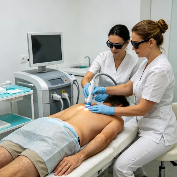
血管内线粒体复苏与血液流变学
激光静脉内治疗会改变循环血液细胞的物理特性。Schikora及其同事发表在《临床光医学》上的一项研究表明,直接的血管内光子递送会改变红细胞膜的表面电荷:
- 提高红细胞变形性:血管内的光子可使红细胞罗勒状聚集解聚,恢复红细胞的柔韧性,使其能够挤过供应缺氧神经鞘的狭窄毛细血管。
- 全身性一氧化氮动员:光刺激可释放与血红蛋白结合的一氧化氮,从而引起全身微血管扩张,并降低外周血管阻力。
- 免疫调节:循环中的免疫细胞吸收光子,从而引发白细胞介素-6和肿瘤坏死因子-α等促炎性细胞因子的下调,这直接降低了神经超敏反应。
临床方案实施:核消融与血管内全身输注的比较
操作高强度IV类设备时,必须明确区分经皮外科消融模式与血管内生物刺激模式。
在经皮椎间盘减压术中,临床医生在直接荧光透视引导下使用细硅石英纤维。 该技术的目标是光热汽化:通过短暂且受控的脉冲间隔,精准输送目标能量剂量(通常每个椎间盘间隙总计1,200至1,800焦耳),以收缩髓核,同时避免椎间盘间隙过热。
对于血管内康复治疗,其临床目标已完全从热气化转变为非破坏性光生物调节。激光连接到无菌的血管内光纤治疗头,以较低的功率水平(每波长50mW至200mW)进行长达30至45分钟的治疗。 交替使用蓝光、绿光和近红外光波段,可在不产生血管内局部热量的同时,刺激线粒体ATP生成和免疫通路的活性。
临床病例记录:介入性脊柱与再生医学门诊
以下临床数据概述了一例患有局限性腰椎间盘突出和持续性神经根病变患者的记录在案的治疗过程,该患者采用双波长(980nm/1470nm)手术激光平台联合术后全身性血管内治疗进行治疗。
患者概况与临床基线
- 病例编号:SPINE-RAD-2026-0941
- 患者年龄/性别:46岁女性
- 主要诊断:L4-L5节段亚韧带内椎间盘突出伴椎间孔狭窄,经腰椎MRI确诊,并发慢性左侧L5神经根病变及继发性小腿微血管缺血。
- 病史:因搬运重物受伤后,持续11个月出现严重的下背部和左腿疼痛。物理治疗、整脊治疗以及两次经椎间孔硬膜外类固醇注射仅能带来短暂的缓解。
- 基线指标:下肢疼痛的数值疼痛评分量表(NPRS)评分为8/10;奥斯韦斯特里残疾指数(ODI)为54%;左侧直腿抬高试验(SLR)在35度时呈阳性。
多阶段治疗方案
该临床方案将单阶段经皮椎间盘气化术与为期三周、共八次的全身性血管内恢复方案相结合。
| 会议编号. | 研究方法与目的 | 波长设置 | 功率输出 | 频率/占空比 | 总能量(焦耳) | 累积剂量 (J) |
| 手术日 | L4-L5 经皮椎间盘消融术 | 1470nm(80%)+ 980nm(20%) | 8.0 瓦 | 1.0秒开 / 1.0秒关(50%) | 1,450焦耳 | 1,450焦耳 |
| 第01节 | 术后静脉内免疫调节 | 650nm (50%) + 405nm (50%) | 0.08 瓦 | 连续波(CW)100% | 144 J | 1,594 J |
| 第02节 | 全身微循环与血管舒张 | 532nm(40%)+ 980nm(60%) | 0.10 瓦 | 连续波(CW)100% | 180 J | 1,774 J |
| 第03节 | 红细胞变形性优化 | 650nm (50%) + 532nm (50%) | 0.10 瓦 | 连续波(CW)100% | 180 J | 1,954 J |
| 第04节 | 神经周围微血管再通 | 980nm (70%) + 650nm (30%) | 0.12 瓦 | 连续波(CW)100% | 216 J | 2,170 J |
| 第05节 | 线粒体细胞色素C的增强 | 650nm (60%) + 810nm (40%) | 0.12 瓦 | 连续波(CW)100% | 216 J | 2,386 J |
| 第06节 | 抗炎细胞因子的平衡 | 405nm (40%) + 650nm (60%) | 0.10 瓦 | 连续波(CW)100% | 180 J | 2,566 J |
| 第07节 | 全身性内皮修复支持 | 532nm(50%)+ 980nm(50%) | 0.12 瓦 | 连续波(CW)100% | 216 J | 2,782 J |
| 第08节 | Final Neuro-Vascular Homeostasis | 650nm (50%) + 810nm (50%) | 0.15 瓦 | 连续波(CW)100% | 270 J | 3,052 J |
临床进展与预后指标
SURGICAL PROCEDURE (Percutaneous Disc Decompression):
- Under fluoroscopic and local anesthesia, an 18G cannula was placed into the postero-lateral L4-L5 disc core.
- A 400-micron quartz fiber delivered 1,450 Joules of gated 1470nm/980nm energy.
- Real-time intradiscal manometry documented an immediate 42% drop in core resting pressure.
- Immediate post-procedure leg pain dropped from 8/10 to 3/10 as neural impingement eased.

POST-OPERATIVE REHABILITATION (Sessions 1-4):
- Patient experienced no post-operative hematoma or wound soreness; ambulatory within 45 minutes.
- Intravenous sessions cleared residual left calf numbness and night spasms.
- Left leg Straight Leg Raise test improved from baseline 35° to 65°.
- Oswestry Disability Index declined from 54% to 26% by the end of week two.

FINAL OUTCOME CONSOLIDATION (Sessions 5-8):
- Complete resolution of left lower extremity motor weakness (dorsiflexion strength 5/5).
- Follow-up dynamic ultrasound showed restored paraspinal capillary perfusion.
- Straight Leg Raise negative bilaterally at 85°.
- Final NPRS scored 0/10 at rest, 1/10 after 30 minutes of continuous treadmill walking.
- ODI reached 6%, showing complete functional return to full work and daily life.
比较分析:高强度激光与传统脊柱介入治疗
Examining minimally invasive laser decompression alongside conventional surgical procedures highlights why modern outpatient centers adopt optical treatments.
Open Microdiscectomy
Open microdiscectomy remains the surgical standard, yet it involves cutting paraspinal musculature, stripping periosteum, and resecting ligamentum flavum to access the canal. This tissue trauma often causes permanent epidural scar tissue that can bind to the dural sac, producing failed back surgery syndrome. In comparison, percutaneous laser disc decompression enters the disc through an 18-gauge needle puncture. The paraspinal muscular sleeve, posterior ligaments, and bony laminar arches remain intact, avoiding structural destabilization.
Radiofrequency Nucleoplasty
Radiofrequency coblation relies on ionic plasma fields to dissolve disc material. However, radiofrequency electrodes require broader tip sweeps inside the disc to ablate tissue, which can damage the annular wall if swept too close to the margins. High-intensity 1470nm lasers deliver concentrated optical energy through thin, flexible fibers with predictable thermal profiles, vaporizing target nuclear fluid without mechanical scraping.
Epidural Steroid Infiltration
Steroid injections flood the epidural space with anti-inflammatory compounds to dull nerve root pain temporarily. They do not alter the physical herniation compressing the nerve. When steroid benefits fade after several weeks, patients remain at risk of structural disc collapse. Minimally invasive laser techniques reduce physical core pressure, providing an anatomical solution rather than simply masking pain signals.
临床实践应用与操作要点
Integrating minimally invasive laser disc ablation and intravenous phototherapy into outpatient clinical routines requires attention to operational technique:
- Real-time energy monitoring: Always calculate cumulative energy totals using built-in system software. Clinicians should pause every 200 Joules during disc procedures to allow heat to dissipate, preventing damage to the vertebral endplates.
- Fluoroscopic positioning: Confirm needle and fiber tip placement using biplanar fluoroscopy. The optical fiber tip must sit within the posterior third of the nucleus pulposus on lateral views, well clear of the posterior annular wall.
- Combining local ablation with systemic support: Pair percutaneous intradiscal decompression with post-procedural systemic microvascular therapy. Decompressing the disc mechanically while supporting microcirculatory blood flow with intravenous sessions helps prevent persistent post-procedural nerve hypersensitivity.
When structured around clear tissue physics, class IV high-intensity laser systems provide outpatient clinics with a reliable, minimally invasive solution for complex disc and nerve conditions.
上一页 应对慢性难治性猫口腔炎下一个 克服犬类脊柱压迫性淤滞