 فوتون ميديكس












التغلب على نقص الأكسجة المفصلي وضعف الحواجز الجلدية
Dual-band coherent optical wavelengths bypass periarticular connective tissue scattering, synchronized microsecond duty cycles prevent epidermal thermal stacking, and targeted photon density triggers mitochondrial respiration in chondrocytes while accelerating granulation tissue closure across ulcerated margins.
A 67-year-old retired heavy-machinery mechanic enters the multidisciplinary outpatient clinic supported by an adult daughter on one side and a rolling walker on the other. Every attempt to bear weight on the left leg elicits a grimace and an audible clicking groan from the knee joint. The knee is visibly distorted by advanced valgus collapse and chronic capsular edema, with a secondary complication complicating standard care: over the pretibial skin and inferior patellar border, chronic venous stasis and skin thinning from prior topical steroid applications have produced a shallow, non-healing ulcerated erosion measuring nearly three centimeters across. The surrounding skin is indurated, purpuric, and fragile. The orthopedic team cannot schedule surgical total knee arthroplasty due to the open cuticular ulceration and elevated infection risks, while the wound care team cannot apply sustained mechanical compression dressings because the patient cannot tolerate contact pressure over the hyperalgesic arthritic joint. Conventional treatments fall short when confronted with this compound pathology. Oral anti-inflammatories exacerbate the patient’s stage-three chronic kidney disease, while direct needle injections risk introducing cutaneous bacteria into an already compromised synovial space. The primary clinical dilemma centers on light attenuation and thermal threshold control: the physician must project therapeutic photon fluxes four to five centimeters into the joint space to decompress capsular tension and stimulate hypoxic subchondral osteocytes, while simultaneously treating the superficial dermal wound bed without inducing thermal necrosis or tearing fragile granulation buds. Solving both conditions non-operatively demands an understanding of optical tissue absorption, precise wave selection, and thermal relaxation kinetics.
Multi-Layered Tissue Penetration Curves and Selective Chromophore Dynamics
Projecting photons through compromised skin into a degenerate joint capsule requires managing differing optical absorption and scattering coefficients. Photons encounter melanin, cellular water, hemoglobin pools, fibrous capsular retinacula, and pockets of joint effusion, with energy dropping along an exponential decay path if wavelengths are poorly matched to tissue targets.
Spectral Separation for Joint Articulation and Dermal Granulation
Achieving targeted tissue saturation without superficial heat damage relies on matching high-intensity class IV light to specific chromophore groups across deep and superficial compartments:
- Microvascular recruitment and hemocoagulation control at 980nm: The 980nm wavelength matches the optical absorption curve of oxygenated hemoglobin and dermal water layers. In degenerative joint capsules and chronic ulcer margins, localized microcirculation is sluggish and clogged with metabolic debris. High-intensity 980nm photons create controlled, sub-ablative thermal gradients within endothelial walls, stimulating endogenous nitric oxide release, opening dormant microcapillary loops, and initiating lymphatic drainage without mechanical trauma.
- Mitochondrial cytochrome c oxidase saturation at 1064nm: The 1064nm band exhibits the lowest scattering coefficient among near-infrared therapeutic spectra in dense human connective tissue. These photons travel straight through dense capsular ligaments, subcutaneous fat, and synovial effusion pockets to reach the subchondral bone plate and articular cartilage. Inside dormant chondrocytes and subchondral osteoblasts, 1064nm light binds to cytochrome c oxidase, displacing inhibitory nitric oxide, rebuilding the mitochondrial proton gradient, and driving adenosine triphosphate (ATP) production required for cellular repair.
- تحفيز المصفوفة الظهارية السطحية عند طول موجة 650 نانومتر: تستهدف فوتونات الطيف الأحمر إنزيم السيتوكروم سي أوكسيديز الموجود داخل الخلايا الكيراتينية السطحية والخلايا الليفية الجلدية، مما يحفز تخليق البروكولاجين من النوع الأول والنوع الثالث لإغلاق القرح المفتوحة دون إحداث اختراق حراري عميق.
تعديل دورة التشغيل وإجراءات الحماية المتعلقة بالاسترخاء الحراري
Projecting fifteen to twenty-five Watts of continuous optical energy across an area with open skin ulceration and an inflamed joint quickly challenges the thermal relaxation time of human skin. Continuous wave delivery causes heat to accumulate rapidly in superficial dermal tissues, setting off epidermal pain receptors, risking skin burns, and destroying fragile capillary buds forming in an open wound bed.
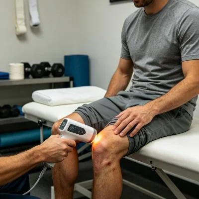
يحل نظام التحكم في دورة النبض المتغيرة هذه المشكلة التقنية. من خلال تعديل إرسال الحزمة إلى نبضات مدتها ميكروثانية (تتراوح من 10 هرتز إلى 20,000 هرتز) مع مدد نبضات وفترات راحة محكومة، تصل الطاقة القصوى العالية إلى الأنسجة العميقة خلال المرحلة النشطة، بينما تعمل الشعيرات الدموية السطحية على تبديد الحرارة خلال مرحلة الراحة. يحافظ هذا التنظيم الحراري على درجات حرارة سطح الجلد بأمان دون 42 درجة مئوية، مما يمكّن الأطباء من تطبيق علاج ليزري مستمر لالتهاب المفاصل مع حماية الجلد المحيط الهش وحواف القرحة.
+--------------------------------------------------------------------------------------------------+
|                    COMPOUND ARTICULAR AND CUTANEOUS PHOTON PROPAGATION                           |
+--------------------------------------------------------------------------------------------------+
| Non-Contact / Contact Fluid-Displacement Probe                                                   |
|   |                                                                                              |
|   v                                                                                              |
| [Epithelial Margin / Ulcer Bed] ---> 650nm accelerates fibroblast migration and closure          |
|   |                                                                                              |
|   v                                                                                              |
| [Dermal Papillary Capillaries]  ---> 980nm resolves stasis and clears pooled inflammatory fluid   |
|   |                                                                                              |
|   v                                                                                              |
| [Prepatellar Bursa & Retinaculum] -> Dynamic pulse duty cycle prevents surface thermal stacking   |
|   |                                                                                              |
|   v                                                                                              |
| [Synovial Joint Capsule]        ---> Suppression of IL-1beta, TNF-alpha, and COX-2 cascades      |
|   |                                                                                              |
|   v                                                                                              |
| [Articular Cartilage & Bone]    ---> 1064nm stimulates Cytochrome C Oxidase, restoring ATP flow   |
+--------------------------------------------------------------------------------------------------+
Biomolecular Mechanics of Chronic Joint Relief and Epithelial Closure
Treating coexisting articular cartilage degeneration and superficial skin loss requires balancing inflammatory cytokine suppression with tissue matrix synthesis. Degenerated chondrocytes and inflamed synovial cells produce catabolic cytokines, including interleukin-1 beta (IL-1β), tumor necrosis factor-alpha (TNF-α), and matrix metalloproteinases (MMPs), which break down type II collagen and aggrecan while sensitizing articular nociceptors.
Applying high-intensity coherent light directly interrupts this degenerative cycle. Targeted near-infrared irradiation downregulates nuclear factor kappa B (NF-κB) transcription inside synovial fibroblasts, decreasing the production of inflammatory mediators like prostaglandin E2 (PGE2) and cyclooxygenase-2 (COX-2) to help protect remaining cartilage integrity. This cellular stabilization underpins the documented long-term effects of laser therapy on arthritis, preventing the rebound stiffness and progressive capsular fibrosis that commonly follow temporary steroid blocks.
وفي الوقت نفسه، تستفيد العناية بالبشرة الهشة فوق مفصل مصاب بالتهاب المفاصل من اتباع تسلسلات تطبيق متخصصة. فبدلاً من وضع الأشرطة اللاصقة التقليدية الثقيلة مباشرةً على البشرة الهشة والمتصلبة، يقوم الأطباء بتنسيق العلاج الضوئي مع بروتوكول منظم للعلاج بالليزر مع تضميد الجروح. إن توجيه ضوء متعدد الأطوال الموجية غير التلامسي مباشرةً على قاع الجرح النظيف قبل وضع ضمادات رغوية من السيليكون غير اللاصقة يُسرّع تكوين النسيج الحبيبي، ويقلل من الحمل البكتيري، ويخلق بيئة محمية لتقدم حواف الظهارة.
وفي الوقت نفسه، فإن تطبيق العلاج بالليزر المنظم لألم التهاب المفاصل يؤثر بشكل مباشر على المسارات العصبية الحسية. حيث يؤدي توصيل جرعات عالية من الفوتونات إلى فرط استقطاب الألياف C غير الميالينية والألياف A-delta ذات الميالين الرقيق داخل الكبسولة المفصلية. وتؤكد الأبحاث التي أجراها هامبلين وزملاؤه في مجال الطب الضوئي أن الإضاءة عالية الطاقة في نطاق الأشعة تحت الحمراء القريبة تقلل من سرعة التوصيل العصبي المحيطي وتحد من تراكم مادة P في الأنسجة المحيطة بالمفصل، مما يوفر تخفيفًا سريعًا ودائمًا للألم دون آثار جانبية جهازية.
Clinical Case Documentation: Outpatient Orthopedic and Tissue Viability Service
The documented case below details the outpatient treatment course of a patient with advanced knee osteoarthritis complicated by an open pretibial stasis ulceration, treated using a multi-wavelength Class IV rehabilitation laser platform equipped with interchangeable quartz contact and non-contact optical heads.
الملف الشخصي للمريض والبيانات السريرية الأساسية
- Case Identifier: OSTEO-WOUND-2026-0924
- Patient Age / Sex: 67-Year-Old Male
- Primary Diagnosis: Advanced Left Knee Osteoarthritis (Kellgren-Lawrence Grade IV) with Severe Medial Joint Space Loss, Tricompartmental Osteophytosis, and Secondary Flexion Deformity, complicated by a Chronic Pretibial Cutaneous Stasis Ulcer measuring 2.8 cm x 1.9 cm x 0.3 cm with indurated peri-wound margins.
- History: 7 years of progressive joint pain and stiffness, with a non-healing lower leg ulcer persisting for 14 weeks. Systemic NSAIDs contraindicated due to baseline renal insufficiency; intra-articular steroid injections declined due to infection risks from the adjacent ulcer; surgical arthroplasty deferred until complete cutaneous closure is achieved.
- Baseline Metrics: Numerical Pain Rating Scale (NPRS) at 9/10 during weight-bearing attempts; Western Ontario and McMaster Universities Osteoarthritis Index (WOMAC) at 78/96; Knee Active Range of Motion (AROM) Flexion restricted to 75°, Extension lacking 16°; Bates-Jensen Wound Assessment Tool (BWAT) score at 34/65.
نظام علاجي متعدد المراحل
The treatment course comprised twelve clinical sessions scheduled over four weeks, progressing from acute wound decontamination and nociceptive gating to deep capsular saturation and functional mobility integration.
| رقم الجلسة. | المرحلة السريرية والهدف | إعداد الطول الموجي | طاقة التشغيل (واط) | تردد النبض (هرتز) | دورة التشغيل (%) | الطاقة المسلمة (J) | الجرعة التراكمية (جول) |
| الجلسة 01 | إزالة التلوث الناتج عن القرحة والتحكم في الإحساس بالألم | 650 نانومتر (30%) + 980 نانومتر (50%) + 1064 نانومتر (20%) | 10.0 واط | 10,000 هرتز | 30% | 3,000 J | 3,000 J |
| الجلسة 02 | Popliteal Drainage & Wound Edge Perfusion | 650 نانومتر (20%) + 980 نانومتر (50%) + 1064 نانومتر (30%) | 12.0 واط | 5,000 هرتز | 40% | 3,600 J | 6,600 جول |
| الجلسة 03 | Synovial Retinacular Flushing & Biofilm Control | 650 نانومتر (20%) + 980 نانومتر (40%) + 1064 نانومتر (40%) | 14.0 واط | 2,500 هرتز | 50% | 4,200 جول | 10,800 J |
| الجلسة 04 | Granulation Bed Activation & Fat Pad Release | 650 نانومتر (25%) + 980 نانومتر (35%) + 1064 نانومتر (40%) | 16.0 واط | 1,000 هرتز | 60% | 4,800 جول | 15,600 J |
| الجلسة 05 | Deep Medial Capsule Softening | 980 نانومتر (40%) + 1064 نانومتر (60%) | 18.0 واط | 300 هرتز | 65% | 5,400 J | 21,000 J |
| الجلسة 06 | Subchondral Plate Perfusion & Epithelial Edge Adv. | 650 نانومتر (20%) + 980 نانومتر (30%) + 1064 نانومتر (50%) | 20.0 واط | 150 هرتز | 70% | 6,000 J | 27,000 J |
| الجلسة 07 | High-Flux Chondrocyte Respiration Induction | 980 نانومتر (25%) + 1064 نانومتر (75%) | 22.0 واط | مستمر (CW) | 100% | 6,600 جول | 33,600 J |
| الجلسة 08 | إعادة تشكيل مصفوفة الغضروف الليفي الكثيفة | 980 نانومتر (20%) + 1064 نانومتر (80%) | 24.0 واط | مستمر (CW) | 100% | 7,200 J | 40,800 J |
| الجلسة 09 | Cutaneous Re-Epithelialization Consolidation | 650 نانومتر (40%) + 980 نانومتر (30%) + 1064 نانومتر (30%) | 18.0 واط | 100 هرتز | 70% | 5,400 J | 46,200 J |
| الجلسة 10 | إعادة التأهيل البيوميكانيكي بالتمديد الديناميكي | 980 نانومتر (30%) + 1064 نانومتر (70%) | 20.0 واط | 200 هرتز | 75% | 6,000 J | 52,200 J |
| الجلسة 11 | التكامل الوظيفي لسلسلة الحركة الحركية للمشي | 980 نانومتر (30%) + 1064 نانومتر (70%) | 20.0 واط | مستمر (CW) | 100% | 6,000 J | 58,200 J |
| الجلسة 12 | التثبيت النهائي للأنسجة والخروج من المستشفى | 650 نانومتر (20%) + 980 نانومتر (40%) + 1064 نانومتر (40%) | 16.0 واط | 500 هرتز | 65% | 4,800 جول | 63,000 J |
مؤشرات التطور السريري والنتائج
WEEK 1 (Sessions 1-3):
- Non-contact scanning over the pretibial ulcer cleared slough; healthy red granulation islands appeared in the wound bed.
- Gentle contact sweeping over the popliteal drainage basin and medial joint line reduced periarticular tension.
- Resting nocturnal pain subsided; NPRS dropped from 9/10 to 5/10.
- Patient slept through the night without waking from positional joint aching after Session 2.

WEEK 2 (Sessions 4-6):
- Pretibial ulcer margins advanced inward; wound surface area shrank from 2.8 x 1.9 cm to 1.6 x 1.1 cm with zero maceration.
- Knee active flexion expanded from 75° to 102°; extension deficit reduced from 16° to 6°.
- Patient stood from a seated position without pulling on assistive furniture arms.
- BWAT wound score improved from 34/65 to 20/65; WOMAC functional score improved from 78 to 44.

WEEK 3 (Sessions 7-9):
- Continuous wave delivery applied over the patellar retinaculum and tibiofemoral margins with firm compression.
- Complete epithelial closure of the pretibial ulcer achieved by Session 9, presenting a fully re-epithelialized skin layer.
- Patient walked 500 meters using only a single-point cane without triggering joint instability or skin breakdown.
- Crepitus during active knee flexion softened considerably as synovial congestion cleared.

WEEK 4 (Sessions 10-12):
- Full active knee extension achieved (0° neutral); active flexion reached 124°.
- Ulcer site completely healed (BWAT score 13/65, representing closed skin); no localized breakdown under direct pressure.
- Final NPRS recorded at 1/10 during extended walking and 0/10 at rest.
- WOMAC score reached 12/96, restoring daily mobility and clearing the patient medically for elective surgical review.
تحليل مقارن: الليزر عالي الكثافة مقابل الأساليب التقليدية
Evaluating high-intensity class IV light alongside traditional clinical options shows why modern physical medicine centers integrate multi-wavelength lasers into daily care:
حقن الكورتيكوستيرويدات داخل المفصل
Steroid injections act as broad chemical immunosuppressants that temporarily mask joint inflammation. However, when an adjacent skin ulcer or vascular breakdown is present, puncturing the joint capsule risks introducing surface pathogens into the synovial cavity, potentially causing septic arthritis. Repeated intra-articular steroid exposure also accelerates cartilage loss, damages native chondrocyte viability, and risks narrowing the joint space over time. High-intensity multi-wavelength laser therapy provides a non-invasive, non-destructive alternative. Rather than damaging chondrocytes, concentrated photons stimulate microvascular circulation, boost cellular ATP synthesis, and reduce joint discomfort without chemical toxicity or infection risks.
Surgical Debridement and Skin Grafting
Surgical debridement of stasis ulcers followed by autologous split-thickness skin grafting requires donor site harvesting and prolonged limb immobilization. In patients with severe arthritis, immobilization causes joint contracture, muscular atrophy, and increased pain upon remobilization. Class IV laser protocols allow simultaneous wound healing and joint mobilization in an outpatient setting, promoting steady re-epithelialization while preserving joint movement without surgical trauma.
الأدوية المضادة للالتهاب غير الستيرويدية (NSAIDs) التي تُؤخذ عن طريق الفم
Systemic anti-inflammatory medications circulate throughout the body, inhibiting protective prostaglandins in the gastric lining and kidneys while delivering limited drug concentrations into poorly vascularized articular cartilage and ulcerated skin margins. Chronic NSAID use carries well-documented risks of gastric ulceration, hypertension, and renal impairment. High-intensity laser therapy targets therapeutic energy directly into the damaged joint capsule and wound bed, relieving joint pain and stiffness locally without systemic adverse effects.
التطبيق السريري العملي والدروس المستفادة على الصعيد التشغيلي
يتطلب تحقيق نتائج سريرية متسقة عند استخدام أنظمة الليزر عالية الكثافة لعلاج التهاب المفاصل المعقد الذي يصاحبه ضعف في الحواف الجلدية اتباع نهج منظم في التنفيذ:
- تقديم العلاج على منطقتين: قم بتقسيم المنطقة السريرية إلى منطقة الجرح المفتوح ومنطقة المحفظة العميقة. استخدم عاكسًا كوارتزيًا لا يلامس الجلد على مسافة تتراوح بين سنتيمترين وثلاثة سنتيمترات فوق القرح الجلدية المفتوحة لتوصيل الضوء الأحمر والأشعة تحت الحمراء القريبة المحفزة بيولوجيًا دون لمس النسيج الحبيبي الهش. ثم انتقل إلى استخدام مسبار ضغط كوارتزي ناعم فوق الأنسجة الرخوة المغلقة المحيطة بالمفصل لإزاحة السوائل السطحية وتوصيل الفوتونات إلى عمق كبسولة المفصل.
- Regional lymphatic clearing: Always treat proximal lymphatic pathways first. Sweeping the popliteal space, femoral triangle, and proximal calf musculature before directing energy into the joint capsule clears fluid pathways and accelerates tissue recovery.
- بروتوكول التدرج الترددي: يبدأ العلاج بترددات نبضية عالية (من 4,000 هرتز إلى 10,000 هرتز) خلال الزيارات الأولية لتهدئة المسارات العصبية الطرفية شديدة الحساسية، ثم الانتقال إلى نبضات معتدلة (من 100 هرتز إلى 500 هرتز) لتحسين التروية المحلية وتخفيف تصلب المحفظة، وانتهي بإرسال موجة مستمرة (CW) لتحفيز التمثيل الغذائي للخلايا الغضروفية وإعادة تشكيلها الهيكلي بمجرد أن يهدأ الألم الحاد.
Applying class IV multi-wavelength laser therapy grounded in tissue optical physics allows outpatient orthopedic and wound care clinics to resolve stubborn, chronic joint degeneration and help patients return to active, pain-free daily lives.