 FotonMedix












Superar la hipoxia articular y el deterioro de las barreras dérmicas
Dual-band coherent optical wavelengths bypass periarticular connective tissue scattering, synchronized microsecond duty cycles prevent epidermal thermal stacking, and targeted photon density triggers mitochondrial respiration in chondrocytes while accelerating granulation tissue closure across ulcerated margins.
A 67-year-old retired heavy-machinery mechanic enters the multidisciplinary outpatient clinic supported by an adult daughter on one side and a rolling walker on the other. Every attempt to bear weight on the left leg elicits a grimace and an audible clicking groan from the knee joint. The knee is visibly distorted by advanced valgus collapse and chronic capsular edema, with a secondary complication complicating standard care: over the pretibial skin and inferior patellar border, chronic venous stasis and skin thinning from prior topical steroid applications have produced a shallow, non-healing ulcerated erosion measuring nearly three centimeters across. The surrounding skin is indurated, purpuric, and fragile. The orthopedic team cannot schedule surgical total knee arthroplasty due to the open cuticular ulceration and elevated infection risks, while the wound care team cannot apply sustained mechanical compression dressings because the patient cannot tolerate contact pressure over the hyperalgesic arthritic joint. Conventional treatments fall short when confronted with this compound pathology. Oral anti-inflammatories exacerbate the patient’s stage-three chronic kidney disease, while direct needle injections risk introducing cutaneous bacteria into an already compromised synovial space. The primary clinical dilemma centers on light attenuation and thermal threshold control: the physician must project therapeutic photon fluxes four to five centimeters into the joint space to decompress capsular tension and stimulate hypoxic subchondral osteocytes, while simultaneously treating the superficial dermal wound bed without inducing thermal necrosis or tearing fragile granulation buds. Solving both conditions non-operatively demands an understanding of optical tissue absorption, precise wave selection, and thermal relaxation kinetics.
Multi-Layered Tissue Penetration Curves and Selective Chromophore Dynamics
Projecting photons through compromised skin into a degenerate joint capsule requires managing differing optical absorption and scattering coefficients. Photons encounter melanin, cellular water, hemoglobin pools, fibrous capsular retinacula, and pockets of joint effusion, with energy dropping along an exponential decay path if wavelengths are poorly matched to tissue targets.
Spectral Separation for Joint Articulation and Dermal Granulation
Achieving targeted tissue saturation without superficial heat damage relies on matching high-intensity class IV light to specific chromophore groups across deep and superficial compartments:
- Microvascular recruitment and hemocoagulation control at 980nm: The 980nm wavelength matches the optical absorption curve of oxygenated hemoglobin and dermal water layers. In degenerative joint capsules and chronic ulcer margins, localized microcirculation is sluggish and clogged with metabolic debris. High-intensity 980nm photons create controlled, sub-ablative thermal gradients within endothelial walls, stimulating endogenous nitric oxide release, opening dormant microcapillary loops, and initiating lymphatic drainage without mechanical trauma.
- Mitochondrial cytochrome c oxidase saturation at 1064nm: The 1064nm band exhibits the lowest scattering coefficient among near-infrared therapeutic spectra in dense human connective tissue. These photons travel straight through dense capsular ligaments, subcutaneous fat, and synovial effusion pockets to reach the subchondral bone plate and articular cartilage. Inside dormant chondrocytes and subchondral osteoblasts, 1064nm light binds to cytochrome c oxidase, displacing inhibitory nitric oxide, rebuilding the mitochondrial proton gradient, and driving adenosine triphosphate (ATP) production required for cellular repair.
- Inducción de la matriz epitelial superficial a 650 nm: los fotones del espectro rojo actúan sobre la citocromo c oxidasa presente en los queratinocitos superficiales y los fibroblastos dérmicos, estimulando la síntesis de procolágeno de tipo I y tipo III para sellar las úlceras abiertas sin generar una penetración térmica profunda.
Modulación del ciclo de trabajo y medidas de seguridad contra la relajación térmica
Projecting fifteen to twenty-five Watts of continuous optical energy across an area with open skin ulceration and an inflamed joint quickly challenges the thermal relaxation time of human skin. Continuous wave delivery causes heat to accumulate rapidly in superficial dermal tissues, setting off epidermal pain receptors, risking skin burns, and destroying fragile capillary buds forming in an open wound bed.
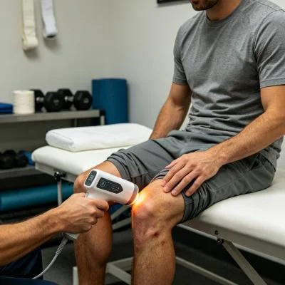
La modulación del ciclo de trabajo variable resuelve este problema técnico. Al modular la emisión del haz en ráfagas de microsegundos (con una frecuencia que oscila entre 10 Hz y 20 000 Hz) con duraciones de pulso e intervalos de reposo controlados, se consigue que una alta potencia de pico llegue a los tejidos profundos durante la fase activa, mientras que los capilares superficiales disipan el calor durante la fase de reposo. Esta regulación térmica mantiene la temperatura superficial de la piel por debajo de los 42 grados Celsius de forma segura, lo que permite a los médicos aplicar un tratamiento con láser prolongado para la artritis, al tiempo que se protege la frágil piel circundante y los bordes de las úlceras.
+--------------------------------------------------------------------------------------------------+
|                    COMPOUND ARTICULAR AND CUTANEOUS PHOTON PROPAGATION                           |
+--------------------------------------------------------------------------------------------------+
| Non-Contact / Contact Fluid-Displacement Probe                                                   |
|   |                                                                                              |
|   v                                                                                              |
| [Epithelial Margin / Ulcer Bed] ---> 650nm accelerates fibroblast migration and closure          |
|   |                                                                                              |
|   v                                                                                              |
| [Dermal Papillary Capillaries]  ---> 980nm resolves stasis and clears pooled inflammatory fluid   |
|   |                                                                                              |
|   v                                                                                              |
| [Prepatellar Bursa & Retinaculum] -> Dynamic pulse duty cycle prevents surface thermal stacking   |
|   |                                                                                              |
|   v                                                                                              |
| [Synovial Joint Capsule]        ---> Suppression of IL-1beta, TNF-alpha, and COX-2 cascades      |
|   |                                                                                              |
|   v                                                                                              |
| [Articular Cartilage & Bone]    ---> 1064nm stimulates Cytochrome C Oxidase, restoring ATP flow   |
+--------------------------------------------------------------------------------------------------+
Biomolecular Mechanics of Chronic Joint Relief and Epithelial Closure
Treating coexisting articular cartilage degeneration and superficial skin loss requires balancing inflammatory cytokine suppression with tissue matrix synthesis. Degenerated chondrocytes and inflamed synovial cells produce catabolic cytokines, including interleukin-1 beta (IL-1β), tumor necrosis factor-alpha (TNF-α), and matrix metalloproteinases (MMPs), which break down type II collagen and aggrecan while sensitizing articular nociceptors.
Applying high-intensity coherent light directly interrupts this degenerative cycle. Targeted near-infrared irradiation downregulates nuclear factor kappa B (NF-κB) transcription inside synovial fibroblasts, decreasing the production of inflammatory mediators like prostaglandin E2 (PGE2) and cyclooxygenase-2 (COX-2) to help protect remaining cartilage integrity. This cellular stabilization underpins the documented long-term effects of laser therapy on arthritis, preventing the rebound stiffness and progressive capsular fibrosis that commonly follow temporary steroid blocks.
Al mismo tiempo, el tratamiento de la piel frágil sobre una articulación artrítica se beneficia de secuencias de aplicación especializadas. En lugar de aplicar las tradicionales cintas adhesivas pesadas directamente sobre la piel friable e indurada, los profesionales sanitarios coordinan la fototerapia con un protocolo estructurado de tratamiento con láser para el vendaje de heridas. La aplicación de luz de múltiples longitudes de onda sin contacto directamente sobre el lecho de la herida limpio, antes de colocar apósitos de espuma de silicona no adherentes, acelera la formación de tejido de granulación, reduce la carga bacteriana y crea un entorno protegido para el avance de los bordes epiteliales.
Al mismo tiempo, la aplicación de la terapia láser estructurada para el dolor de la artritis actúa directamente sobre las vías nerviosas sensoriales. La administración de altas dosis de fotones hiperpolariza las fibras C no mielinizadas y las fibras A-delta con mielina fina dentro de la cápsula articular. Las investigaciones de Hamblin y sus colegas en el campo de la fotomedicina confirman que la iluminación con infrarrojo cercano de alta energía reduce la velocidad de conducción nerviosa periférica y frena la acumulación de sustancia P en los tejidos periarticulares, lo que proporciona un alivio rápido y duradero del dolor sin efectos secundarios sistémicos.
Clinical Case Documentation: Outpatient Orthopedic and Tissue Viability Service
The documented case below details the outpatient treatment course of a patient with advanced knee osteoarthritis complicated by an open pretibial stasis ulceration, treated using a multi-wavelength Class IV rehabilitation laser platform equipped with interchangeable quartz contact and non-contact optical heads.
Perfil del paciente y datos clínicos iniciales
- Case Identifier: OSTEO-WOUND-2026-0924
- Patient Age / Sex: 67-Year-Old Male
- Primary Diagnosis: Advanced Left Knee Osteoarthritis (Kellgren-Lawrence Grade IV) with Severe Medial Joint Space Loss, Tricompartmental Osteophytosis, and Secondary Flexion Deformity, complicated by a Chronic Pretibial Cutaneous Stasis Ulcer measuring 2.8 cm x 1.9 cm x 0.3 cm with indurated peri-wound margins.
- History: 7 years of progressive joint pain and stiffness, with a non-healing lower leg ulcer persisting for 14 weeks. Systemic NSAIDs contraindicated due to baseline renal insufficiency; intra-articular steroid injections declined due to infection risks from the adjacent ulcer; surgical arthroplasty deferred until complete cutaneous closure is achieved.
- Baseline Metrics: Numerical Pain Rating Scale (NPRS) at 9/10 during weight-bearing attempts; Western Ontario and McMaster Universities Osteoarthritis Index (WOMAC) at 78/96; Knee Active Range of Motion (AROM) Flexion restricted to 75°, Extension lacking 16°; Bates-Jensen Wound Assessment Tool (BWAT) score at 34/65.
Régimen de tratamiento en varias fases
The treatment course comprised twelve clinical sessions scheduled over four weeks, progressing from acute wound decontamination and nociceptive gating to deep capsular saturation and functional mobility integration.
| N.º de sesión. | Fase clínica y objetivo | Configuración de la longitud de onda | Potencia de funcionamiento (W) | Frecuencia de impulsos (Hz) | Ciclo de trabajo (%) | Energía suministrada (J) | Dosis acumulada (J) |
| Sesión 01 | Descontaminación de úlceras y filtrado nociceptivo | 650 nm (30%) + 980 nm (50%) + 1064 nm (20%) | 10,0 W | 10.000 Hz | 30% | 3,000 J | 3,000 J |
| Sesión 02 | Popliteal Drainage & Wound Edge Perfusion | 650 nm (20%) + 980 nm (50%) + 1064 nm (30%) | 12,0 W | 5.000 Hz | 40% | 3,600 J | 6,600 J |
| Sesión 03 | Synovial Retinacular Flushing & Biofilm Control | 650 nm (20%) + 980 nm (40%) + 1064 nm (40%) | 14,0 W | 2.500 Hz | 50% | 4 200 J | 10,800 J |
| Sesión 04 | Granulation Bed Activation & Fat Pad Release | 650 nm (25%) + 980 nm (35%) + 1064 nm (40%) | 16,0 W | 1.000 Hz | 60% | 4 800 J | 15,600 J |
| Sesión 05 | Deep Medial Capsule Softening | 980 nm (40%) + 1064 nm (60%) | 18,0 W | 300 Hz | 65% | 5,400 J | 21,000 J |
| Sesión 06 | Subchondral Plate Perfusion & Epithelial Edge Adv. | 650 nm (20%) + 980 nm (30%) + 1064 nm (50%) | 20,0 W | 150 Hz | 70% | 6,000 J | 27,000 J |
| Sesión 07 | High-Flux Chondrocyte Respiration Induction | 980 nm (25%) + 1064 nm (75%) | 22,0 W | Continuo (CW) | 100% | 6,600 J | 33,600 J |
| Sesión 08 | Remodelación de la matriz del fibrocartílago denso | 980 nm (20%) + 1064 nm (80%) | 24,0 W | Continuo (CW) | 100% | 7,200 J | 40,800 J |
| Sesión 09 | Cutaneous Re-Epithelialization Consolidation | 650 nm (40%) + 980 nm (30%) + 1064 nm (30%) | 18,0 W | 100 Hz | 70% | 5,400 J | 46,200 J |
| Sesión 10 | Reeducación biomecánica mediante extensión dinámica | 980 nm (30%) + 1064 nm (70%) | 20,0 W | 200 Hz | 75% | 6,000 J | 52,200 J |
| Sesión 11 | Integración funcional de la cadena cinética de la marcha | 980 nm (30%) + 1064 nm (70%) | 20,0 W | Continuo (CW) | 100% | 6,000 J | 58,200 J |
| Sesión 12 | Consolidación final del tejido y alta | 650 nm (20%) + 980 nm (40%) + 1064 nm (40%) | 16,0 W | 500 Hz | 65% | 4 800 J | 63,000 J |
Métricas de progresión clínica y resultados
WEEK 1 (Sessions 1-3):
- Non-contact scanning over the pretibial ulcer cleared slough; healthy red granulation islands appeared in the wound bed.
- Gentle contact sweeping over the popliteal drainage basin and medial joint line reduced periarticular tension.
- Resting nocturnal pain subsided; NPRS dropped from 9/10 to 5/10.
- Patient slept through the night without waking from positional joint aching after Session 2.

WEEK 2 (Sessions 4-6):
- Pretibial ulcer margins advanced inward; wound surface area shrank from 2.8 x 1.9 cm to 1.6 x 1.1 cm with zero maceration.
- Knee active flexion expanded from 75° to 102°; extension deficit reduced from 16° to 6°.
- Patient stood from a seated position without pulling on assistive furniture arms.
- BWAT wound score improved from 34/65 to 20/65; WOMAC functional score improved from 78 to 44.

WEEK 3 (Sessions 7-9):
- Continuous wave delivery applied over the patellar retinaculum and tibiofemoral margins with firm compression.
- Complete epithelial closure of the pretibial ulcer achieved by Session 9, presenting a fully re-epithelialized skin layer.
- Patient walked 500 meters using only a single-point cane without triggering joint instability or skin breakdown.
- Crepitus during active knee flexion softened considerably as synovial congestion cleared.

WEEK 4 (Sessions 10-12):
- Full active knee extension achieved (0° neutral); active flexion reached 124°.
- Ulcer site completely healed (BWAT score 13/65, representing closed skin); no localized breakdown under direct pressure.
- Final NPRS recorded at 1/10 during extended walking and 0/10 at rest.
- WOMAC score reached 12/96, restoring daily mobility and clearing the patient medically for elective surgical review.
Análisis comparativo: láser de alta intensidad frente a modalidades convencionales
Evaluating high-intensity class IV light alongside traditional clinical options shows why modern physical medicine centers integrate multi-wavelength lasers into daily care:
Inyecciones intraarticulares de corticosteroides
Steroid injections act as broad chemical immunosuppressants that temporarily mask joint inflammation. However, when an adjacent skin ulcer or vascular breakdown is present, puncturing the joint capsule risks introducing surface pathogens into the synovial cavity, potentially causing septic arthritis. Repeated intra-articular steroid exposure also accelerates cartilage loss, damages native chondrocyte viability, and risks narrowing the joint space over time. High-intensity multi-wavelength laser therapy provides a non-invasive, non-destructive alternative. Rather than damaging chondrocytes, concentrated photons stimulate microvascular circulation, boost cellular ATP synthesis, and reduce joint discomfort without chemical toxicity or infection risks.
Surgical Debridement and Skin Grafting
Surgical debridement of stasis ulcers followed by autologous split-thickness skin grafting requires donor site harvesting and prolonged limb immobilization. In patients with severe arthritis, immobilization causes joint contracture, muscular atrophy, and increased pain upon remobilization. Class IV laser protocols allow simultaneous wound healing and joint mobilization in an outpatient setting, promoting steady re-epithelialization while preserving joint movement without surgical trauma.
Medicamentos antiinflamatorios no esteroideos (AINE) de administración oral
Systemic anti-inflammatory medications circulate throughout the body, inhibiting protective prostaglandins in the gastric lining and kidneys while delivering limited drug concentrations into poorly vascularized articular cartilage and ulcerated skin margins. Chronic NSAID use carries well-documented risks of gastric ulceration, hypertension, and renal impairment. High-intensity laser therapy targets therapeutic energy directly into the damaged joint capsule and wound bed, relieving joint pain and stiffness locally without systemic adverse effects.
Aplicación clínica práctica y conclusiones operativas
Para obtener resultados clínicos consistentes al utilizar sistemas láser de alta intensidad en casos de artritis articular compleja con márgenes cutáneos comprometidos, es necesario seguir un procedimiento estructurado:
- Aplicación del tratamiento en dos zonas: divide el área clínica en una zona de herida abierta y una zona capsular profunda. Utilice un deflector de cuarzo sin contacto a una distancia de entre dos y tres centímetros sobre las úlceras cutáneas abiertas para aplicar luz bioestimulante roja y del infrarrojo cercano sin tocar el frágil tejido de granulación. Pase a utilizar una sonda de compresión de cuarzo lisa sobre los tejidos blandos periarticulares cerrados para desplazar el líquido superficial y hacer llegar los fotones a lo más profundo de la cápsula articular.
- Regional lymphatic clearing: Always treat proximal lymphatic pathways first. Sweeping the popliteal space, femoral triangle, and proximal calf musculature before directing energy into the joint capsule clears fluid pathways and accelerates tissue recovery.
- Protocolo de progresión de frecuencia: Comenzar con frecuencias de pulso altas (de 4.000 Hz a 10.000 Hz) durante las primeras visitas para calmar las vías nerviosas periféricas hipersensibles; pasar a pulsos moderados (de 100 Hz a 500 Hz) para mejorar la perfusión local y eliminar la rigidez capsular, y terminar con la emisión de onda continua (CW) para estimular el metabolismo de los condrocitos y la remodelación estructural una vez que el dolor agudo haya remitido.
Applying class IV multi-wavelength laser therapy grounded in tissue optical physics allows outpatient orthopedic and wound care clinics to resolve stubborn, chronic joint degeneration and help patients return to active, pain-free daily lives.