 FotonMedix












Overcoming Articular Hypoxia and Compromised Dermal Barriers
Dual-band coherent optical wavelengths bypass periarticular connective tissue scattering, synchronized microsecond duty cycles prevent epidermal thermal stacking, and targeted photon density triggers mitochondrial respiration in chondrocytes while accelerating granulation tissue closure across ulcerated margins.
A 67-year-old retired heavy-machinery mechanic enters the multidisciplinary outpatient clinic supported by an adult daughter on one side and a rolling walker on the other. Every attempt to bear weight on the left leg elicits a grimace and an audible clicking groan from the knee joint. The knee is visibly distorted by advanced valgus collapse and chronic capsular edema, with a secondary complication complicating standard care: over the pretibial skin and inferior patellar border, chronic venous stasis and skin thinning from prior topical steroid applications have produced a shallow, non-healing ulcerated erosion measuring nearly three centimeters across. The surrounding skin is indurated, purpuric, and fragile. The orthopedic team cannot schedule surgical total knee arthroplasty due to the open cuticular ulceration and elevated infection risks, while the wound care team cannot apply sustained mechanical compression dressings because the patient cannot tolerate contact pressure over the hyperalgesic arthritic joint. Conventional treatments fall short when confronted with this compound pathology. Oral anti-inflammatories exacerbate the patient’s stage-three chronic kidney disease, while direct needle injections risk introducing cutaneous bacteria into an already compromised synovial space. The primary clinical dilemma centers on light attenuation and thermal threshold control: the physician must project therapeutic photon fluxes four to five centimeters into the joint space to decompress capsular tension and stimulate hypoxic subchondral osteocytes, while simultaneously treating the superficial dermal wound bed without inducing thermal necrosis or tearing fragile granulation buds. Solving both conditions non-operatively demands an understanding of optical tissue absorption, precise wave selection, and thermal relaxation kinetics.
Multi-Layered Tissue Penetration Curves and Selective Chromophore Dynamics
Projecting photons through compromised skin into a degenerate joint capsule requires managing differing optical absorption and scattering coefficients. Photons encounter melanin, cellular water, hemoglobin pools, fibrous capsular retinacula, and pockets of joint effusion, with energy dropping along an exponential decay path if wavelengths are poorly matched to tissue targets.
Spectral Separation for Joint Articulation and Dermal Granulation
Achieving targeted tissue saturation without superficial heat damage relies on matching high-intensity class IV light to specific chromophore groups across deep and superficial compartments:
- Microvascular recruitment and hemocoagulation control at 980nm: The 980nm wavelength matches the optical absorption curve of oxygenated hemoglobin and dermal water layers. In degenerative joint capsules and chronic ulcer margins, localized microcirculation is sluggish and clogged with metabolic debris. High-intensity 980nm photons create controlled, sub-ablative thermal gradients within endothelial walls, stimulating endogenous nitric oxide release, opening dormant microcapillary loops, and initiating lymphatic drainage without mechanical trauma.
- Mitochondrial cytochrome c oxidase saturation at 1064nm: The 1064nm band exhibits the lowest scattering coefficient among near-infrared therapeutic spectra in dense human connective tissue. These photons travel straight through dense capsular ligaments, subcutaneous fat, and synovial effusion pockets to reach the subchondral bone plate and articular cartilage. Inside dormant chondrocytes and subchondral osteoblasts, 1064nm light binds to cytochrome c oxidase, displacing inhibitory nitric oxide, rebuilding the mitochondrial proton gradient, and driving adenosine triphosphate (ATP) production required for cellular repair.
- Induzione della matrice epiteliale superficiale a 650 nm: i fotoni dello spettro rosso agiscono sulla citocromo c ossidasi presente nei cheratinociti superficiali e nei fibroblasti dermici, stimolando la sintesi del procollagene di tipo I e di tipo III per sigillare le ulcere aperte senza generare una penetrazione termica profonda.
Modulazione del ciclo di lavoro e misure di sicurezza relative al rilassamento termico
Projecting fifteen to twenty-five Watts of continuous optical energy across an area with open skin ulceration and an inflamed joint quickly challenges the thermal relaxation time of human skin. Continuous wave delivery causes heat to accumulate rapidly in superficial dermal tissues, setting off epidermal pain receptors, risking skin burns, and destroying fragile capillary buds forming in an open wound bed.
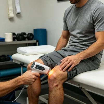
Il gating a ciclo di lavoro variabile risolve questo problema tecnico. Modulando l’emissione del fascio in impulsi della durata di microsecondi (con una frequenza compresa tra 10 Hz e 20.000 Hz) con durate degli impulsi e intervalli di riposo controllati, l’elevata potenza di picco raggiunge i tessuti profondi durante la fase attiva, mentre i capillari superficiali dissipano il calore durante la fase di riposo. Questa regolazione termica mantiene la temperatura superficiale della pelle al di sotto dei 42 gradi Celsius in modo sicuro, consentendo ai medici di applicare un trattamento laser prolungato per l’artrite, salvaguardando al contempo la fragile cute circostante e i bordi delle ulcere.
+--------------------------------------------------------------------------------------------------+
|                    COMPOUND ARTICULAR AND CUTANEOUS PHOTON PROPAGATION                           |
+--------------------------------------------------------------------------------------------------+
| Non-Contact / Contact Fluid-Displacement Probe                                                   |
|   |                                                                                              |
|   v                                                                                              |
| [Epithelial Margin / Ulcer Bed] ---> 650nm accelerates fibroblast migration and closure          |
|   |                                                                                              |
|   v                                                                                              |
| [Dermal Papillary Capillaries]  ---> 980nm resolves stasis and clears pooled inflammatory fluid   |
|   |                                                                                              |
|   v                                                                                              |
| [Prepatellar Bursa & Retinaculum] -> Dynamic pulse duty cycle prevents surface thermal stacking   |
|   |                                                                                              |
|   v                                                                                              |
| [Synovial Joint Capsule]        ---> Suppression of IL-1beta, TNF-alpha, and COX-2 cascades      |
|   |                                                                                              |
|   v                                                                                              |
| [Articular Cartilage & Bone]    ---> 1064nm stimulates Cytochrome C Oxidase, restoring ATP flow   |
+--------------------------------------------------------------------------------------------------+
Biomolecular Mechanics of Chronic Joint Relief and Epithelial Closure
Treating coexisting articular cartilage degeneration and superficial skin loss requires balancing inflammatory cytokine suppression with tissue matrix synthesis. Degenerated chondrocytes and inflamed synovial cells produce catabolic cytokines, including interleukin-1 beta (IL-1β), tumor necrosis factor-alpha (TNF-α), and matrix metalloproteinases (MMPs), which break down type II collagen and aggrecan while sensitizing articular nociceptors.
Applying high-intensity coherent light directly interrupts this degenerative cycle. Targeted near-infrared irradiation downregulates nuclear factor kappa B (NF-κB) transcription inside synovial fibroblasts, decreasing the production of inflammatory mediators like prostaglandin E2 (PGE2) and cyclooxygenase-2 (COX-2) to help protect remaining cartilage integrity. This cellular stabilization underpins the documented long-term effects of laser therapy on arthritis, preventing the rebound stiffness and progressive capsular fibrosis that commonly follow temporary steroid blocks.
Allo stesso tempo, la gestione della cute fragile su un’articolazione artritica trae beneficio da sequenze di applicazione specializzate. Anziché applicare i tradizionali nastri adesivi pesanti direttamente sulla cute friabile e indurita, gli operatori sanitari coordinano la fototerapia con un protocollo strutturato di trattamento laser con medicazione della ferita. L’emissione di luce a lunghezze d’onda multiple senza contatto direttamente sul letto della ferita pulito, prima dell’applicazione di medicazioni in schiuma di silicone non aderenti, accelera la formazione del tessuto di granulazione, riduce la carica batterica e crea un ambiente protetto per l’avanzamento dei margini epiteliali.
Allo stesso tempo, l’applicazione della terapia laser strutturata per il dolore da artrite agisce direttamente sulle vie nervose sensoriali. L’erogazione di elevate dosi di fotoni iperpolarizza le fibre C non mielinizzate e le fibre A-delta con mielina sottile all’interno della capsula articolare. Una ricerca condotta da Hamblin e colleghi nel campo della fotomedicina conferma che l’illuminazione ad alta energia nel vicino infrarosso riduce la velocità di conduzione dei nervi periferici e frena l’accumulo di sostanza P nei tessuti periarticolari, fornendo un sollievo dal dolore rapido e duraturo senza effetti collaterali sistemici.
Clinical Case Documentation: Outpatient Orthopedic and Tissue Viability Service
The documented case below details the outpatient treatment course of a patient with advanced knee osteoarthritis complicated by an open pretibial stasis ulceration, treated using a multi-wavelength Class IV rehabilitation laser platform equipped with interchangeable quartz contact and non-contact optical heads.
Profilo del paziente e dati clinici di base
- Case Identifier: OSTEO-WOUND-2026-0924
- Patient Age / Sex: 67-Year-Old Male
- Primary Diagnosis: Advanced Left Knee Osteoarthritis (Kellgren-Lawrence Grade IV) with Severe Medial Joint Space Loss, Tricompartmental Osteophytosis, and Secondary Flexion Deformity, complicated by a Chronic Pretibial Cutaneous Stasis Ulcer measuring 2.8 cm x 1.9 cm x 0.3 cm with indurated peri-wound margins.
- History: 7 years of progressive joint pain and stiffness, with a non-healing lower leg ulcer persisting for 14 weeks. Systemic NSAIDs contraindicated due to baseline renal insufficiency; intra-articular steroid injections declined due to infection risks from the adjacent ulcer; surgical arthroplasty deferred until complete cutaneous closure is achieved.
- Baseline Metrics: Numerical Pain Rating Scale (NPRS) at 9/10 during weight-bearing attempts; Western Ontario and McMaster Universities Osteoarthritis Index (WOMAC) at 78/96; Knee Active Range of Motion (AROM) Flexion restricted to 75°, Extension lacking 16°; Bates-Jensen Wound Assessment Tool (BWAT) score at 34/65.
Regime terapeutico multifase
The treatment course comprised twelve clinical sessions scheduled over four weeks, progressing from acute wound decontamination and nociceptive gating to deep capsular saturation and functional mobility integration.
| Numero di sessione. | Fase clinica e obiettivo | Configurazione della lunghezza d'onda | Potenza operativa (W) | Frequenza d'impulso (Hz) | Ciclo di funzionamento (%) | Energia erogata (J) | Dose cumulativa (J) |
| Sessione 01 | Decontaminazione delle ulcere e controllo nocicettivo | 650 nm (30%) + 980 nm (50%) + 1064 nm (20%) | 10,0 W | 10.000 Hz | 30% | 3,000 J | 3,000 J |
| Sessione 02 | Popliteal Drainage & Wound Edge Perfusion | 650 nm (20%) + 980 nm (50%) + 1064 nm (30%) | 12,0 W | 5.000 Hz | 40% | 3,600 J | 6,600 J |
| Sessione 03 | Synovial Retinacular Flushing & Biofilm Control | 650 nm (20%) + 980 nm (40%) + 1064 nm (40%) | 14,0 W | 2.500 Hz | 50% | 4.200 J | 10,800 J |
| Sessione 04 | Granulation Bed Activation & Fat Pad Release | 650 nm (25%) + 980 nm (35%) + 1064 nm (40%) | 16,0 W | 1.000 Hz | 60% | 4.800 J | 15,600 J |
| Sessione 05 | Deep Medial Capsule Softening | 980 nm (40%) + 1064 nm (60%) | 18,0 W | 300 Hz | 65% | 5,400 J | 21,000 J |
| Sessione 06 | Subchondral Plate Perfusion & Epithelial Edge Adv. | 650 nm (20%) + 980 nm (30%) + 1064 nm (50%) | 20,0 W | 150 Hz | 70% | 6,000 J | 27,000 J |
| Sessione 07 | High-Flux Chondrocyte Respiration Induction | 980 nm (25%) + 1064 nm (75%) | 22,0 W | Continuo (CW) | 100% | 6,600 J | 33,600 J |
| Sessione 08 | Rimodellamento della matrice fibrocartilaginea densa | 980 nm (20%) + 1064 nm (80%) | 24,0 W | Continuo (CW) | 100% | 7,200 J | 40,800 J |
| Sessione 09 | Cutaneous Re-Epithelialization Consolidation | 650 nm (40%) + 980 nm (30%) + 1064 nm (30%) | 18,0 W | 100 Hz | 70% | 5,400 J | 46,200 J |
| Sessione 10 | Rieducazione biomeccanica con estensione dinamica | 980 nm (30%) + 1064 nm (70%) | 20,0 W | 200 Hz | 75% | 6,000 J | 52,200 J |
| Sessione 11 | Integrazione funzionale della catena cinetica dell'andatura | 980 nm (30%) + 1064 nm (70%) | 20,0 W | Continuo (CW) | 100% | 6,000 J | 58,200 J |
| Sessione 12 | Consolidamento finale dei tessuti e dimissione | 650 nm (20%) + 980 nm (40%) + 1064 nm (40%) | 16,0 W | 500 Hz | 65% | 4.800 J | 63,000 J |
Progressione clinica e indicatori di esito
WEEK 1 (Sessions 1-3):
- Non-contact scanning over the pretibial ulcer cleared slough; healthy red granulation islands appeared in the wound bed.
- Gentle contact sweeping over the popliteal drainage basin and medial joint line reduced periarticular tension.
- Resting nocturnal pain subsided; NPRS dropped from 9/10 to 5/10.
- Patient slept through the night without waking from positional joint aching after Session 2.

WEEK 2 (Sessions 4-6):
- Pretibial ulcer margins advanced inward; wound surface area shrank from 2.8 x 1.9 cm to 1.6 x 1.1 cm with zero maceration.
- Knee active flexion expanded from 75° to 102°; extension deficit reduced from 16° to 6°.
- Patient stood from a seated position without pulling on assistive furniture arms.
- BWAT wound score improved from 34/65 to 20/65; WOMAC functional score improved from 78 to 44.

WEEK 3 (Sessions 7-9):
- Continuous wave delivery applied over the patellar retinaculum and tibiofemoral margins with firm compression.
- Complete epithelial closure of the pretibial ulcer achieved by Session 9, presenting a fully re-epithelialized skin layer.
- Patient walked 500 meters using only a single-point cane without triggering joint instability or skin breakdown.
- Crepitus during active knee flexion softened considerably as synovial congestion cleared.

WEEK 4 (Sessions 10-12):
- Full active knee extension achieved (0° neutral); active flexion reached 124°.
- Ulcer site completely healed (BWAT score 13/65, representing closed skin); no localized breakdown under direct pressure.
- Final NPRS recorded at 1/10 during extended walking and 0/10 at rest.
- WOMAC score reached 12/96, restoring daily mobility and clearing the patient medically for elective surgical review.
Analisi comparativa: laser ad alta intensità vs. modalità convenzionali
Evaluating high-intensity class IV light alongside traditional clinical options shows why modern physical medicine centers integrate multi-wavelength lasers into daily care:
Iniezioni intra-articolari di corticosteroidi
Steroid injections act as broad chemical immunosuppressants that temporarily mask joint inflammation. However, when an adjacent skin ulcer or vascular breakdown is present, puncturing the joint capsule risks introducing surface pathogens into the synovial cavity, potentially causing septic arthritis. Repeated intra-articular steroid exposure also accelerates cartilage loss, damages native chondrocyte viability, and risks narrowing the joint space over time. High-intensity multi-wavelength laser therapy provides a non-invasive, non-destructive alternative. Rather than damaging chondrocytes, concentrated photons stimulate microvascular circulation, boost cellular ATP synthesis, and reduce joint discomfort without chemical toxicity or infection risks.
Surgical Debridement and Skin Grafting
Surgical debridement of stasis ulcers followed by autologous split-thickness skin grafting requires donor site harvesting and prolonged limb immobilization. In patients with severe arthritis, immobilization causes joint contracture, muscular atrophy, and increased pain upon remobilization. Class IV laser protocols allow simultaneous wound healing and joint mobilization in an outpatient setting, promoting steady re-epithelialization while preserving joint movement without surgical trauma.
Farmaci antinfiammatori non steroidei (FANS) per via orale
Systemic anti-inflammatory medications circulate throughout the body, inhibiting protective prostaglandins in the gastric lining and kidneys while delivering limited drug concentrations into poorly vascularized articular cartilage and ulcerated skin margins. Chronic NSAID use carries well-documented risks of gastric ulceration, hypertension, and renal impairment. High-intensity laser therapy targets therapeutic energy directly into the damaged joint capsule and wound bed, relieving joint pain and stiffness locally without systemic adverse effects.
Applicazioni cliniche pratiche e spunti operativi
Per ottenere risultati clinici costanti nell’utilizzo di sistemi laser ad alta intensità nel trattamento dell’artrite articolare complessa con margini cutanei compromessi è necessario seguire un approccio strutturato:
- Somministrazione del trattamento a due zone: suddividere l’area clinica in una zona di ferita aperta e una zona capsulare profonda. Utilizzare un deflettore in quarzo senza contatto a una distanza di due o tre centimetri sopra le ulcere cutanee aperte per erogare luce biostimolante rossa e nel vicino infrarosso senza toccare il fragile tessuto di granulazione. Passare poi a una sonda di compressione liscia in quarzo sui tessuti molli periarticolari chiusi per spostare il liquido superficiale ed erogare fotoni in profondità nella capsula articolare.
- Regional lymphatic clearing: Always treat proximal lymphatic pathways first. Sweeping the popliteal space, femoral triangle, and proximal calf musculature before directing energy into the joint capsule clears fluid pathways and accelerates tissue recovery.
- Protocollo di progressione della frequenza: iniziare con frequenze di impulso elevate (da 4.000 Hz a 10.000 Hz) durante le prime sedute per calmare le vie nervose periferiche ipersensibili, passare poi a impulsi moderati (da 100 Hz a 500 Hz) per migliorare la perfusione locale e alleviare la rigidità capsulare, e terminare con l’emissione in onda continua (CW) per stimolare il metabolismo dei condrociti e il rimodellamento strutturale una volta che il dolore acuto si è attenuato.
Applying class IV multi-wavelength laser therapy grounded in tissue optical physics allows outpatient orthopedic and wound care clinics to resolve stubborn, chronic joint degeneration and help patients return to active, pain-free daily lives.