 FotonMedix











Bare Tip Laser Perforation Stalls Vascular Surgery
Circumferential optical emission delivers uniform endovenous photothermal ablation while eliminating vein wall perforation and post-procedural ecchymosis.
Vascular surgeons running busy endovenous outpatient clinics regularly face the procedural headache of vein wall carbonization and sudden structural perforation during thermal ablation. The surgical theater reality is tense and inefficient: an operating specialist advances a basic forward-firing bare fiber into an incompetent great saphenous vein, only to witness excessive peak heat burn a direct hole through the venous adventitia into surrounding subcutaneous fat, triggering immediate pain and severe hematoma. Grooms of surgical stock struggle with brittle fiber coatings that fracture inside tortuous vessels, forcing mid-procedure catheter extractions and expensive device disposals. When surgical procurement managers review the recurring costs of endovenous consumables against extended operative times, postoperative pain complaints, and delayed patient discharge, standard light delivery designs demonstrate obvious structural flaws. Delivering seamless, controlled circumferential thermal closure along a collapsing vessel wall demands an advanced medical optical fiber capable of scattering energy evenly instead of concentrating a destructive forward-facing spike.
Forward Firing Energy Spikes Rupture Venous Tunica Layers
The architectural mechanics of human and animal venous trunks present a challenging thermodynamic environment for thermal ablation. The saphenous vein wall is composed of thin, distensible layers: the inner endothelial intima, the muscular media, and the outer collagenous adventitia.
Delivering high-energy coherent light through a standard flat-cut fiber creates severe optical and thermal hot spots. According to thermal propagation frameworks documented by Welch and van Gemert in optical-thermal tissue interactions, a bare forward-projecting optical beam concentrates maximum photon flux across a minimal forward target area. As the vein undergoes tumescent spasm around the tip, the forward beam directly contacts the vascular endothelium rather than dispersing throughout the luminal circumference.
This concentrated forward blast instantly exceeds the vaporization threshold of tissue water, producing rapid carbonization along the fiber face. The resulting high-temperature blackbody absorber acts as an open heating element, exceeding 300 degrees Celsius in milliseconds. The thin muscular tunica media melts, puncturing the adventitia and releasing extravasated blood into perivascular compartments. Surrounding saphenous nerve branches experience thermal injury, leaving patients with long-term paresthesia, skin burns, and painful thrombophlebitis.
Dual Wave Interactions Inside Incompetent Vascular Lumens
Preventing catastrophic structural perforation requires pairing specific absorption mechanics with the optical profile of collapsing vascular targets: eliminating luminal blood pockets while shrinking collagenous vein walls.
The 1470nm wavelength interacts directly with intracellular and interstitial water molecules within the venous endothelial wall. Water exhibits an absorption peak at 1470nm that is roughly forty times higher than traditional 980nm outputs. When this wavelength is deployed through an endovascular delivery system, photon absorption concentrates within the inner 100 to 200 micrometers of the endothelial layer. This controlled, water-specific energy absorption initiates rapid, low-temperature thermal denaturation of collagen fibrils within the vein wall at roughly 75 to 85 degrees Celsius, triggering immediate luminal shrinkage without burning through to adjacent nerve bundles.
Concurrently, the 980nm wavelength provides targeted interaction with residual intra-luminal deoxygenated and oxygenated hemoglobin. While 1470nm seals the vessel wall, residual venous blood within dilated, tortuous segments must be coagulated without generating explosive vapor bubbles. The 980nm wavelength penetrates the thin blood film, stabilizing fibrin deposition and creating a firm, organized endoluminal seal that completely closes the refluxing pathway.
Combining precise water absorption at 1470nm with controlled hemoglobin interaction at 980nm transforms endovascular treatment, achieving total venous occlusion without the carbonization risks of single-wavelength forward beams.
Circumferential Emission Dynamics of Advanced Radial Fiber Optics
Eliminating point-source heat concentration requires an optical design that redirects the laser beam away from the forward axis into a uniform 360-degree cylindrical ring. This is achieved by utilizing an advanced radial fiber assembly.
A circular glass prism or precision-fused silica quartz cap mounted at the distal tip of the medical laser fiber reflects the axial beam outward. As light travels down the silica core, the engineered optical geometry deflects photons radially across the entire circumference of the inner vein wall.
By expanding the beam into a smooth circular band, the radial configuration drops the peak power density across any single contact point while ensuring 360-degree wall contact. When the vein contracts around the quartz ring under tumescent fluid compression, the energy distributes evenly across the vascular endothelium. No localized carbonization forms on the tip, preventing the fiber from sticking to the melting vascular wall during pullback. Surgeons maintain a steady withdrawal speed of one millimeter per second, leaving behind a smooth, uniformly contracted, non-perforated fibrotic cord.
Radial Optical Delivery Versus Standard Bare Fiber Systems
Surgical centers and hospital procurement teams must distinguish between legacy bare forward-firing fibers and advanced circumferential radial systems. The operational contrast dictates whether vein procedures yield seamless recoveries or painful clinical complications.
| Operating Parameter | 360-Degree Radial Fiber System | Standard Bare Flat-Cut Fiber |
| Beam Geometry | 360-degree cylindrical ring emission | Direct forward axial focal point |
| Wall Perforation Risk | Near zero due to distributed surface fluence | High due to direct endothelial contact spikes |
| Tip Carbonization | Absent; fused quartz cap prevents adherence | Severe; rapid blood and tissue charring |
| Post-Op Pain & Ecchymosis | Minimal; intact perivenous tissue planes | Extensive; frequent hematoma and nerve pain |
| Operating Power Needed | 8W to 12W with dual 1470nm/980nm emission | 15W to 25W single-band high energy |
| Pullback Consistency | Smooth gliding without tissue snagging | Frequent tip sticking, dragging, and tearing |
Attempting endovascular closure with an old-style bare glass strand increases the risk of surgical complications. Delivering twelve hundred joules along a saphenous trunk with a bare fiber requires high operational wattage to ensure closure, vastly increasing the chances of burning through the vessel wall into the subcutaneous layer. In contrast, an advanced circumferential medical optical fiber operates efficiently at much lower wattages, delivering smooth, uniform energy that shrinks the vessel wall without compromising adventitial integrity.
Integrating radial technology across hospital surgical suites upgrades both clinical outcomes and financial performance. Clinics minimize postoperative follow-ups for extensive bruising, prevent costly nerve litigation, eliminate mid-procedure device failures, and shorten turnaround times between outpatient surgical blocks.
Clinical Case Protocol: Great Saphenous Vein Insufficiency with CEAP C4a Stasis Dermatitis
Documented clinical metrics confirm the performance and recovery benefits of 360-degree circumferential radial delivery during endovenous thermal ablation.
Case History and Pathological Staging
A 54-year-old female patient presented with severe chronic venous insufficiency of the left lower extremity, classified as CEAP clinical stage C4a. The patient reported intense throbbing pain, evening lower-leg edema, and progressive hyperpigmentation with stasis dermatitis across the medial supramalleolar region. Duplex ultrasound mapping demonstrated marked saphenofemoral junction incompetence with continuous retrograde reflux exceeding 3.5 seconds during calf deflation maneuvers.
The left great saphenous vein displayed severe ectasia, measuring 11.2 millimeters in diameter at the proximal thigh and 8.6 millimeters at the mid-calf level. Extensive tortuosity was noted along the distal third of the thigh.
Previous conservative treatment consisting of medical-grade 30-40 mmHg graduated compression stockings and oral venoactive flavonoids over eight months failed to prevent dermatological worsening and persistent pain. Endovenous thermal ablation utilizing an advanced radial delivery platform was scheduled as the primary surgical intervention.
Laser Intervention Dosimetry and Operational Matrix
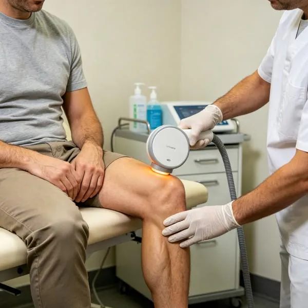
The patient underwent ultrasound-guided endovenous thermal ablation using synchronized 1470nm and 980nm wavelengths delivered through an advanced circumferential radial assembly under cold perivenous tumescent anesthesia.
| Case Identifier | Patient Profile | Diagnosis and Lesion Extent | Operating Parameters | Dosimetric Delivery | Clinical Horizon |
| SURG-VAS-2026-0419 | 54yo Female (68 kg) | CEAP C4a GSV Incompetence (11.2mm proximal diameter) | 1470nm (10W) + 980nm (2W); 12W Continuous; Radial 360 | Linear Endovenous Energy Density: 65 J/cm; Total 4,200 J (65 cm treated) | 24 Hours: Complete occlusion, zero ecchymosis; Day 30: Fibrotic cord, stasis dermatitis resolved |
Measured Recovery Trajectory
Following ultrasound-guided micropuncture access at the distal calf and catheter placement, the delivery system was advanced to two centimeters below the superficial epigastric vein junction. Chilled tumescent solution was infused along the saphenous compartment under continuous ultrasound visualization, compressing the large 11.2-millimeter lumen onto the quartz ring.
A total length of 65 centimeters of the great saphenous vein was treated using a continuous automated pullback speed of 1.0 millimeter per second. The 1470nm wavelength provided 85 percent of the output, focusing directly on the water-rich endothelial wall, while the 980nm component stabilized luminal micro-clotting. Intraoperative duplex imaging confirmed immediate vein wall coaptation with zero bubbling or acoustic cavitation beyond the adventitia. Total energy delivered was 4,200 joules, yielding an average linear endovenous energy density of 65 joules per centimeter.
The patient walked out of the recovery suite within thirty minutes without narcotic analgesics. Follow-up duplex ultrasound at 24 hours revealed complete, smooth occlusion of the treated trunk without deep vein thrombosis, skin discoloration, or perivascular hematoma. By day thirty, the saphenous trunk had transformed into an impalpable fibrous cord, medial calf eczema cleared completely, and the patient reported total relief from lower limb heaviness.
Circumferential Ablation Replaces Aggressive High Stripping Surgeries
Advanced radial photothermal ablation provides surgical departments with clear clinical and economic advantages over traditional high ligation and stripping.
Surgical stripping of the great saphenous vein requires hospital admission, general or spinal anesthesia, groin incisions, and mechanical evulsion of the vascular trunk. This mechanical tearing inevitably avulses tributary veins, lacerates the adjacent saphenous nerve, and leaves patients with extensive hematomas, incision infections, and weeks of painful immobility. In contrast, endovenous radial light delivery achieves permanent anatomical closure through a tiny single-puncture access site under local tumescent anesthesia.
By emitting light in a circular band directly onto the vessel wall, the radial design drives collagen denaturation without charring or tearing vascular tissue. Endothelial cells undergo controlled apoptosis, while the medial muscular layers shrink smoothly into a stable connective tissue matrix that the body reabsorbs naturally. Patients avoid post-surgical drainage, preserve regional lymph nodes, and return to full work and daily activities within forty-eight hours.
Where traditional stripping and outdated flat-cut fibers lead to surgical trauma, tissue burns, and prolonged disability, advanced circumferential medical laser fiber technology provides clean, predictable vascular closure. Surgical departments expand procedural volume, eliminate post-operative complications, and set a modern clinical standard using precision optical delivery.