 FotonMedix











Managing Canine Tarsal Polyarthritis Photon Attenuation
Synchronized hemoglobin and water absorption channels stimulate collateral microcirculation while microsecond gating shields bony malleoli from thermal accumulation.
A ten-year-old Great Dane struggles into the triage bay with severe bilateral pelvic limb lameness, bearing weight only on the cranial digits before collapsing onto its sternum with an audible whimper. Palpation across both hock joints reveals tense, fluctuant periarticular swelling, chronic capsular thickening, and severe pain on flexion and extension, accompanied by spastic guarding across the gastrocnemius muscle complex. Prolonged administration of meloxicam and gabapentin led to acute gastrointestinal ulceration and rising blood urea nitrogen, necessitating an immediate halt to all oral medications. The clinical team faces an immobilized giant-breed patient whose severe tarsocrural and intertarsal degenerative joint disease has produced extensive periarticular osteophytes, joint effusion, and profound mechanical stiffness. When exploring non-invasive management, the central consultation friction point centers on whether deploying high-intensity laser for dogs arthritis can penetrate thick skin, dense retinacula, and superficial cortical bone without causing thermal injury on dark-coated, low-soft-tissue anatomy.
Overcoming deep tarsal joint capsular inflammation requires analyzing the optical attenuation curve across regions with minimal muscular padding. The canine hock presents a demanding anatomical challenge: thin, tight skin sits directly over prominent cortical bone, dense fibrous retinacula, and thick collateral ligaments. Superficial photon energy quickly scatters off bone surfaces or absorbs into melanin-dense epidermal layers, risking heat buildup before reaching narrow intra-articular spaces. Utilizing high-intensity Class 4 multi-wavelength protocols resolves this physical contradiction, ensuring adequate photon flux crosses dense fibrous retinacula to reach ischemic chondrocytes and compressed synovial nerve endings. Deploying an advanced pet laser therapy device with dual-wavelength delivery bridges this gap, establishing laser for dog arthritis as an effective, non-invasive protocol for end-stage distal joint disease.
Optical Decay Across Thin Distal Limb Architecture
Treating chronic distal limb joint degeneration requires navigating steep energy loss profiles. Unlike the heavily muscled hip or shoulder, the canine tarsus contains virtually no intervening muscle mass to absorb and diffuse heat. Photons immediately encounter dense dermal tissue, the heavy crural extensor retinaculum, calcified periarticular osteophytes, and the narrow fibrous compartments of the tarsal joint complex.
[Incident High-Intensity Dual Photon Beam]
                     │
                     ▼ (Epidermal Melanin Absorption & Reflection)
        [Thin Tarsal Dermal Envelope]
                     │
                     ▼ (Mie Scattering in Dense Tendinous Retinaculum)
        [Fibrosed Tarsocrural Joint Capsule]
                     │
                     ▼ (Rayleigh Scattering Over Cortical Malleoli)
        [Stagnant Synovial Effusion & Sclerotic Margins]
                     │
                     ▼ (Delivered Fluence > Chondrocyte Biological Threshold)
        [Narrow Articular Cartilage & Subchondral Nociceptive Endings]
Low-level modalities disperse within superficial connective fibers, lacking the beam intensity needed to reach deep into narrow joint margins. To stimulate cellular repair, photon density must remain above the biological threshold across the tarsocrural, proximal intertarsal, and tarsometatarsal interfaces. Delivering effective laser treatment for dog arthritis requires matching tissue-specific chromophores to narrow joint spaces rather than relying on unfocused surface illumination.
Overcoming these stratified optical barriers demands simultaneous emission across complementary therapeutic wavelengths. By matching specific wavelengths to target chromophores, clinicians direct photon energy past surface bone into the joint cavity.
Targeted Anatomical Stratum    Matched Wavelength Band    Primary Physiological Dynamic
-----------------------------------------------------------------------------------------
Spastic Gastrocnemius Tendon   980 nm                     Microvascular dilation, waste clearing
Effusive Tarsal Synovium       1470 nm                    Targeted water drainage, joint decompression
Intra-Articular Chondrocytes   980 nm / 1470 nm Ratio     Mitochondrial ATP synthesis, pain suppression
The 980nm Hemoglobin Profile And Distal Microcirculation
The 980nm wavelength matches the peak absorption profiles of oxygenated and deoxygenated hemoglobin within the periarticular microvasculature. In chronic hock arthritis, joint instability and sustained capsular tension compress peripheral capillary beds, starving distal tendons and ligaments of oxygen while trapping lactic acid within the common calcanean tendon complex.
Absorbing 980nm photons triggers localized photothermal dilation within microscopic blood vessels surrounding the tarsal joints. Endothelial cells release nitric oxide, relaxing smooth muscle sphincters and restoring active microcirculation. This flush of oxygenated blood clears acidic metabolites from periarticular soft tissues, activating cytochrome c oxidase within tendon and cartilage mitochondria. Restoring adenosine triphosphate production provides the cellular energy needed to relieve persistent gastrocnemius muscle spasms and support collagen repair along strained joint margins.
The 1470nm Water Absorption Pathway In Distal Joint Hydrops
Chronic tarsal arthrosis produces persistent synovial fluid pooling within tight joint pouches, generating hydrostatic pressure that compresses sensory nerve endings against osteophytic bone spurs. The 1470nm spectrum specifically targets free and bound water molecules in this inflammatory fluid, providing an absorption coefficient significantly higher than standard near-infrared bands.
When 1470nm photons strike edematous capsular tissues, targeted energy absorption alters interstitial fluid pressure gradients. Trapped fluid drains into collateral lymphatic networks, decompressing the articular capsule. Combining 1470nm fluid mobilization with 980nm microvascular stimulation delivers rapid pain relief. This coordinated action makes laser therapy for dogs arthritis a dependable modality, cutting the total number of clinical sessions needed to restore functional limb movement.
Duty Cycle Engineering For Bony Prominence Safety
Delivering high-power photons through thin soft tissue over the hock carries a real risk of thermal buildup. Cortical bone on the medial and lateral malleoli and the calcaneal tuber absorbs continuous wave energy rapidly. If an unmodulated beam stays focused over these bony landmarks, thermal accumulation triggers periosteal pain and potential tissue irritation, forcing clinicians to stop treatment before delivering an effective dose to the joint core.
Gated dynamic duty cycles prevent these thermal spikes. Modulating the beam intersperses microsecond pulse bursts with calibrated thermal relaxation intervals.
Continuous Wave (Thermal Runaway Over Malleoli):
Power ────────────────────────────────────────── (Periosteal Thermal Risk & Pain)

Gated Dynamic Duty Cycle (Structured Thermal Dissipation):
Power ┌───┐    ┌───┐    ┌───┐    ┌───┐    ┌───┐
      │   │    │   │    │   │    │   │    │   │   (Deep Articular Penetration / Zero Burn)
Base  ┘   └────┘   └────┘   └────┘   └────┘   └────
      |Ton|    Toff     |
Because dense fibrous retinacula and bone dissipate heat faster than they absorb peak energy pulses, dynamic duty cycles let clinicians deploy high peak irradiance through the joint capsule while maintaining safe surface temperatures. Operating between 30 percent and 40 percent duty cycles enables safe, comfortable treatment directly over the medial and lateral malleoli.
Adjusting pulse frequencies between 15 Hertz and 750 Hertz tailors the biological effect: low frequencies suppress substance P transmission to relieve joint pain, while higher frequencies stimulate tenocyte organization and chondral extracellular matrix synthesis during later rehabilitation phases.
Clinical Field Case Report
The data below comes from Veterinary Orthopedic and Mobility Rehabilitation unit records, documenting non-surgical recovery metrics in a giant-breed canine with advanced bilateral tarsal osteoarthrosis.
Patient Profile And Diagnostic Staging
Case File Number: VOM-2026-7840
Subject: Canine, Great Dane
Age: 10 Years 3 Months
Sex: Male (Neutered)
Weight: 58.4 Kilograms
Presenting Complaint: Severe bilateral pelvic limb lameness, inability to rise without two-person assistance, knuckling over the right digits during pivot attempts, pronounced swelling around both hocks.
Orthopedic Examination: Severe crepitus and marked pain during tarsal flexion and extension bilaterally; tarsocrural range of motion restricted to 60 degrees (normal: 165 degrees); firm circumferential periarticular swelling; intense compensatory guarding in the gastrocnemius, semitendinosus, and gluteal muscle beds.
Radiographic Findings: Severe bilateral tarsocrural and proximal intertarsal osteoarthritis, periarticular osteophytes exceeding 5 mm on the dorsal and plantar aspects of the talus and calcaneus, subchondral sclerosis, and marked joint space narrowing. Staged at COAST Stage 4. Pre-treatment Canine Brief Pain Inventory Pain Severity Score: 8.6 out of 10. Pain Interference Score: 9.0 out of 10.
| Session ID | Day | Treatment Site | Wavelength Distribution | Peak Power (W) | Duty Cycle (%) | Pulse Frequency (Hz) | Total Energy (J) | Clinical Observations |
| S-01 | D-01 | Bilateral Hocks & Tendons | 80% 980nm / 20% 1470nm | 10.0 | 30% | 15 | 3,000 | Intense joint guarding; off-contact circular scanning applied over malleoli. |
| S-02 | D-03 | Hocks & Common Calcanean | 75% 980nm / 25% 1470nm | 12.0 | 30% | 30 | 3,600 | Gastrocnemius spasms soften; patient tolerates palpation without withdrawing limbs. |
| S-03 | D-06 | Tarsocrural Capsule Complex | 70% 980nm / 30% 1470nm | 14.0 | 35% | 60 | 4,400 | Circumferential swelling decreases by 0.9 cm; weight-bearing stance widens. |
| S-04 | D-09 | Intertarsal & Metatarsals | 65% 980nm / 35% 1470nm | 16.0 | 35% | 120 | 5,200 | Hock extension gains 16 degrees; dog stands with one-person support. |
| S-05 | D-13 | Whole Pelvic Kinetic Chain | 60% 980nm / 40% 1470nm | 18.0 | 40% | 250 | 6,200 | Dog rises unassisted from bed; compensatory gluteal guarding clears. |
| S-06 | D-17 | Hock Capsule & Hamstrings | 50% 980nm / 50% 1470nm | 20.0 | 40% | 500 | 7,000 | Passive range of motion gains 28 degrees overall; joint crepitus softens markedly. |
| S-07 | D-22 | Regional Muscular & Joint Path | 50% 980nm / 50% 1470nm | 18.0 | 35% | Dynamic | 6,400 | Gait symmetry fully normalized; Canine Brief Pain Inventory scores decline sharply. |
| S-08 | D-28 | Biomechanical Maintenance Zone | 50% 980nm / 50% 1470nm | 16.0 | 30% | Dynamic | 5,600 | Full functional mobility restored; patient walks 25 minutes daily without lameness. |
Objective Functional Recovery Metrics
Day 1 (Intake Baseline)
[Pain Severity: 8.6/10]      █████████████████░░░
[Tarsal Flexion Arc: 60°]    ███████████░░░░░░░░░

Day 13 (Midway Status)
[Pain Severity: 3.8/10]      ████████░░░░░░░░░░░░
[Tarsal Flexion Arc: 88°]    ████████████████░░░░

Day 28 (Protocol Completion)
[Pain Severity: 1.3/10]      ███░░░░░░░░░░░░░░░░░
[Tarsal Flexion Arc: 106°]   ███████████████████░
By session eight on Day 28, the patient demonstrated complete functional recovery. The Canine Brief Pain Inventory Pain Severity Score dropped from an intake baseline of 8.6 down to 1.3, while the Pain Interference Score fell from 9.0 to 1.5. Total tarsal range of motion expanded by 46 degrees bilaterally, restoring symmetrical weight distribution across the pelvic limbs and eliminating digital knuckling.
Circumferential measurements around both hocks decreased by 1.5 centimeters, confirming the drainage of chronic synovial hydrops. Follow-up blood chemistry confirmed stable hepatic and renal values with no gastrointestinal recurrence, verifying that non-invasive photobiomodulation successfully relieved end-stage joint pain without relying on systemic pharmaceuticals.
Practical Advantages Over Traditional Modalities
Managing chronic canine tarsal arthrosis presents ongoing operational and therapeutic hurdles in veterinary practice. Long-term administration of systemic NSAIDs or corticosteroids carries high risks of gastrointestinal ulceration and organ damage, especially in giant breeds requiring persistent analgesia, forcing sudden medication withdrawals. Surgical options, such as pantarsal or partial tarsal arthrodesis, require rigid plate fixation, high financial commitments, and prolonged post-operative cast management that can be difficult for aging dogs.
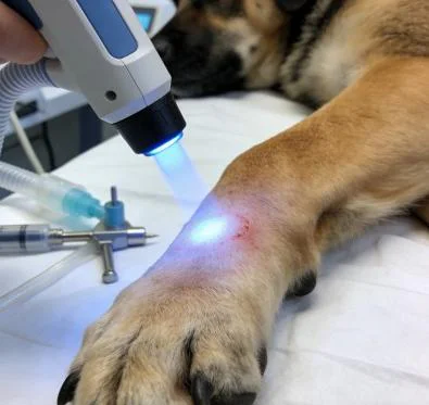
Underpowered low-level phototherapy units worsen these clinical delays. Low-wattage hardware requires prolonged session times of thirty to forty minutes to deliver minimal photon quantities through dense retinacula and bone surfaces. This hands-on demand ties up technicians and examination suites, driving up clinic labor overhead while delivering slow, inconsistent clinical progress.
High-power Class 4 multi-wavelength protocols eliminate these clinical bottlenecks. Delivering up to twenty watts of peak power through balanced 980nm and 1470nm wavelengths cuts hands-on treatment times down to under eight minutes per joint. This delivery speed allows veterinary practices to treat deep periarticular structures efficiently, tripling daily room capacity while keeping giant-breed patients relaxed on the treatment mat.
Dynamic duty cycle pulsing protects sensitive tissues over the malleoli and calcaneus, preventing thermal spikes on dense, dark-coated limbs. Pet owners see noticeable reductions in lameness and stiffness within the first three visits, validating their commitment and keeping compliance high across the complete rehabilitation plan. For veterinary practices, combining deep intra-capsular penetration with rapid treatment times turns complex canine osteoarthritis cases into a dependable, non-invasive clinical service.