 FotonMedix











Overcoming Dense Fibrocartilage Attenuation in Thumb Basal Joint Rhizarthrosis
Multi-spectral coherent photons penetrate the dense trapezial fibrocartilage matrix, downregulate localized synovial cytokine synthesis, and suppress mechanoreceptor nociceptive firing via microsecond duty cycle modulation without cutaneous thermal stacking.
A 61-year-old dental hygienist enters the hand and upper extremity clinic resting her dominant right forearm on the examination cushion, unable to pinch a key or open a tight screw cap without wincing. Every attempt to oppose the thumb to the small finger triggers an acute, grinding ache deep within the base of the first metacarpal. Palpation over the carpometacarpal (CMC) junction reveals localized bogginess, dorsal osteophytic shelf formation, and a positive grind test that reproduces sickening joint crepitus. Over four years, the patient has worn rigid thumb spica splints, consumed daily celecoxib, and received three fluoroscopically guided intra-articular corticosteroid injections. While the initial injection afforded three months of manageable comfort, subsequent rounds yielded diminishing returns, culminating in continuous aching during nocturnal rest and progressive radial subluxation of the metacarpal base. Surgical trapeziectomy with ligament reconstruction and tendon interposition (LRTI) was presented as the final recourse, carrying a twelve-week immobilization and rehabilitation arc. The fundamental technical contradiction in treating advanced basal joint degeneration non-surgically centers on soft-tissue optics: the first carpometacarpal articulation is compact, enclosed by dense, thickened palmar and dorsoradial stabilizing ligaments that scatter incident light beams. Superficial phototherapy devices and low-output emitters scatter within the first few millimeters of the thenar dermis, failing to reach the subchondral bone plate. Delivering sustained clinical relief requires driving calibrated photon flux through dense connective tissues to downregulate the localized inflammatory cascade while restoring microvascular perfusion across the compressed joint capsule.
Optical Penetration Across Thick Thenar Ligamentous Barriers
Delivering therapeutic photon densities into the first carpometacarpal cavity requires overcoming high reflection and scattering coefficients across compact connective tissues. Thenar skin, dense collagenous retinacula, and the anterior oblique ligament scatter non-coherent light beams, converting standard therapeutic devices into superficial heating pads.
Wavelength-Selective Chromophore Dynamics in Basal Joint Cartilage
The clinical efficacy of high-intensity class IV optical therapy depends on selecting wavelengths that bypass superficial dermal scatter while targeting specific biological components within the degenerate joint capsule:
- Microvascular recruitment and capillary clearing at 980nm: The 980nm wavelength matches the optical absorption profile of oxygenated hemoglobin and interstitial water. In arthritic carpometacarpal synovium, chronic capsular contracture and osteophytic impingement produce microvascular stagnation and tissue hypoxia. Photons at 980nm induce localized microvascular warming, triggering endothelial nitric oxide release, opening compressed capillary beds, and promoting the reabsorption of inflammatory effusion within periarticular tissues.
- Mitochondrial enzyme stimulation and tissue repair at 1064nm: The 1064nm wavelength displays the lowest scattering coefficient among therapeutic near-infrared bands in dense connective tissue. These photons penetrate directly through the thick dorsoradial ligament to reach the subchondral bone of the trapezium and first metacarpal base. Inside dormant chondrocytes and capsular fibroblasts, 1064nm light binds to cytochrome c oxidase, displacing inhibitory nitric oxide, restoring the mitochondrial proton gradient, and increasing adenosine triphosphate (ATP) production for cellular repair.
Preventing Cutaneous Heat Accumulation via Duty Cycle Modulation
Emitting high-power continuous energy over the thin dorsal and thenar skin of the hand quickly challenges the thermal relaxation time of human skin. Continuous wave delivery causes heat to accumulate rapidly in superficial dermal tissues, setting off epidermal pain receptors and risking surface burns before the deep carpometacarpal joint receives a therapeutic dose.
Class IV clinical systems overcome this barrier using variable pulse duty cycle gating. By pulsing the beam between 20 Hz and 15,000 Hz with precise on/off ratios, the laser delivers high peak power during active microsecond pulses, followed by brief rest intervals where local capillary blood flow carries surface heat away. This thermal regulation keeps skin temperatures safely below 42 degrees Celsius, enabling clinicians to apply sustained laser therapy for joint conditions without risking photothermal burns.
+--------------------------------------------------------------------------------------------------+
|                          FIRST CMC BASAL JOINT PHOTON PROPAGATION                                |
+--------------------------------------------------------------------------------------------------+
| Conical Quartz Contact Handpiece Compression                                                     |
|   |                                                                                              |
|   v                                                                                              |
| [Thenar Cutaneous Layer]    ---> Dermal heat dissipated via microsecond duty cycle pauses        |
|   |                                                                                              |
|   v                                                                                              |
| [Thenar Fascia & Tendons]   ---> Physical probe pressure clears interstitial water barrier      |
|   |                                                                                              |
|   v                                                                                              |
| [Dorsoradial Ligament]      ---> 980nm dilates compressed vessels, clearing inflammatory waste   |
|   |                                                                                              |
|   v                                                                                              |
| [Synovial Cavity Margin]    ---> Downregulation of IL-1beta, TNF-alpha, and COX-2 cascades       |
|   |                                                                                              |
|   v                                                                                              |
| [Trapezial Cartilage & Bone]---> 1064nm activates Cytochrome C Oxidase, restoring ATP synthesis   |
+--------------------------------------------------------------------------------------------------+
Biochemical Pathways: Suppressing Synovial Catabolism and Hand Nociception
Managing degenerative basal joint arthritis requires addressing progressive joint space loss, chronic biochemical inflammation, and persistent peripheral nerve sensitization. Degenerated chondrocytes and inflamed synovial cells produce catabolic cytokines, including interleukin-1 beta (IL-1β), tumor necrosis factor-alpha (TNF-α), and matrix metalloproteinases (MMPs), which break down type II collagen and aggrecan while sensitizing articular nociceptors.
Applying a targeted laser for arthritis interrupts this destructive cycle. Coherent near-infrared irradiation downregulates nuclear factor kappa B (NF-κB) transcription inside synovial fibroblasts, decreasing the production of inflammatory mediators like prostaglandin E2 (PGE2) and cyclooxygenase-2 (COX-2) to help protect remaining cartilage integrity.
Simultaneously, implementing structured laser arthritis treatment acts directly on sensory nerve pathways. Delivering high photon doses hyperpolarizes unmyelinated C-fibers and thinly myelinated A-delta fibers within the articular capsule. Research by Bjordal and colleagues in physical rehabilitation literature demonstrates that high-energy near-infrared illumination reduces peripheral nerve conduction velocity and curbs substance P accumulation in periarticular tissues, providing rapid and lasting pain relief without systemic side effects.
Clinical Case Documentation: Outpatient Hand Rehabilitation
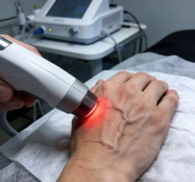
The documented case below outlines the outpatient rehabilitation course of a patient with advanced thumb carpometacarpal osteoarthritis, managed using a multi-wavelength Class IV rehabilitation laser platform equipped with a specialized conical quartz massage applicator.
Patient Profile and Clinical Baseline
- Case Identifier: HAND-RHIZ-2026-0412
- Patient Age / Sex: 61-Year-Old Female
- Primary Diagnosis: Advanced Thumb Basal Joint Osteoarthritis (Eaton-Littler Stage III) with Dorsal Subluxation, Marked Trapezial Sclerosis, and Secondary Adductor Pollicis Spasm, confirmed via Stress Radiographs and Musculoskeletal Ultrasound.
- History: 48 months of worsening right thumb base pain, loss of key pinch strength, and dropping household objects. Failed conservative management including custom thermoplastic splints, topical NSAIDs, oral analgesics, and three cortisone injections. Patient unable to hold dental instruments for more than ten minutes without severe pain.
- Baseline Metrics: Numerical Pain Rating Scale (NPRS) at 8/10 during key pinch tasks; Functional Index for Hand Osteoarthritis (FIHOA) at 24/30; Key Pinch Strength measured at 2.4 kg (contralateral unaffected thumb measured at 6.8 kg); Palmar Abduction restricted to 28°.
Multi-Stage Treatment Regimen
The treatment course comprised ten clinical sessions scheduled over four weeks, progressing from early pain management and lymphatic drainage to deep joint biostimulation and functional grip integration.
| Session No. | Clinical Phase & Objective | Wavelength Setup | Operating Power (W) | Pulse Frequency (Hz) | Duty Cycle (%) | Energy Delivered (J) | Cumulative Dose (J) |
| Session 01 | Nociceptive Gating & Radial Styloid Drainage | 980nm (65%) + 1064nm (35%) | 8.0 W | 8,000 Hz | 35% | 2,400 J | 2,400 J |
| Session 02 | Thenar Muscular Decompression | 980nm (60%) + 1064nm (40%) | 9.0 W | 4,000 Hz | 45% | 2,700 J | 5,100 J |
| Session 03 | Palmar Oblique Ligament Capillary Flush | 980nm (50%) + 1064nm (50%) | 10.0 W | 2,000 Hz | 50% | 3,000 J | 8,100 J |
| Session 04 | Dorsoradial Capsular Saturation | 980nm (40%) + 1064nm (60%) | 11.0 W | 500 Hz | 60% | 3,300 J | 11,400 J |
| Session 05 | Deep Trapezial Bed Photon Infusion | 980nm (35%) + 1064nm (65%) | 12.0 W | 250 Hz | 65% | 3,600 J | 15,000 J |
| Session 06 | Synovial Fibroblast Cytokine Suppression | 980nm (30%) + 1064nm (70%) | 13.0 W | 100 Hz | 70% | 3,900 J | 18,900 J |
| Session 07 | High-Flux Chondrocyte Respiration Push | 980nm (20%) + 1064nm (80%) | 14.0 W | Continuous (CW) | 100% | 4,200 J | 23,100 J |
| Session 08 | Subchondral Trapezial Plate Saturation | 980nm (20%) + 1064nm (80%) | 14.0 W | Continuous (CW) | 100% | 4,200 J | 27,300 J |
| Session 09 | Dynamic Pinch Resistance Loading | 980nm (30%) + 1064nm (70%) | 12.0 W | 150 Hz | 70% | 3,600 J | 30,900 J |
| Session 10 | Final Tissue Consolidation & Discharge | 980nm (40%) + 1064nm (60%) | 10.0 W | 400 Hz | 60% | 3,000 J | 33,900 J |
Clinical Progression and Outcome Metrics
WEEK 1 (Sessions 1-3):
- Gentle contact sweeping over the anatomical snuffbox and thenar eminence relieved periarticular tension.
- Resting nocturnal pain subsided; NPRS dropped from 8/10 to 4/10.
- Patient slept through the night without waking from thumb base throbbing after Session 2.
- Adductor pollicis spasm relaxed noticeably during manual palpation.

WEEK 2 (Sessions 4-6):
- Palmar abduction expanded from 28° to 42°; first web space contracture softened.
- Patient opened doorknobs and held eating utensils without sharp pinching sensations.
- FIHOA functional score improved from 24/30 to 12/30.
- Key pinch strength increased from 2.4 kg to 4.2 kg.

WEEK 3 (Sessions 7-8):
- Continuous wave delivery applied over the first CMC joint line with firm conical quartz contact.
- Patient held light dental scaling tools for 30 minutes without post-activity inflammation.
- Ultrasound evaluation showed reduced capsular thickening and resolution of local synovial hyperemia.

WEEK 4 (Sessions 9-10):
- Full active palmar abduction restored (48°), matching contralateral baseline.
- Key pinch strength reached 6.2 kg (within 90% of the unaffected hand).
- Final NPRS recorded at 1/10 during firm sustained pinching and 0/10 at rest.
- FIHOA score reached 2/30, enabling the patient to resume professional dental hygiene duties full-time.
Comparative Analysis: High-Intensity Laser vs. Conventional Modalities
Evaluating high-intensity class IV light alongside traditional clinical options shows why modern physical medicine centers integrate multi-wavelength lasers into daily hand therapy:
Intra-Articular Corticosteroid Injections
While steroid injections offer temporary anti-inflammatory relief, repeated use inside the small carpometacarpal joint suppresses chondrocyte metabolism, weakens stabilizing capsular ligaments, and accelerates progressive radial subluxation of the first metacarpal. In contrast, multi-wavelength high-intensity laser therapy works non-invasively. Rather than damaging cartilage, concentrated photons stimulate microvascular circulation, boost cellular ATP synthesis, and reduce joint discomfort without chemical toxicity.
Trapeziectomy with Ligament Reconstruction (LRTI Surgery)
Surgical excision of the trapezium eliminates bone-on-bone contact, but the procedure requires tendon harvesting, long immobilization windows, and often results in persistent weakness in pinch grip. Class IV laser therapy provides an effective non-surgical treatment that preserves native joint anatomy, reduces capsular inflammation, and restores mobility without operative trauma.
Oral Non-Steroidal Anti-Inflammatory Drugs (NSAIDs)
Systemic anti-inflammatory medications circulate throughout the body, inhibiting protective prostaglandins in the gastric lining and kidneys while delivering low drug concentrations into dense, poorly vascularized hand ligaments. Chronic NSAID use carries well-documented risks of gastric ulceration, hypertension, and renal impairment. High-intensity laser therapy targets therapeutic energy directly into the damaged joint capsule, relieving joint pain and stiffness locally without systemic adverse effects.
Practical Clinical Application and Operational Takeaways
Achieving consistent clinical results when using high-intensity laser systems for thumb basal joint degeneration requires structured execution:
- Dynamic compression with conical quartz probes: Clinicians should apply firm, steady pressure with the conical quartz contact probe directly into the anatomical snuffbox and palmar margins of the first metacarpal base. Physical compression displaces superficial water and venous blood away from the beam path, creating an optimal optical pathway that delivers higher photon counts into deep capsule structures.
- Regional lymphatic clearing: Always treat proximal lymphatic pathways first. Sweeping the radial styloid, distal forearm, and thenar web space before directing energy into the joint capsule clears fluid pathways and accelerates tissue recovery.
- Frequency progression protocol: Begin with high pulse frequencies (4,000 Hz to 8,000 Hz) during the initial visits to calm hypersensitive peripheral nerve pathways, transition to moderate pulsing (100 Hz to 500 Hz) to improve local perfusion and clear capsular stiffness, and finish with continuous wave (CW) delivery to stimulate chondrocyte metabolism and structural remodeling once acute pain has subsided.
Applying class IV multi-wavelength laser therapy grounded in tissue optical physics allows outpatient orthopedic and hand therapy clinics to resolve stubborn basal joint osteoarthritis and help patients return to precise, pain-free daily hand function.