 FotonMedix



Overcoming Distal Ischemic Ulceration and Recalcitrant Bed Stasis
Targeted dual-chromophore photon absorption stimulates stagnant endothelial beds, suppresses microbial colonization, and preserves fragile granulating margins without thermal sloughing.
Managing chronic non-healing wounds and compromised peripheral vascular networks in lower extremities poses a brutal challenge in outpatient podiatric and vascular surgical suites. When dealing with ischemic diabetic ulcers, non-draining venous stasis lesions, and recalcitrant microangiopathy, clinicians repeatedly run into the same physical dead end: conventional debridement and topical dressings manage surface bioburden but fail to restart stagnant cellular respiration in deeply starved, hypoperfused tissue beds. Delivering therapeutic photon energy to wake up dormant fibroblastic margins without inducing collateral thermal injury demands fine spectral control across distinctive tissue chromophores.
Optical Penetration Mechanics and Chromophore Affinity across Ischemic Bases
Biological wound margins and surrounding hyperkeratotic dermal boundaries act as dense optical scatter shields. When photon energy enters chronically inflamed, edematous, or fibrotic lower-limb tissues, path lengths become irregular due to tissue heterogeneity, intercellular edema, and disordered collagen matrices.
Energy transfer efficiency within these damaged anatomical structures depends strictly on selective optical absorption. Melanin, intravascular hemoglobin derivatives, and interstitial water exhibit distinct extinction coefficients across the near-infrared spectrum. In poorly granulating plantar ulcers, surface exudate and dense necrotic debris rapidly scatter uncollimated beams, causing low-irradiance devices to lose their biological punch within the first millimeter of tissue.
Overcoming this severe photon extinction requires high-intensity multi-wavelength architectures. An 810nm output directly targets the oxidized copper centers of cytochrome c oxidase in mitochondrial membranes, shifting stagnant local cells out of metabolic dormancy and boosting adenosine triphosphate synthesis.
Combining 810nm with a 980nm wavelength introduces strong absorption by oxygenated hemoglobin and moderate absorption by interstitial water. This combination triggers instant microvascular dilation, promoting active neoangiogenesis and washing away localized ischemic neurotoxins.
Where vascular pathology involves tortuous feeder channels, chronic venous insufficiency, or telangiectasias around ulcer beds, integrating 1470nm energy changes the tissue reaction entirely. Water inside vascular endothelial cell walls absorbs 1470nm light roughly forty times more readily than 980nm light. This high water affinity allows clinicians using professional laser vascular treatment devices to seal incompetent feeder micro-vessels or ablate non-healing sinus margins with minimal energy, keeping thermal spread tightly contained within 1 to 1.5 millimeters of the emitter tip.
This multi-spectrum synergy makes high-power laser therapy for slow-healing injuries a reliable clinical strategy for jump-starting stalled granulating tissue beds that fail to respond to standard dressing protocols.
Mitigating Lateral Thermal Accumulation via Pulsed Gating
Continuous-wave energy delivery poses significant clinical risks when working over compromised distal extremities with impaired sensory feedback or marginal arterial supply. Continuous emission continuously adds thermal energy to tissue beds. If the rate of photon absorption exceeds microvascular heat clearance, local baseline temperatures climb rapidly past 43 degrees Celsius, risking protein denaturation, microvascular thrombosis, and secondary tissue necrosis.
Clinical protocols neutralize this danger by matching delivery directly to the Thermal Relaxation Time of the targeted structures. The thermal relaxation time is the duration required for a targeted vascular structure or cellular envelope to lose half of its accumulated heat via passive capillary conduction.
Applying calibrated pulse duty cycles breaks up energy delivery into microsecond bursts:
Duty Cycle (%) = [Pulse On Duration / (Pulse On Duration + Pulse Off Duration)] x 100
Restricting the active pulse duration to a fraction of the tissue cooling window confines thermal impact strictly to targeted microbial biofilms, stagnant collagen walls, or diseased vascular channels. The intervening pulse-off intervals provide enough quiet time for surrounding viable capillaries to dissipate residual heat safely into surrounding tissues.
When treating non-healing distal wounds or recalcitrant musculoskeletal attachments, selecting duty cycles between 20% and 40% permits high peak outputs up to 25W. These short, high-energy pulses break through dense calloused boundaries and deep fascial layers, triggering reparative photobiomodulation while keeping cutaneous temperatures comfortable and safe.
Clinical Realities within the Evolving Podiatry Lasers Market
Lower-extremity clinical facilities handle some of the most stubborn pathologies in modern medicine, including severe plantar ulcerations, recalcitrant plantar fasciitis, and deep nail-bed onychomycosis with secondary tissue breakdown.
The broader podiatry lasers market has historically been crowded with low-power, single-purpose units that deliver superficial thermal effects without enough irradiance to alter deep cellular activity. Modern clinics are shifting away from these underpowered systems in favor of high-power Class IV multi-wavelength platforms capable of moving seamlessly between delicate wound stimulation and high-precision microvascular coagulation.
In non-healing neuro-ischemic ulcers, standard sharp debridement repeatedly removes newly formed, fragile epithelial cells. By contrast, non-contact laser stimulation decontaminates the ulcer bed without mechanical disruption. Delivering calibrated photon flux suppresses anaerobic bacterial biofilms while activating tenocytes, fibroblasts, and vascular endothelial cells along the wound perimeter.
For chronic plantar fasciitis and deep tendon entrapment, high photon densities travel straight through thick plantar fascia and dense calcaneal fat pads. This restores mitochondrial energy generation in strained connective tissues, breaks down chronic fascial scar bands, and relieves persistent heel pain without relying on corticosteroid injections that risk fascial rupture.
Restoring Microvascular Integrity in Stagnant Tissue Beds
Chronic wound stasis is driven by persistent microvascular shutdown. Endothelial cells inside long-standing diabetic wounds or chronic venous ulcers enter a state of functional senescence, losing their ability to organize into patent tubular capillaries. As a result, the wound center remains anoxic, acidic, and trapped in an unresolving inflammatory phase.
Deploying Class IV medical laser systems breaks this ischemic stalemate through three distinct biological steps:
- Endothelial Proliferation and Tubulogenesis: High photon irradiance triggers the release of vascular endothelial growth factor (VEGF) and basic fibroblast growth factor (bFGF) from perivascular cells. This initiates organized capillary sprouting from healthy peripheral margins into the ischemic ulcer core.
- Biofilm Decontamination: Targeted near-infrared wavelengths generate mild, localized photodynamic reactions within bacterial cell walls. This suppresses Pseudomonas aeruginosa and Staphylococcus aureus colonies without generating resistant bacterial strains or causing chemical trauma to fragile granulating margins.
- Extracellular Matrix Reorganization: Photon absorption accelerates procollagen mRNA transcription in local fibroblasts, helping the wound transition from disorganized provisional granulation to dense, well-aligned Type I collagen matrices that resist shear stress during ambulation.
Standardized Clinical Practice and Detailed Treatment Record
The following treatment log outlines an integrated clinical protocol addressing a chronic non-healing plantar neuropathic ulcer paired with severe medial marginal telangiectatic venous pooling.
Clinical Registry Record: FMX-2026-PU6612
| Parameter Group | Specification Detail | Clinical Target Value |
| Case Identification | Clinical Case ID | FMX-2026-PU6612 |
| Patient Demographics | Age / Biological Sex | 58 Years / Male (Type 2 Diabetes Mellitus) |
| Primary Pathologies | Ulceration Diagnosis | Stagnant Plantar Neuropathic Ulcer (University of Texas Grade 1, Stage B; 14-Month Duration) |
| Secondary Pathologies | Microvascular Diagnosis | Severe Medial Plantar Venous Reflux with Surrounding Corona Phlebectatica |
| Device Configuration | Operational Classification | Multi-Wavelength High-Intensity Class IV Medical Laser |
| Delivery Mechanics (Ulcer Bed) | Non-Contact Handpiece | Defocused Collimated Handpiece with Thermal Distance Ring (20mm spot size) |
| Delivery Mechanics (Vascular) | Precision Vascular Probe | 1.0mm Focused Micro-Spot Handpiece with Contact Sapphire Tip |
| Wavelength Balance (Ulcer Bed) | Tri-Spectrum Ratio | 810nm (50%) + 980nm (30%) + 1064nm (20%) |
| Wavelength Balance (Vascular) | Dual-Spectrum Ratio | 1470nm (70%) + 980nm (30%) |
| Operational Power (Ulcer Bed) | Wattage Setting | 8.0W Average Power (Pulsed Mode) |
| Operational Power (Vascular) | Wattage Setting | 6.0W Micro-Pulse Coagulation Mode |
| Pulse Frequency (Ulcer Bed) | Repetition Rate | 10.0 Hz modulated down to 2.5 Hz |
| Pulse Frequency (Vascular) | Repetition Rate | 1.0 Hz Single-Pulse Delivery (100ms pulse width) |
| Energy Dose Per Ulcer Session | Total Delivered Joules | 1,200J across ulcer base and peri-wound margins |
| Energy Dose Per Vascular Session | Total Delivered Joules | 450J along incompetent feeder micro-vessels |
| Surface Preservation System | Cutaneous Safeguard | Continuous Infrared Non-Contact Thermal Monitoring (Target < 39°C) |
Treatment Progression and Objective Clinical Metrics
| Treatment Stage | Ulcer Surface Area (Square Centimeters) | Granulation Tissue Coverage (%) | Periwound Transcutaneous Oxygen Pressure (TcPO2, mmHg) | Pain / Paresthesia Score (VAS 0-10) | Clinical Observations and Tissue Morphology |
| Baseline (Pre-Treatment) | 4.8 cm² (Atonic, pale, fibrotic base) | 10% (Minimal sluggish granulation) | 22 mmHg (Severe local ischemia) | 7.0 / 10 (Deep burning dysesthesia) | Macerated edges, zero wound edge migration, incompetent venous pooling at medial margin |
| Day 7 (Session 3) | 4.2 cm² | 35% (Healthy pink islands forming) | 29 mmHg | 5.5 / 10 | Bioburden reduced, purulent exudate cleared, micro-vessels successfully photocoagulated |
| Week 3 (Session 7) | 2.6 cm² | 75% (Robust hyperemic bed) | 38 mmHg | 3.0 / 10 | Distinct concentric epithelial advancement, peri-wound edema completely resolved |
| Week 6 (Session 12) | 0.8 cm² | 95% (Fully granulating margins) | 44 mmHg (Normalized local perfusion) | 1.0 / 10 | Fragile wound center contracting smoothly; zero hyperkeratotic calloused rim |
| Week 10 (Final Evaluation) | 0.0 cm² (Complete epidermal closure) | 100% Structural Epithelialization | 47 mmHg | 0.0 / 10 | Durable skin barrier with restored sensory feedback; patient walking fully in normal footwear |
Scientific Citations and Biophysical Frameworks
The clinical efficacy of multi-wavelength photobiomodulation in treating non-healing lower-extremity wounds is supported by research led by Dr. Raymond J. Lanzafame and colleagues, published in Photomedicine and Laser Surgery. Their clinical evaluations demonstrated that near-infrared laser irradiation stimulates cellular mitochondrial output, accelerates wound closure rates, and upregulates microvascular perfusion in chronically hypoperfused tissue beds.
Targeted closure of incompetent microvascular channels builds on biophysical models established by Dr. R. Rox Anderson and Dr. John A. Parrish in their landmark study on Selective Photothermolysis, published in Science. Their work proved that matching laser pulse duration directly to the thermal relaxation time of targeted vascular structures produces selective endothelial photocoagulation without damaging surrounding parenchymal cells.
Further confirmation of cellular repair pathways comes from research by Dr. Tiina Karu, published in the Journal of Photochemistry and Photobiology B: Biology. Her research confirmed that near-infrared photon absorption activates cytochrome c oxidase within mitochondrial membranes, increasing adenosine triphosphate synthesis and initiating downstream gene expressions that drive cell proliferation and tissue repair.
In lower-extremity vascular medicine, clinical trials conducted by Dr. Thomas E. Serena and associates in the Journal of Wound Care showed that applying Class IV multi-wavelength laser therapy significantly suppresses local bacterial bioburden, accelerates wound edge migration, and shortens complete healing times in recalcitrant diabetic foot ulcers compared to standard moist wound therapy.
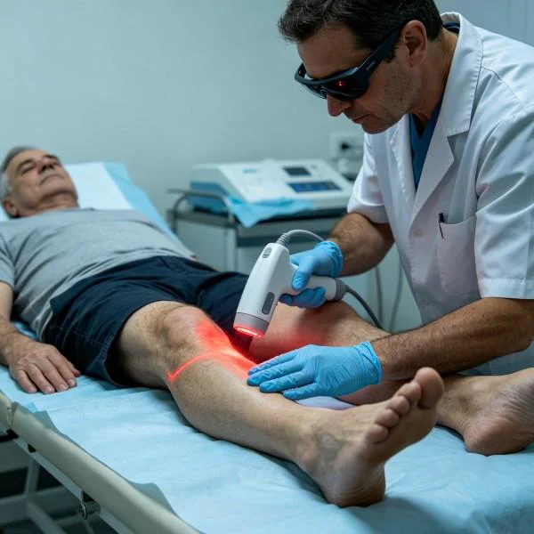
Clinical Realities: High Intensity Laser vs Conventional Approaches
Moving away from conventional wound care toward high-power Class IV laser therapy improves outcomes across both podiatric and outpatient vascular clinics.
Standard management of chronic lower-extremity wounds often relies on passive symptom care. Patients undergo repetitive mechanical debridement, receive rotating topical antimicrobial dressings, and face long courses of systemic antibiotics, carrying risks of drug-resistant infections, gastrointestinal upset, and persistent wound breakdown. When conservative measures fail, patients face aggressive surgical revascularization, extensive skin grafting, or progressive toe and partial-foot amputations that permanently impair mobility.
High-intensity Class IV laser therapy provides a tissue-preserving alternative that treats the underlying ischemia directly. By delivering dense photon flux through calloused wound perimeters, it restores microcirculatory blood flow, suppresses bacterial colonization, and stimulates dormant repair cells without mechanical trauma or chemical toxicity. Patients achieve steady, measurable wound closure and regain durable skin integrity while avoiding repetitive, painful debridements.
In managing chronic peripheral vascular lesions, standard options often default to chemical sclerotherapy or electrocautery. Chemical sclerotherapy can trigger localized tissue necrosis or allergic reactions, while electrocautery generates high temperatures exceeding 300 degrees Celsius that leave deep, carbonized wounds prone to secondary infection.
Utilizing dedicated high-power vascular laser systems provides a clean, precise approach. Matching wavelength absorption directly to hemoglobin and water enables instantaneous microvascular closure without open skin breakdown, post-procedural pain, or extended downtime.
Modern podiatry and vascular centers adopt these advanced laser systems to elevate their standard of care. By resolving the true physiological causes of poor wound healing and microvascular stasis, they deliver reliable, accelerated functional recovery that restores patient mobility and prevents limb loss.