 FotonMedix



Overcoming Severe Joint Capsule Effusion and Synovial Hypoxia
Simultaneous multi-spectral photonic saturation drives synovial capillary neo-perfusion, suppresses inflammatory cytokine cascades at the articular baseline, and prevents cutaneous thermal accumulation through dynamic pulse duty cycle gating.
An active middle-aged patient hobbles into an orthopedic sports medicine clinic, supporting their weight on the good leg while the affected knee remains locked in twenty degrees of protective flexion. The infrapatellar space is visibly swollen, presenting a tense, fluid-filled suprapatellar pouch warm to the touch. The patient explains that every stair climbed sends a sharp, sickening throbbing ache straight through the retropatellar joint line. They have already undergone repeated needle arthrocentesis to aspirate synovial fluid, followed by intra-articular corticosteroid injections. Within four weeks, the effusion inevitably returns, leaving the patient dependent on systemic NSAIDs that irritate the stomach lining. The underlying clinical barrier facing the rehabilitation specialist is rooted in deep tissue optics: articular cartilage and dense capsular ligaments sit four to five centimeters beneath cutaneous layers, shielded by fluid bags and adipose tissues that scatter and diffuse non-focused light beams. Reversing chronic synovial inflammation without surgical lavage demands driving calibrated photon fluxes past the fluid effusion barrier to downregulate pro-inflammatory cascades while restoring local lymphatic drainage.
Optical Attenuation Curves and Depth-Targeted Chromophore Coupling
Delivering therapeutic photon densities into an inflamed joint capsule requires navigating differing absorption and scattering coefficients across stratified biological layers. Cutaneous melanin, subcutaneous fat, and intra-articular effusion fluid alter light propagation paths, turning standard low-output light devices into superficial heat sources.
Wavelength-Selective Chromophore Dynamics in Inflamed Synovium
The biological efficacy of high-intensity class IV optical therapy hinges on matching specific wavelengths to targeted chromophores within the inflamed joint compartment. Using single-spectrum emitters creates an operational compromise, whereas combining distinct near-infrared wavelengths achieves structural and biochemical modulation simultaneously:
- Hemoglobin absorption and microvascular recruitment via 980nm: The 980nm wavelength coincides with the optical absorption peak of oxygenated hemoglobin and local water. In chronic synovitis, capillary networks are congested and hypoxic. Energy delivered at 980nm generates localized microthermal gradients within endothelial walls, opening dormant capillary sphincters, washing out metabolic waste products, and jumpstarting stagnant lymphatic drainage channels.
- Cytochrome c oxidase activation and metabolic restoration via 1064nm: The 1064nm wavelength demonstrates the lowest optical scattering coefficient across dense fibrotic tissues. Photons penetrate straight through the joint capsule to reach unit IV of the mitochondrial respiratory chain, known as cytochrome c oxidase. Stimulating this enzyme displaces inhibitory nitric oxide, restores the transmembrane proton gradient, and fuels tenocytes and chondrocytes with adenosine triphosphate (ATP) necessary to maintain cellular homeostasis.
Duty Cycle Modulation and Thermal Dissipation Kinetics
Delivering ten to twenty Watts of continuous laser energy into a joint space quickly challenges the thermal relaxation time of human skin. Continuous wave delivery causes rapid heat accumulation in superficial dermal layers, activating epidermal pain fibers and risking photothermal blistering long before the synovial membrane receives an adequate biostimulative dose.
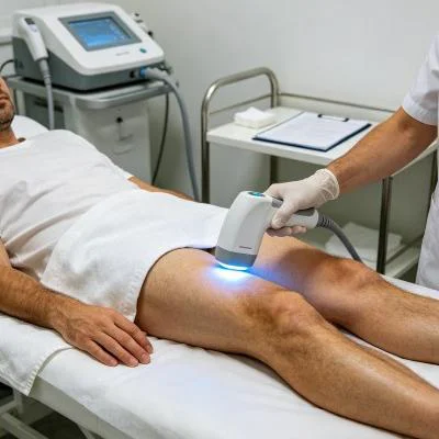
Modern high-intensity class IV clinical platforms solve this limitation by integrating variable pulse duty cycles. By modulating emission frequencies between 10 Hz and 20,000 Hz with precise on/off ratios, the laser emits high peak power during active microsecond pulses, followed by quiescent intervals where vascular blood flow conducts surface heat away. This allows clinicians to maintain direct skin contact and deliver substantial cumulative Joules directly into the deep joint capsule, establishing effective laser therapy for inflammation while safeguarding cutaneous boundaries.
+--------------------------------------------------------------------------------------------------+
|                            ARTICULAR CAPSULE PHOTON PENETRATION                                  |
+--------------------------------------------------------------------------------------------------+
| Quartz Glass Handpiece Compression Application                                                   |
|   |                                                                                              |
|   v                                                                                              |
| [Epidermis & Dermis]        ---> Superficial scatter bypassed via duty cycle cooling rest        |
|   |                                                                                              |
|   v                                                                                              |
| [Prepatellar Adipose]       ---> Mechanical contact displaces interstitial fluid volume          |
|   |                                                                                              |
|   v                                                                                              |
| [Synovial Membrane]         ---> 980nm targets congested hemoglobin, draining chronic effusion   |
|   |                                                                                              |
|   v                                                                                              |
| [Infrapatellar Fat Pad]     ---> Downregulation of IL-1beta, TNF-alpha, and COX-2 enzymes         |
|   |                                                                                              |
|   v                                                                                              |
| [Subchondral Bone Margin]   ---> 1064nm photons trigger mitochondrial Cytochrome C Oxidase       |
+--------------------------------------------------------------------------------------------------+
Biomolecular Pathways: Inflammatory Cytokines vs. Peripheral Nociception
Managing joint capsular degeneration requires addressing both chemical inflammation and sensory hyperalgesia. Damaged synovial cells release substance P, bradykinin, and pro-inflammatory cytokines such as interleukin-1 beta (IL-1β) and tumor necrosis factor-alpha (TNF-α), creating a toxic microenvironment that degrades cartilage matrices and sensitizes local C-fiber nociceptors.
When clinicians utilize a high-intensity laser for inflammation, the treatment suppresses nuclear factor kappa B (NF-κB) transcription pathways inside synovial fibroblasts. This downregulation reduces prostaglandin E2 (PGE2) synthesis and halts matrix metalloproteinase production.
Concurrently, implementing high-intensity laser therapy for pain relief alters peripheral neurological conduction. High-density photon bursts temporarily hyperpolarize primary sensory afferent fibers, slowing conduction velocities along non-myelinated pain pathways. Research documented by Chow and colleagues in clinical pain literature demonstrates that high-irradiance near-infrared light induces reversible neural conduction blockades in peripheral nerves, clearing neurogenic edema and interrupting chronic pain loops without pharmaceutical agents.
Clinical Case Documentation: Outpatient Orthopedic Rehabilitation
The documented record below represents an outpatient rehabilitation course for an active patient presenting with chronic knee synovitis, recurrent joint effusion, and secondary patellofemoral pain, managed with a multi-wavelength Class IV rehabilitation laser system equipped with an optical quartz massage applicator.
Patient Profile and Clinical Baseline
- Case Identifier: ORTHO-SYNOV-2026-1045
- Patient Age / Sex: 54-Year-Old Female
- Primary Diagnosis: Chronic Recurrent Knee Synovitis with Grade III Suprapatellar Effusion and Outerbridge Grade II Patellofemoral Cartilage Fissuring, confirmed via Musculoskeletal Ultrasound and MRI.
- History: 9 months of persistent joint swelling and anterior knee pain after a hiking accident. Underwent two prior joint aspirations and one cortisone injection with symptom recurrence inside three weeks; unable to descend stairs or walk without a limp.
- Baseline Metrics: Numerical Pain Rating Scale (NPRS) at 8/10 during weight-bearing flexion; Western Ontario and McMaster Universities Osteoarthritis Index (WOMAC) at 64/96; Knee Active Range of Motion (AROM) Flexion limited to 85° with marked suprapatellar fullness; Mid-patellar joint circumference measured at 41.5 cm.
Multi-Stage Treatment Regimen
The rehabilitation schedule consisted of ten comprehensive clinical sessions divided across four consecutive weeks, transitioning from acute fluid drainage to deep cellular regeneration.
| Session No. | Clinical Phase & Objective | Wavelength Setup | Operating Power (W) | Pulse Frequency (Hz) | Duty Cycle (%) | Energy Delivered (J) | Cumulative Dose (J) |
| Session 01 | Nociceptive Blocking & Popliteal Drainage | 980nm (65%) + 1064nm (35%) | 12.0 W | 8,000 Hz | 40% | 3,600 J | 3,600 J |
| Session 02 | Suprapatellar Lymphatic Pumping | 980nm (60%) + 1064nm (40%) | 14.0 W | 4,000 Hz | 50% | 4,200 J | 7,800 J |
| Session 03 | Retropatellar Synovial Perfusion | 980nm (50%) + 1064nm (50%) | 16.0 W | 2,000 Hz | 60% | 4,800 J | 12,600 J |
| Session 04 | Infrapatellar Fat Pad Desensitization | 980nm (40%) + 1064nm (60%) | 18.0 W | 500 Hz | 70% | 5,400 J | 18,000 J |
| Session 05 | Deep Capsular Effusion Clearance | 980nm (40%) + 1064nm (60%) | 20.0 W | 250 Hz | 75% | 6,000 J | 24,000 J |
| Session 06 | Synovial Fibroblast Modulation | 980nm (30%) + 1064nm (70%) | 22.0 W | 100 Hz | 80% | 6,600 J | 30,600 J |
| Session 07 | Subchondral Bone Matrix Saturation | 980nm (20%) + 1064nm (80%) | 24.0 W | Continuous (CW) | 100% | 7,200 J | 37,800 J |
| Session 08 | Chondrocyte ATP Resynthesis Push | 980nm (20%) + 1064nm (80%) | 24.0 W | Continuous (CW) | 100% | 7,200 J | 45,000 J |
| Session 09 | Functional Eccentric Loading Support | 980nm (30%) + 1064nm (70%) | 20.0 W | 150 Hz | 75% | 6,000 J | 51,000 J |
| Session 10 | Final Tissue Homeostasis & Consolidation | 980nm (40%) + 1064nm (60%) | 16.0 W | 500 Hz | 65% | 4,800 J | 55,800 J |
Clinical Progression and Outcome Metrics
WEEK 1 (Sessions 1-3):
- Mechanical lymphatic tracing over the popliteal fossa drained accumulated interstitial fluid.
- Mid-patellar circumference reduced from 41.5 cm to 39.2 cm.
- Patient experienced the first night without throbbing knee ache; resting NPRS dropped from 8/10 to 4/10.

WEEK 2 (Sessions 4-6):
- Active flexion range increased from 85° to 115° without resistance or patellar tracking pinch.
- Suprapatellar fluid pocket decreased on ultrasound, with the hypoechoic fluid layer shrinking from 14mm to 4mm.
- WOMAC functional score improved from 64 to 36.

WEEK 3 (Sessions 7-8):
- High-density continuous wave delivery applied directly around the patellar retinaculum with deep contact compression.
- Patient started step-down exercises and stationary cycling with zero swelling flare-up following sessions.
- Synovial membrane thickening showed marked regression during sonographic re-evaluation.

WEEK 4 (Sessions 9-10):
- Full active knee range of motion achieved (135° flexion, 0° extension).
- Mid-patellar circumference stabilized at 37.8 cm, matching the unaffected contralateral limb.
- Final NPRS scored 0/10 at rest and 1/10 following prolonged stair descent.
- WOMAC score reached 5/96, indicating full return to uninhibited physical activity.
Comparative Analysis: High-Intensity Laser vs. Conventional Modalities
Comparing high-intensity class IV light with traditional treatment paths clarifies why outpatient orthopedic clinics incorporate this technology into their daily workflow.
Intra-Articular Corticosteroid Injections
Steroid injections act as broad chemical immunosuppressants that temporarily halt inflammatory symptoms. However, frequent corticosteroid exposure accelerates cartilage breakdown, damages native chondrocyte viability, and risks joint space narrowing over time. High-intensity multi-wavelength laser therapy provides an anatomical, non-destructive alternative. Rather than shutting down cellular machinery, high-energy photons stimulate natural microvascular perfusion, encourage lymphatic fluid drainage, and replenish cellular ATP pools without degrading structural cartilage.
Oral Non-Steroidal Anti-Inflammatory Drugs (NSAIDs)
Systemic NSAIDs inhibit COX-1 and COX-2 enzymes across the entire body, muting pain signals while exposing the patient to risks of gastrointestinal ulceration, renal stress, and cardiovascular strain. They do little to improve local joint perfusion or drain stagnant synovial effusion. Class IV laser treatments work at the exact anatomical site of injury, targeting concentrated energy into inflamed capsular tissues to accelerate tissue recovery without systemic biochemical toxicity.
Traditional Cryotherapy and Passive Ice Packs
Applying ice packs to an inflamed knee induces surface vasoconstriction, which provides temporary pain relief through thermal numbing. Unfortunately, prolonged cold application restricts capillary blood flow, reduces lymphatic clearance, slows tissue metabolism, and stiffens capsular collagen. High-intensity laser therapy takes the opposite physiological approach: by using pulsed near-infrared wavelengths, it boosts capillary circulation, flushes inflammatory cytokines, and relaxes surrounding muscle guarding without superficial heat damage.
Practical Clinical Application and Operational Takeaways
Achieving reproducible clinical outcomes when using high-intensity laser systems for capsular joint inflammation depends on structured procedural execution:
- Dynamic compression with quartz massage probes: Clinicians should firmly press the optical contact probe into peri-articular soft tissues. Applying physical pressure temporarily displaces superficial blood and water layers away from the light path, creating a clear optical channel that allows higher photon densities to reach the deeper synovial lining.
- Centripetal lymphatic clearance: Never start by targeting the center of an inflamed joint. Begin by sweeping proximal lymphatic drainage basins (such as the femoral triangle and popliteal fossa) to clear downstream fluid pathways before treating the inflamed capsule directly.
- Frequency-matched treatment progression: Use high pulse frequencies (5,000 Hz to 10,000 Hz) during the initial phases to calm hypersensitive peripheral nerve endings, transition to moderate pulsing (200 Hz to 500 Hz) to clear fluid effusions, and finish with continuous wave (CW) delivery to stimulate chondrocyte and tenocyte remodeling once acute swelling has resolved.
Grounding clinical protocols in the optical principles of human tissue allows physical medicine practices and sports rehabilitation centers to reliably resolve chronic joint inflammation and restore patient mobility.