 FotonMedix



Solving Cervical Radiculopathy and Chronic Microvascular Stasis
Targeted 1470nm water vaporization reduces intradiscal herniation volume without structural collapse, intravascular photonic perfusion eliminates erythrocyte aggregation, and gated pulse intervals eliminate periradicular thermal risk.
A patient presents to an interventional spine and pain practice holding their neck rigid, unable to rotate their head without triggering sharp electrical shocks radiating down the C6-C7 dermatome into the thumb and forefinger. Months of selective nerve root blocks, traction, and neuropathic medications have failed to restore motor strength. When spinal surgery is presented as the next step, patients frequently investigate alternatives and ask what is the cost of laser disc ablation compared to an anterior cervical discectomy and fusion (ACDF) or artificial disc replacement. In the operating suite, the clinical challenge is anatomical density: the cervical intervertebral space is compact, surrounded by vertebral arteries, the spinal cord, and sympathetic nerve plexuses. Standard surgical intervention destabilizes adjacent segments, while mechanical decompression risks nerve root traction. Delivering targeted optical energy through a micro-cannula decompresses the impinged spinal nerve root while resolving systemic and perineural microvascular stasis.
Biophysical Mechanisms of Micro-Volume Cervical Disc Decompression
Cervical disc architecture contains smaller hydraulic volumes than lumbar segments. A minor nuclear displacement of even 1.5 to 2 millimeters within the neuroforamen is sufficient to trigger severe root ischemia. Decompressing this space requires precise photo-ablation that does not compromise disc height or vertebral endplate vascularization.
High-Affinity Water Absorption vs. Structural Annular Protection
The cervical nucleus pulposus relies heavily on residual hydration to maintain viscoelastic shock absorption. Using non-specific wavelengths carries the risk of lateral heat conduction into the posterior longitudinal ligament.
Employing precision-guided high-intensity class IV energy utilizes specific tissue absorption characteristics:
- Micro-localized water vaporization at 1470nm: Water molecules in the degenerated nuclear matrix exhibit high absorption at 1470nm. Optical energy delivered along this peak converts directly into rapid thermal vaporization confined to a sub-millimeter interaction zone. This creates a micro-cavity within the nucleus pulposus without conducting destructive heat into the sensitive vertebral endplates.
- Controlled microvascular coagulation at 980nm: The 980nm band targets hemoglobin pools in inflamed, hypervascularized tear margins. Delivering this wavelength coagulates ingrown sinuvertebral nerve fibers and micro-vessels, eliminating localized chemical pain generators without damaging structural annular rings.
Pulsed Energy Delivery and Thermal Boundary Control
Operating in close proximity to the cervical spinal cord demands strict thermal control. Continuous wave exposure within a confined disc can elevate surrounding tissue temperatures above the threshold for nerve root injury.
Pulsed duty cycles solve this problem through calibrated thermal relaxation intervals. Delivering optical bursts lasting 0.2 to 0.4 seconds interspersed with 0.8-second rest intervals ensures that heat dissipates through surrounding intradiscal water before it can reach adjacent neural structures. This duty cycle gating maintains boundary temperatures below 43 degrees Celsius, enabling safe percutaneous disc decompression within narrow cervical margins.
+--------------------------------------------------------------------------------------------------+
|                            CERVICAL PERCUTANEOUS ABLATION MODEL                                  |
+--------------------------------------------------------------------------------------------------+
| 20G Spinal Cannula Placement (Anterolateral Approach)                                           |
|   |                                                                                              |
|   v                                                                                              |
| [Anterior Annulus Fibrosus]   ---> Atraumatic needle puncture preserves spinal stability         |
|   |                                                                                              |
|   v                                                                                              |
| [Central Nucleus Pulposus]    ---> 1470nm precisely photo-vaporizes nuclear water volume         |
|   |                                                                                              |
|   v                                                                                              |
| [Intradiscal Pressure Relief] ---> Immediate hydraulic suction retracts foraminal bulge         |
|   |                                                                                              |
|   v                                                                                              |
| [Periradicular Safety Margin] ---> Duty cycle rest intervals prevent heat reaching nerve roots   |
+--------------------------------------------------------------------------------------------------+
Systemic Endothelial and Microvascular Restoration via Intravascular Light
Structural nerve decompression addresses mechanical impingement, but prolonged compression often leaves chronic perineural edema, localized tissue hypoxia, and capillary sludge in its wake.
Introducing systemic intravascular phototherapy, known as iv laser therapy, treats this downstream vascular compromise. When coherent light is introduced directly into a peripheral vein, circulating blood components absorb photons, initiating systemic circulatory and immunological shifts.
Intravascular Hemorheology and Cellular Respiration
Research conducted by Mittermayr and colleagues on photonic blood conditioning confirms that endovascular illumination alters the physical properties of circulating blood:
- Erythrocyte membrane repolarization: Delivering laser iv therapy restores electrical zeta potentials across red blood cell membranes. This separates clustered erythrocytes, allowing single-file red cell passage through compressed capillaries supplying ischemic nerve sheaths.
- Endothelial nitric oxide release: Photons stimulate endothelial cells to release nitric oxide, widening downstream micro-vessels and restoring capillary perfusion pressures.
- Mitochondrial reactivation: Cytochrome c oxidase inside circulating leukocytes and platelets absorbs near-infrared and visible light, elevating adenosine triphosphate (ATP) reserves and supporting microvascular repair.
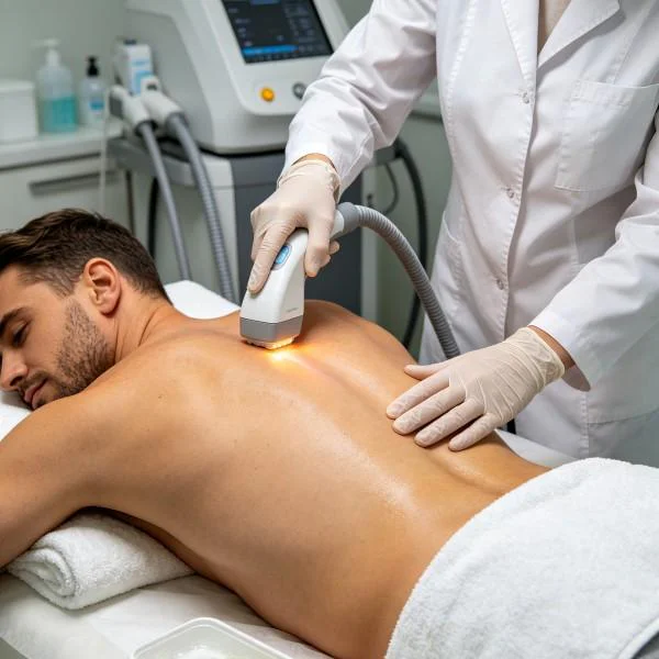
Clinical Protocol Implementation: Percutaneous Disc Ablation vs. Systemic Blood Conditioning
Applying class IV high-intensity systems in cervical spine care requires balancing surgical vaporization with non-thermal systemic modulation.
Percutaneous cervical disc ablation requires sterile, fluoroscopically guided placement of a 360-micron or 400-micron silica optical fiber. The clinical objective is controlled tissue vaporization: delivering precise energy totals (typically 600 to 900 Joules for cervical discs) using short duty cycles to ensure structural stability.
Systemic blood conditioning operates on a non-thermal photobiomodulation protocol. Sterile intravenous catheters deliver multi-wavelength light (such as green, red, and near-infrared) at 60mW to 180mW for 30 to 45 minutes. This systemic approach addresses circulating inflammation and supports local recovery after decompression.
Clinical Case Documentation: Interventional Spine and Neuro-Rehabilitation
The clinical data below outlines the documented treatment course for a patient presenting with cervical disc herniation and persistent radiculopathy, managed with percutaneous laser decompression followed by systemic intravascular therapy.
Patient Profile and Clinical Baseline
- Case Identifier: CERV-SPINE-2026-1218
- Patient Age / Sex: 49-Year-Old Male
- Primary Diagnosis: Contained C6-C7 Foraminal Disc Protrusion with Unilateral C7 Nerve Root Compression confirmed via Cervical MRI, associated with chronic ipsilateral Upper Extremity Neurogenic Dysesthesia and Brachial Capillary Stasis.
- History: 8 months of radiating left neck, shoulder, and index-finger pain following an industrial accident. Cervical epidural steroid injections and physical therapy provided no sustainable improvement.
- Baseline Metrics: Numerical Pain Rating Scale (NPRS) at 8/10 for radiating arm pain; Neck Disability Index (NDI) at 52%; Triceps reflex diminished (1+/4); Grip strength on left side measured at 21 kg (versus 44 kg on right).
Multi-Stage Treatment Regimen
The management protocol integrated an outpatient percutaneous disc vaporization procedure with eight subsequent intravascular photo-perfusion sessions over three weeks.
| Session No. | Modality & Objective | Wavelength Setup | Operating Power | Frequency / Duty Cycle | Total Energy (J) | Cumulative Dose (J) |
| Procedure Day | Percutaneous C6-C7 Disc Decompression | 1470nm (75%) + 980nm (25%) | 6.0 W | 0.2s ON / 0.8s OFF (20%) | 780 J | 780 J |
| Session 01 | Endovenous Microvascular Recanalization | 650nm (50%) + 532nm (50%) | 0.08 W | Continuous (CW) 100% | 144 J | 924 J |
| Session 02 | Erythrocyte Deformability Optimization | 532nm (40%) + 980nm (60%) | 0.10 W | Continuous (CW) 100% | 180 J | 1,104 J |
| Session 03 | Periradicular Edema Drainage Support | 650nm (50%) + 405nm (50%) | 0.10 W | Continuous (CW) 100% | 180 J | 1,284 J |
| Session 04 | Mitochondrial Respiratory Chain Activation | 980nm (70%) + 650nm (30%) | 0.12 W | Continuous (CW) 100% | 216 J | 1,500 J |
| Session 05 | Endothelial Nitric Oxide Stimulation | 532nm (50%) + 980nm (50%) | 0.12 W | Continuous (CW) 100% | 216 J | 1,716 J |
| Session 06 | Anti-Inflammatory Cytokine Balancing | 405nm (40%) + 650nm (60%) | 0.12 W | Continuous (CW) 100% | 216 J | 1,932 J |
| Session 07 | Neuro-Muscular Transmission Support | 650nm (60%) + 810nm (40%) | 0.15 W | Continuous (CW) 100% | 270 J | 2,202 J |
| Session 08 | Final Systemic Tissue Consolidation | 650nm (50%) + 980nm (50%) | 0.15 W | Continuous (CW) 100% | 270 J | 2,472 J |
Clinical Progression and Outcome Metrics
OUTPATIENT SURGICAL OUTCOME (Cervical Percutaneous Decompression):
- Under local anesthesia and biplanar fluoroscopy, a 20G needle entered the anterolateral C6-C7 space.
- A 360-micron quartz optical fiber delivered 780 Joules of gated 1470nm/980nm energy.
- Real-time intradiscal manometry recorded a 38% decrease in resting hydrostatic pressure.
- Radiating dermatomal arm pain decreased from NPRS 8/10 to 2/10 within 60 minutes post-procedure.

REHABILITATION & ENDOVENOUS RECOVERY (Sessions 1-4):
- Patient ambulated 30 minutes following procedure completion without cervical collar support.
- Intravenous sessions resolved index finger paresthesias and forearm hypersensitivity.
- Left triceps deep tendon reflex returned to symmetric baseline (2+/4).
- NDI score improved from 52% to 22% by day ten.

FINAL CONSOLIDATION & FUNCTIONAL RETURN (Sessions 5-8):
- Left hand grip strength increased from 21 kg to 42 kg, demonstrating complete motor recovery.
- Full active cervical range of motion restored across flexion, extension, and bilateral rotation.
- Final NPRS recorded at 0/10 at rest and 0/10 during maximum cervical extension and rotation.
- NDI score reached 4%, confirming complete functional recovery without neurological deficits.
Comparative Analysis: High-Intensity Laser vs. Conventional Spinal Interventions
Reviewing minimally invasive laser decompression alongside conventional cervical spine surgery highlights several clinical distinctions.
Anterior Cervical Discectomy and Fusion (ACDF)
ACDF involves full discectomy, bone graft placement, and anterior titanium plating. While it resolves nerve compression, fusing the segment permanently eliminates motion at that level and increases mechanical stress on adjacent vertebrae, frequently leading to adjacent segment disease. Percutaneous laser disc decompression preserves native disc height, ligamentous structures, and normal cervical biomechanics without implants.
Cervical Epidural Injections
Fluoroscopic epidural steroid injections deliver short-term anti-inflammatory relief to compressed nerve sheaths. However, steroids do not reduce the physical volume of the protruding disc material. When steroid efficacy declines, mechanical root impingement remains. Minimally invasive laser ablation provides an anatomical reduction in disc volume through targeted photo-vaporization, addressing the underlying mechanical cause.
Cervical Microforaminotomy
Surgical microforaminotomy requires bony resection of the facet joint margins to free the compressed nerve root. Resecting facet bone can cause cervical instability and chronic postoperative neck pain. Percutaneous laser decompression avoids bone resection entirely, utilizing an anterior micro-puncture to decompress the root from within the disc core.
Practical Clinical Application and Operational Takeaways
Safely implementing percutaneous cervical disc ablation and intravascular photo-perfusion requires standardized clinical practices:
- Fiber positioning verification: Maintain strict fluoroscopic needle verification. In the cervical spine, the optical fiber tip must remain in the central-to-posterior third of the nucleus, avoiding placement beyond the posterior vertebral margin.
- Controlled thermal intervals: Deliver energy in short intervals. In small cervical discs, optical delivery should pause every 100 Joules to allow thermal relaxation, protecting vertebral endplates and adjacent neural tissue.
- Post-procedural systemic microvascular therapy: Pair intradiscal decompression with intravascular phototherapy. Relieving mechanical pressure while improving red blood cell flexibility and microvascular flow accelerates recovery and reduces persistent neurogenic discomfort.
Applying targeted optical physics allows outpatient surgical centers and interventional spine practices to resolve complex cervical disc and nerve root conditions with minimal tissue disruption.