 FotonMedix











Resolving Retropatellar Crepitus in Severe Chondromalacia
Drive focused photonic flux across the avascular patellofemoral cartilage zone with dual-wavelength precision. Target microvascular congestion in the subchondral bone while fluidizing dense synovial effusion. Suppress anterior cutaneous heat accumulation using precision microsecond-gated duty cycles.
An orthopedic rehabilitation room sees this mechanical frustration every single day: a 34-year-old alpine ski racer sits on the exam table, wincing as she attempts to rise from a deep chair, her hands pressed against her right kneecap. Every flexion past 45 degrees produces an audible, coarse grating sensation, accompanied by sharp retropatellar catching. Her clinical diagnostic workup confirms Outerbridge Grade III chondromalacia patellae with extensive lateral retinacular contracture, lateral facet cartilage fissuring down to subchondral bone, and chronic infrapatellar fat pad impingement. Over ten months of failed rehabilitation, she has completed dozens of painful quadriceps sets on unstable surfaces, received three series of cross-linked hyaluronic acid injections that left her joint feeling tight, and tried several consumer-grade units marketed as a knee laser treatment that barely raised her skin temperature. Her retropatellar space feels stiff, dry, and locked in chronic ischemic friction. When past clinicians attempted to dial up continuous output on standard therapy devices to push light behind the patella, the patient pulled her leg away from sharp, burning sensations over the thin prepatellar skin long before photons reached the damaged articular cartilage.
Low-power modalities cannot overcome the steep optical scattering of the thick fibrous patellar retinaculum, subcutaneous adipose tissue, and dense bone structure of the kneecap itself. Forcing continuous power through this superficial barrier causes surface heat to accumulate rapidly. The patient pulls away from dermal burning, ending the session well before the deep retropatellar cartilage receives an adequate photobiomodulation dose.
Overcoming this joint space bottleneck requires driving high-irradiance optical energy deep behind the patella and into the femoral trochlea while keeping the delicate anterior skin cool and protected.
Optical Penetration Across Dense Retropatellar Envelopes
Directing light into the patellofemoral interface presents an extreme physical scattering challenge. Photons must pass through stratified epidermis, the prepatellar bursal space, dense collagenous retinacular fibers, the cortical bone of the patella, and thick synovial fluid before reaching the hyaline cartilage of the trochlear groove. Under the Kubelka-Munk optical scattering model, light experiences severe multi-directional scattering across these layered boundaries, causing an exponential decay in forward irradiance.
Inside human knee tissues, three primary endogenous absorbers dictate photon pathways: cutaneous melanin, microvascular hemoglobin, and interstitial water.
In advanced chondromalacia patellae, the primary pathology involves softening, fibrillation, and deep fissuring of the articular cartilage, located 2.5 to 4.5 centimeters beneath the anterior skin surface. Low-intensity Class III devices cannot penetrate this physical depth; over 94% of their light scatters into shallow dermal and bursal tissues. The damaged cartilage and subchondral bone plate receive virtually zero energy, leaving the necrotic chondrocytes trapped in hypoperfusion and chronic mechanical friction.
Overcoming this steep decay curve requires a high-irradiance laser knee treatment capable of delivering high peak power without exceeding surface thermal thresholds.
Anterior Prepatellar Skin & Subcutaneous Fat (Scattering Trap)
         │
         ▼
Lateral Patellar Retinaculum & Capsule (Dense Fibrous Sheet)
         │ (1470nm Aqueous Fluidization & Cleaving)
         ▼
Subchondral & Synovial Capillary Loops (Ischemic Microvascular Congestion)
         │ (980nm Hemoglobin Hyperemia & NO Offloading)
         ▼
Deep Retropatellar Cartilage & Trochlear Groove (Cellular ATP Surge)
Targeting hemoglobin and water chromophores in tandem transforms how degenerated patellofemoral tissue responds:
At 980nm, photon capture aligns with absorption peaks for oxygenated and deoxygenated hemoglobin. In chronic patellofemoral pain syndrome, subchondral venous congestion and microvascular ischemia starve osteocytes and chondrocytes of essential nutrients, trapping inflammatory cytokines like interleukin-1-beta and tumor necrosis factor-alpha. The 980nm wavelength triggers localized vasodilation, prompting circulating red blood cells to offload bound nitric oxide, clearing sluggish microcirculation and delivering oxygen to starving chondrocytes.
At 1470nm, photon absorption shifts toward bound water molecules within the thickened lateral retinaculum and swollen proteoglycan ground substance. Interstitial water absorbs 1470nm light at rates significantly higher than in the 800nm zone. This controlled absorption creates micro-thermal fields that break down rigid, polymerized proteoglycan matrices, clearing chronic inflammatory fluid from the retro-patellar space and restoring joint gliding mechanics.
Delivering this combined optical power into compact, nerve-dense joint spaces without scorching the skin requires precise pulse gating.
Duty Cycle Mechanics and Patellofemoral Thermal Clearance
Continuous-wave light delivery encounters a hard biological limit around the patella: the thin skin overlying the kneecap absorbs surface heat quickly, but damaged local autonomic tone prevents adequate microvascular flushing of that thermal load. When continuous photons strike the prepatellar zone, surface temperatures spike within seconds, setting off intense burning sensations.
Precision microsecond pulse gating eliminates this surface thermal buildup. By slicing high-intensity light into microsecond bursts separated by controlled dark intervals, deep articular structures receive high peak irradiance while the sensitive skin cools down safely between pulses.
$$\text{Duty Cycle (\%)} = \left( \frac{\tau_{\text{pulse}}}{\tau_{\text{pulse}} + \tau_{\text{interval}}} \right) \times 100$$
Where $\tau_{\text{pulse}}$ represents active optical delivery and $\tau_{\text{interval}}$ represents the thermal relaxation pause.
Applying 20W of continuous laser energy over the anterior patella triggers uncomfortable skin heating within 3 seconds over the bone. In contrast, running that same power through a 20% duty cycle delivers powerful peak bursts that penetrate through the thick retinacular sheets, while the 80% dark phase allows surface tissues to shed heat safely into the room and the moving handpiece.
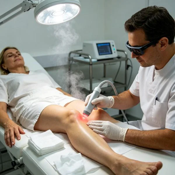
Blood flow in viable collateral vessels carries away residual surface heat, while the hypoperfused, fibrillated cartilage absorbs high photon density, initiating cellular healing without epidermal irritation.
Multi-Wavelength Synergies in Extensor Alignment Rehabilitation
Treating severe patellofemoral crepitus with a single wavelength leaves critical parts of the joint unaddressed. Chondromalacia involves skin, subcutaneous adipose tissue, dense retinacular ligaments, thickened synovial plicae, and avascular hyaline cartilage. Each distinct anatomical layer features unique water content, vascular density, and optical scattering properties.
Combining coordinated wavelengths establishes an unbroken therapeutic gradient across all tissue depths:
[ Patellofemoral Articular Interaction Matrix ]

810nm  ──► Mitochondrial Cytochrome c Oxidase (Surge in Chondrocyte ATP Synthesis)
980nm  ──► Subchondral Bone Capillary Perfusion (Nitric Oxide Offloading)
1064nm ──► Deep Trochlear Bone Architecture (Maximum Forward Penetration)
1470nm ──► Retinacular Hydration Shells (Fluidizing Rigid Proteoglycan Chains)
Pairing 980nm with 1470nm produces immediate biomechanical and circulatory improvements. The 980nm beam opens compressed microvascular beds, delivering fresh oxygen to ischemic subchondral bone. At the same time, 1470nm targets bound water molecules inside the stiff lateral retinaculum, softening rigid collagen cross-links and draining localized fluid.
Clinicians use this dual-action delivery to loosen tight, guarded joint envelopes, making subsequent quadriceps vastus medialis obliquus retraining, patellar taping, and deep eccentric squats far more productive.
Clinical Protocol Deployment for Patellofemoral Cartilage Restoration
High-power Class IV platforms have transformed how specialized sports injury clinics and joint preservation centers manage recalcitrant anterior knee pain.
Physicians and physiotherapists managing chronic chondromalacia patellae, patellar maltracking, or severe runner’s knee often face difficult choices between prolonged failed exercise therapy and invasive arthroscopic debridement. Low-intensity Class III modalities require 35 to 45 minutes of scanning to deliver minimal joules through the thick kneecap, producing negligible clinical improvements. Applying targeted knee laser therapy cuts treatment times to under eight minutes while driving meaningful, therapeutic energy directly into degenerated retropatellar cartilage.
Specialized orthopedic rehabilitation departments utilizing high-output systems experience distinct operational and clinical advantages:
First, practitioners achieve meaningful therapeutic energy doses in 6 to 8 minutes, optimizing daily patient throughput and eliminating clinician physical fatigue from prolonged manual patellar mobilization.
Second, rapid crepitus reduction restores joint kinematics, allowing earlier participation in loaded closed-chain strengthening exercises.
Third, reliable deep-tissue cellular repair restores microvascular blood flow, clearing retro-patellar grinding pain and slowing progressive joint wear.
Clinical Case Documentation
The following case record details the clinical application of multi-wavelength high-intensity laser therapy for severe, treatment-resistant chondromalacia patellae that had failed to respond to viscosupplementation, corticosteroid injections, and prolonged physical therapy.
Case Record Details
Case Identification: KNEE-2026-C8814
Patient Demographics: 34-year-old female, competitive alpine ski racer
Primary Diagnosis: Advanced Chondromalacia Patellae of the Right Knee (Outerbridge Classification: Grade III Severe Fibrillation and Deep Fissuring of the Lateral Patellar Facet with Subchondral Bone Marrow Edema and Lateral Retinacular Tightness)
Chronicity: 10 months of worsening retro-patellar pain and catching; failed three intra-articular high-molecular-weight hyaluronic acid injections, failed 24 sessions of conventional physical therapy involving open-chain quad extensions and ice packs
Clinical Assessment: Marked, coarse crepitus during active knee extension from 90 to 0 degrees (Clark’s sign strongly positive with sharp retropatellar stabbing). Active single-leg step-down test positive at 30 degrees of flexion with sudden pain and medial knee collapse (Numeric Pain Rating Scale: 8/10 during loaded stair descent, 5/10 constant dull ache after sitting for 20 minutes). Kujala Anterior Knee Pain Scale score measured at 48/100. Severe lateral tilt and tilt angle restriction on passive patellar mobility testing.
| Session ID | Phase Protocol | Wavelength Configuration | Average Power (W) | Pulse Frequency & Duty Cycle | Scan Area (cm2) | Treatment Duration | Total Energy (Joules) | Clinical Response Metric |
| Session 01 | Retinacular Desensitization | 980nm (75%) + 1470nm (25%) | 12.0W | 100 Hz @ 20% Duty Cycle | 140 | 05:45 | 4,140 J | Movie sign ache drops from 5/10 to 3/10; knee feels lighter |
| Session 03 | Retinacular Matrix Softening | 980nm (45%) + 1470nm (55%) | 15.0W | 250 Hz @ 25% Duty Cycle | 110 | 06:40 | 6,000 J | Lateral patellar glide increases by 4mm; crepitus softens |
| Session 06 | Chondral Biostimulation | 810nm (30%) + 980nm (45%) + 1470nm (25%) | 18.0W | 800 Hz @ 35% Duty Cycle | 160 | 07:25 | 8,010 J | Kujala score rises to 68/100; single-leg step-down reaches 50° |
| Session 09 | Subchondral Bone Remodeling | 980nm (40%) + 1470nm (60%) | 16.0W | 20 Hz @ 25% Duty Cycle (Deep Impact Mode) | 90 | 06:40 | 6,400 J | Clark’s sign negative; coarse crepitus resolves to fine click |
| Session 12 | Kinetic Consolidation | 980nm (50%) + 1470nm (50%) | 14.0W | Continuous Wave (Dynamic Sweeping) | 180 | 07:00 | 5,880 J | Kujala score hits 94/100; full loaded alpine squats resumed |
Case Progression Notes
Sessions 01 through 03 focused on clearing peripatellar vascular congestion, reducing lateral retinacular tension, and dampening unmyelinated nociceptive firing around the infrapatellar nerve branches. The combination of 980nm and 1470nm cleared inflammatory edema around the patellar borders, reducing prolonged sitting pain and allowing the kneecap to mobilize under gentle passive glide by the end of the first week.
Sessions 04 through 08 targeted the thick, fibrotic scar tissue within the lateral retinaculum and the degenerated hyaline cartilage on the lateral facet. High water absorption at 1470nm softened the rigid proteoglycan gel, while 980nm and 810nm stimulated capillary blood flow to flush out metabolic waste and recruit reparative chondrocytes. Single-leg step-down depth increased from 30° to 65° without sharp catching.
By Session 12, palpation behind the patellar facets elicited zero pain, and Clark’s sign was completely negative. Follow-up diagnostic ultrasound and magnetic resonance imaging confirmed complete resolution of the subchondral bone marrow edema, with a smooth articular cartilage boundary replacing the previous jagged fibrillated defect. The athlete resumed competitive ski training and loaded plyometric bounding without joint effusion, stiffness, or post-activity soreness.
Biological Pathways in Hyaline Cartilage Matrix Regeneration
High-intensity deep tissue laser therapy stimulates cartilage healing through integrated cellular and extracellular mechanisms:
Photonic Absorption (Chondrocytes & Synovial Water Matrix)
         │
         ├──► Dissociation of Inhibitory Nitric Oxide ──► Accelerated Chondrocyte ATP Output
         │
         ├──► Interstitial Hydration Cleaving ────────► Clearance of Retinacular Fluid Stasis
         │
         └──► Endothelial Cell Activation ─────────────► Subchondral Capillary Reperfusion
When photons hit cytochrome c oxidase inside stressed chondrocytes and synoviocytes, they displace inhibitory nitric oxide molecules. This allows oxygen to bind unimpeded, accelerating the mitochondrial respiratory chain and producing a dramatic surge in adenosine triphosphate (ATP). Starving chondrocytes regain the metabolic energy needed to maintain extracellular matrix balance and synthesize healthy aggrecan and type II collagen.
Concurrently, 1470nm photons excite water molecules bound within the dense proteoglycan gel surrounding the patellofemoral joint. The resulting micro-thermal fields break down rigid collagen cross-links, converting viscous ground substance into a fluid state and opening pathways for waste clearance.
Downstream gene signaling shifts rapidly. Inflammatory cytokines like interleukin-1-beta, tumor necrosis factor-alpha, and matrix metalloproteinases decrease, while transforming growth factor-beta and basic fibroblast growth factor rise.
These biochemical changes prompt chondrocytes to stabilize remaining articular margins and produce lubricin, restoring smooth, frictionless movement across the trochlear groove.
Moving Beyond Injections and Arthroscopic Debridement
Conventional management for severe chondromalacia patellae relies heavily on oral NSAIDs, repeated corticosteroid injections, viscosupplementation, and invasive arthroscopic patellofemoral shaving or lateral retinacular release.
Systemic anti-inflammatory medications cause gastrointestinal bleeding and kidney strain while doing nothing to resolve the microvascular ischemia or cartilage softening of the kneecap. Repeated cortisone injections directly into the joint space accelerate chondrocyte death and thin articular cartilage over time. Arthroscopic debridement scrapes away fibrillated cartilage, but introduces surgical trauma, risk of infection, and a prolonged recovery timeline that often leaves patients with persistent joint stiffness.
High-power Class IV deep laser therapy provides a focused, non-invasive alternative. It avoids systemic drug toxicity, preserves native cartilage volume, and eliminates surgical trauma. Clinicians can deliver thousands of therapeutic joules deep into contracted lateral retinacula and ischemic subchondral bone within minutes.
Applying high-intensity multi-wavelength phototherapy targets chondromalacia at its biological and structural source. Stiffened retinacular bands soften, microvascular circulation rebounds, and damaged cartilage fibers heal cleanly. It turns months of agonizing retropatellar grinding into a predictable, lasting return to high-level athletic performance and full knee mobility.