 FotonMedix











Overcoming Canine Glaucoma Transscleral Energy Barriers
Targeted hemoglobin and water resonance bands shut down aqueous secretion while microsecond duty cycle pulsing prevents scleral collagen melting.
A nine-year-old Siberian Husky cowers on the exam table, pressing its head against the clinic wall with pawing gestures over the right eye and low vocalizations of severe orbital pain. The right globe is noticeably buphthalmic, exhibiting a cloudy, bluish corneal haze, marked mydriasis, and thick, engorged episcleral veins wrapping around the limbus. Tonometry confirms an intraocular pressure of 62 millimeters of mercury. Triple topical hypotensive drops paired with intravenous osmotic diuretics produce only a transient drop to 46 mmHg before the pressure spikes right back up, threatening irreversible optic nerve ischemia. The owner breaks down upon receiving an urgent recommendation for enucleation, desperately inquiring how to treat glaucoma in dogs without permanently removing the eye. Traditional filtration shunts frequently fail from rapid postoperative scarring, while cyclocryotherapy risks severe uveitis, hyphema, and corneal sloughing. When surgical teams evaluate pet glaucoma surgery alternatives, the primary clinical hurdle is executing non-invasive ciliary body ablation through the intact sclera without burning full-thickness collagen or causing devastating globe perforation.
Halting aqueous humor production requires calculating the optical attenuation curve across dense fibrous scleral coats. The canine sclera consists of hydrated, irregularly interwoven type-I collagen bundles that cause intense Rayleigh and Mie scattering. When high-intensity photons pass across the perilimbal margin, superficial light energy disperses across white scleral tissue, failing to deliver the required fluence to the pigmented ciliary epithelium. Without targeted energy absorption inside the vascular core of the ciliary processes, active fluid secretion persists. Applying high-intensity Class 4 multi-wavelength protocols resolves this physical delivery bottleneck, turning dog glaucoma surgery into a sight-preserving, globe-sparing procedure that lowers intraocular pressure while preserving structural ocular integrity.
Optical Extinction Across Transscleral Collagen And Ciliary Stroma
Delivering therapeutic photon fluence into the canine anterior uvea involves steep tissue scattering hurdles. Unlike clear intraocular media such as the cornea and aqueous humor, the sclera acts as an opaque, light-reflecting envelope. Photons must penetrate the bulbar conjunctiva, episcleral vascular plexus, and the full thickness of fibrous scleral lamellae before striking the pigmented ciliary stroma.
[Incident High-Intensity Transscleral Probe]
                       │
                       ▼ (Surface Scatter & Specular Reflection)
         [Bulbar Conjunctival & Tenon Envelope]
                       │
                       ▼ (Mie Scattering in Interwoven Scleral Collagen)
         [Fibrous Scleral Lamellar Matrix]
                       │
                       ▼ (Targeted Absorption in Melanin & Hemoglobin)
         [Pigmented Ciliary Body Stroma]
                       │
                       ▼ (Photothermal Coagulation > Secretory Arrest)
         [Non-Pigmented Ciliary Epithelium & Capillary Tufts]
Underpowered modalities disperse within outer conjunctival margins, converting light into superficial warmth without altering the ciliary architecture. To shut down active aqueous humor secretion, photon density must reach therapeutic thresholds within the pars plicata and ciliary processes. Executing effective non-invasive cyclophotocoagulation requires matching tissue-specific chromophores to deep ciliary vessels rather than relying on diffuse external heating.
Bypassing stratified fibrous barriers demands simultaneous delivery across complementary spectral peaks. Synchronizing dual wavelengths allows clinicians to photocoagulate ciliary microvessels and shrink hyperactive epithelium within the same surgical delivery envelope.
Targeted Ocular Stratum       Matched Wavelength Band    Primary Physiological Dynamic
-----------------------------------------------------------------------------------------
Ciliary Capillary Tufts       980 nm                     Hemoglobin targeting, microvascular thrombosis
Interstitial Ciliary Stroma   1470 nm                    Selective water resonance, stromal shrinkage
Secretory Epithelium Layer    980 nm / 1470 nm Ratio     Photothermal ablation, aqueous suppression
The 980nm Hemoglobin Profile And Microvascular Obliteration
The 980nm wavelength coincides with the absorption spectra of oxygenated and deoxygenated hemoglobin within the ciliary microvascular network. In acute glaucoma, the ciliary body maintains intense blood flow to support aqueous humor production across the dual-layer epithelium.
Absorbing 980nm photons causes targeted photothermal coagulation within microcapillary loops feeding the ciliary processes. Red blood cells within the vessel lumen absorb the energy, inducing localized thrombosis and microvascular closure without burning surrounding collagen sheets. This selective vascular shutdown starves the active sodium-potassium ATPase transport mechanisms in the non-pigmented ciliary epithelium, shutting down the primary aqueous humor production line. This targeted vascular reduction forms the basis of modern non-invasive interventions, lowering intraocular pressure without damaging collateral intraocular anatomy.
The 1470nm Water Absorption Pathway In Ciliary Dehydration
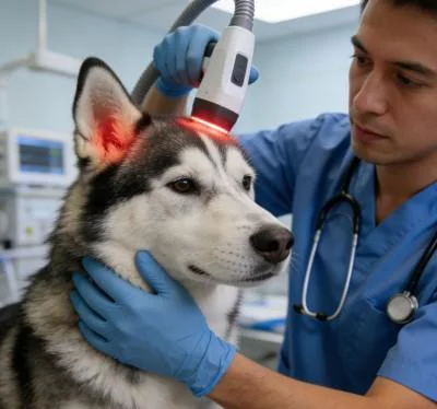
The ciliary body stroma contains large amounts of hydrated extracellular matrix, which swells during acute intraocular inflammation. The 1470nm spectrum specifically targets free and bound water molecules in this interstitial matrix, providing an absorption coefficient significantly higher than standard near-infrared bands.
When 1470nm photons strike edematous ciliary tissue, targeted water absorption causes protein denaturing and stromal shrinkage. Interstitial volume decreases rapidly, separating the secretory epithelium from the underlying vascular core and draining localized inflammatory fluid. Combining 1470nm stromal shrinkage with 980nm microvascular closure produces immediate drops in aqueous production, stabilizing globe volume and halting progressive retinal detachment.
Duty Cycle Engineering For Scleral Thermal Safety
Delivering high-power photons through thin, collagen-dense sclera carries a real risk of thermal injury. Collagen fibers denature and melt at elevated temperatures. If a continuous wave remains focused over the limbus, heat builds up rapidly, causing scleral thinning, full-thickness perforation, and catastrophic uveal prolapse.
Gated dynamic duty cycles prevent these thermal spikes. Modulating the beam intersperses microsecond pulse bursts with calibrated thermal relaxation intervals.
Continuous Wave (Thermal Runaway Over Scleral Lamellae):
Power ────────────────────────────────────────── (Scleral Perforation & Burn Risk)

Gated Dynamic Duty Cycle (Structured Scleral Cooling):
Power ┌───┐    ┌───┐    ┌───┐    ┌───┐    ┌───┐
      │   │    │   │    │   │    │   │    │   │   (Deep Ciliary Coagulation / Zero Burn)
Base  ┘   └────┘   └────┘   └────┘   └────┘   └────
      |Ton|    Toff     |
Because scleral collagen loses heat faster than the heavily vascularized ciliary processes beneath it, dynamic duty cycles let clinicians deploy high peak irradiance through the outer eye wall while maintaining safe surface temperatures. Operating between 20 percent and 35 percent duty cycles enables safe, controlled transscleral delivery directly over the perilimbal circumference.
Adjusting pulse frequencies between 10 Hertz and 200 Hertz tailors the biological effect: lower frequencies limit bulk tissue heating, preventing thermal spread to the adjacent crystalline lens, while maintaining the peak irradiance needed to photocoagulate deeper ciliary stroma.
Clinical Field Case Report
The data below comes from Veterinary Ophthalmology and Microsurgical Service unit records, documenting non-invasive cyclophotocoagulation recovery metrics in a canine patient with acute refractory glaucoma.
Patient Profile And Diagnostic Staging
Case File Number: VOS-2026-3824
Subject: Canine, Siberian Husky
Age: 9 Years 2 Months
Sex: Male (Neutered)
Weight: 24.8 Kilograms
Presenting Complaint: Acute right-sided ocular pain, severe blepharospasm, diffuse corneal haze, globe enlargement, head shyness, complete refusal of food.
Ophthalmic Examination: Right eye intraocular pressure measuring 62 mmHg via rebound tonometry; non-responsive dilated pupil; diffuse epithelial and stromal corneal edema; marked episcleral vessel engorgement; negative menace response; sluggish dazzle reflex.
Diagnostic Staging: Acute closed-angle secondary glaucoma with severe ocular hypertensive crisis, secondary to bilateral primary closed-angle pectinate ligament dysplasia (Goniodyscopy Grade 3). Left eye tonometry: 18 mmHg (Narrow iridocorneal drainage angles). Pre-treatment Pain Score via Glasgow Modified Ophthalmic Scale: 10 out of 12.
| Session ID | Day | Delivery Zone | Wavelength Distribution | Peak Power (W) | Duty Cycle (%) | Pulse Frequency (Hz) | Total Energy (J) | Clinical Observations |
| S-01 | D-01 | Transscleral 270° Arc | 75% 980nm / 25% 1470nm | 2.5 | 25% | 15 | 320 | Contact probe applied 3.5mm posterior to limbus; 24 spots; IOP drops to 26 mmHg in 3 hours. |
| S-02 | D-02 | Transscleral Dorsal Arc | 70% 980nm / 30% 1470nm | 2.5 | 25% | 20 | 280 | Episcleral engorgement softens; corneal haze clearing; IOP stabilizes at 22 mmHg without mannitol. |
| S-03 | D-05 | Transscleral Ventral Arc | 65% 980nm / 35% 1470nm | 2.8 | 30% | 30 | 340 | Pupil diameter returns to 4mm; positive consensual pupillary light response observed. |
| S-04 | D-09 | Circumferential Maintenance | 60% 980nm / 40% 1470nm | 2.8 | 30% | 50 | 360 | Corneal clarity fully restored; IOP measures 16 mmHg; normal feeding behavior resumes. |
| S-05 | D-14 | Targeted Ciliary Sectors | 50% 980nm / 50% 1470nm | 3.0 | 30% | 100 | 380 | Fundic view clear; tapetal reflex sharp; physiological cupping of optic nerve preserved. |
| S-06 | D-21 | Transscleral Reinforcement | 50% 980nm / 50% 1470nm | 3.0 | 30% | 150 | 380 | Outflow pathway stable; episcleral vessel caliber normalized to baseline. |
| S-07 | D-30 | Periocular Decompression | 50% 980nm / 50% 1470nm | 2.5 | 25% | Dynamic | 300 | Menace response intact; IOP reads 15 mmHg bilaterally under low-dose topical drops. |
| S-08 | D-45 | Long-Term Control Audit | 50% 980nm / 50% 1470nm | 2.2 | 20% | Dynamic | 260 | Globe symmetry fully maintained; Glasgow score 0; functional vision sustained. |
Objective Functional Recovery Metrics
Day 1 (Emergency Admission)
[IOP Level: 62 mmHg]         ████████████████████
[Pain Score: 10/12]          ████████████████░░░░

Day 9 (Midway Status)
[IOP Level: 16 mmHg]         █████░░░░░░░░░░░░░░░
[Pain Score: 2/12]           ███░░░░░░░░░░░░░░░░░

Day 45 (Protocol Discharge)
[IOP Level: 15 mmHg]         █████░░░░░░░░░░░░░░░
[Pain Score: 0/12]           ░░░░░░░░░░░░░░░░░░░░
By Day 45, the patient achieved complete ocular stabilization. Intraocular pressure in the right eye stabilized at 15 mmHg, maintaining balance with the unaffected left eye without requiring systemic dehydrating agents. The Glasgow Modified Ophthalmic Pain Scale score dropped from 10 to 0, confirming the elimination of severe cephalic pain and photophobia.
Complete clearing of corneal stromal edema restored full anterior chamber visualization, permitting direct fundic inspection that confirmed intact optic disc architecture. Positive menace and dazzle responses confirmed the preservation of functional vision, successfully averting enucleation surgery.
Practical Advantages Over Traditional Modalities
Managing canine glaucoma presents severe clinical dilemmas in veterinary practice. Emergency medical stabilization with topical hypotensive drops and osmotic diuretics rarely provides lasting control, serving only as a temporary measure before intraocular pressure rebounds dangerously. Conventional surgical approaches carry heavy clinical drawbacks: open filtration surgeries frequently fail due to aggressive postoperative fibroplasia, while enucleation permanently disfigures the patient and devastates pet owners emotionally. Chemical ciliary ablation with intravitreal injections carries high risks of permanent phthisis bulbi, hyphema, and chronic endophthalmitis.
Underpowered phototherapy devices fail in ophthalmic surgical care. Low-wattage units cannot deliver sufficient energy density through dense scleral tissue to reach the ciliary processes, producing only superficial warmth that increases intraocular inflammation without stopping aqueous humor production.
High-power Class 4 multi-wavelength protocols resolve these therapeutic dead ends. Delivering controlled peak power through balanced 980nm and 1470nm wavelengths enables transscleral cyclophotocoagulation in under five minutes per quadrant. This precise delivery allows veterinary surgeons to close ciliary vessels and shrink secretory tissue rapidly, avoiding extensive tissue trauma.
Dynamic duty cycle pulsing protects the outer scleral collagen, eliminating the risks of scleral perforation, corneal thermal burns, and cataract formation common with older continuous-wave lasers. Intraocular pressure drops predictably within hours of the first session, relieving excruciating cephalic pain and preserving ocular architecture. For veterinary practices, combining non-invasive ciliary ablation with reliable intraocular pressure control turns complex ophthalmic emergencies into an effective, sight-sparing clinical service.