 FotonMedix



Resolving Equine Superficial Digital Flexor Tendonitis
Dual-wavelength Class IV photon deposition accelerates tenocyte realignment, clearing intratendinous edema and fibrotic lesions without thermal injury through duty cycle modulated energy delivery.
A 6-year-old Thoroughbred gelding stands with the right forelimb placed forward, displaying classic warmth, focal bowing, and intense sensitivity over the mid-metacarpal region. The diagnostic ultrasound confirms the trainer’s worst fear: an acute core lesion involving twenty-eight percent of the cross-sectional area in the superficial digital flexor tendon. Traditional conservative management means six to nine months of strict box stall rest, cold hosing, and systemic anti-inflammatory drugs that blunt early pain but do little to organize collagen deposition. When these athletes return to training, disorganized type III collagen creates brittle scar tissue, making reinjury rates notoriously high.
Tendon tissue presents a harsh clinical hurdle: it is hypovascular, brims with tightly woven connective fibrils, and experiences prolonged metabolic latency. Low-output phototherapy units lack the raw photonic thrust to penetrate through the flexor retinaculum, peri-tendinous fluid cuffs, and dense fibrous sheaths. Addressing deep-seated soft tissue architecture demands robust veterinary laser equipment designed to project deep energy into hypovascular zones, driving immediate metabolic rescue without inducing destructive thermal buildup along the equine cannon bone.
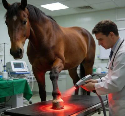
Photon Scattering and Depth Dynamics in Dense Fascial Architecture
Equine distal limb anatomy features practically no subcutaneous fat; the skin lies directly over the deep digital flexor tendon, the suspensory ligament, and cortical bone. This geometry creates a severe optical trap for uncalibrated emitters. Skin surface pigments and dense hair follicles cause intense backscattering, while the high collagen density of healthy tendon margins reflects up to seventy percent of incident uncollimated light.
To alter cellular respiration within the center of a dense tendon lesion, emitted light must penetrate deep into the hypovascular core:
- 980 nm Microvascular Revascularization: Intratendinous core lesions cause localized ischemia, producing hypoxic cellular death. Light at 980 nm targets hemoglobin chromophores, producing controlled photothermal micro-gradients that open spastic capillaries, accelerate macrophage clearance of necrotic cellular debris, and restore interstitial oxygenation without causing surface blisters.
- 1064 nm and 1470 nm Extracellular Matrix Regulation: The 1064 nm wavelength penetrates straight through dense fibrous bundles, while 1470 nm selectively interacts with free and bound interstitial water molecules. Modulating the hydration shell around swollen collagen fibrils reduces excessive hydrostatic pressure, relieving tension on sensory nerve endings embedded within the peritenon.
- Type I vs. Type III Collagen Synthesis Shift: Tendons repair naturally by depositing weak, haphazard type III collagen fibers. Deep photonic saturation triggers transforming growth factor beta pathways, inducing tenocytes to shift from disorganized scar matrix formation toward high-tensile, longitudinal type I collagen alignment.
Relying on low-power devices forces the clinician to hold a stationary probe against a sensitive equine limb for prolonged intervals, risking uncalibrated surface overheating. Operating an advanced veterinary laser machine allows the equine practitioner to treat broad metacarpal zones with continuous dynamic motion, bathing the injured core in uniform photon density in minutes.
Eliminating Epidermal Heat Peaks with Duty Cycle Architecture
Treating distal equine extremities with high continuous power introduces genuine safety challenges. Because thin skin lies immediately over periosteal margins, continuous wave emission can rapidly elevate periosteal temperature, causing discomfort, sudden limb withdrawal, or epidermal burns.
Dynamic pulse duration control mitigates this risk by interleaving high-intensity bursts with thermal relaxation intervals.
Continuous Emission:  [============= Heat Build-up =============] -> Periosteal Irritation
Duty Cycle Modulated: [=== Burst ===] [ Cooldown ] [=== Burst ===] -> Steady Deep Flux
By dialing a fifty percent duty cycle, peak power delivers deep photon penetration, while the intermittent dark cycle lets blood flow in the dermal plexuses carry away excess heat. Clinicians using modern veterinary lasers can safely deliver upward of 6,000 joules per treatment zone, driving energy deep into stubborn connective tissue lesions without distressing the animal.
Clinical Protocol and Objective Ultrasonographic Outcomes
Peer-reviewed equine sports medicine literature underscores that tendon healing depends on early mechanical tension and elevated cellular metabolism. Supplying high photon flux during early proliferation increases Tenascin-C and decorin synthesis, which orchestrate collagen fibrillogenesis and enhance tensile integrity under weight-bearing loads.
The following clinical case record outlines the rehabilitation of an acute, performance-limiting tendon lesion in a competition horse.
Case Record: Equine Acute Superficial Digital Flexor Tendonitis
- Case Identifier: EQUINE-2026-SPORTS-0319
- Patient Profile: 6-year-old Thoroughbred gelding, flat racing / eventing prospect
- Body Mass: 485 kg
- Diagnosis: Acute right forelimb mid-metacarpal superficial digital flexor tendonitis (Zone 2B), Grade 3 lameness on the AAEP scale, cross-sectional core lesion defect measuring 28% under diagnostic ultrasound
- Prior Conservative Therapy: Phenylbutazone at 2.2 mg/kg orally every 12 hours for five days, ice boots twice daily; moderate swelling remained, with marked pain on palpation and refusal to bear full weight on the right forelimb
Comprehensive Treatment Protocol Parameters
| Session Number | Clinical Phase | Output Power (W) | Operating Mode & Duty Cycle | Target Wavelength Balance | Frequency (Hz) | Total Joules (J) | Energy Density (J/cm²) |
| 01 | Acute Anti-inflammatory | 10.0 W | Pulsed (40% Duty Cycle) | High 980 nm / Capillary Focus | 3,000 Hz | 3,000 J | 7.5 J/cm² |
| 02 | Edema Clearance & Drain | 12.0 W | Pulsed (50% Duty Cycle) | Balanced 980 nm / 1064 nm | 1,500 Hz | 4,200 J | 10.5 J/cm² |
| 03 | Core Lesion Debridement | 15.0 W | Pulsed (50% Duty Cycle) | High 1064 nm / Deep Fibrils | 800 Hz | 5,400 J | 13.5 J/cm² |
| 04 | Tenocyte Proliferation | 18.0 W | Superpulsed (50% Duty Cycle) | Mixed Penetration Spectrum | 400 Hz | 6,000 J | 15.0 J/cm² |
| 05 | Collagen Alignment Induction | 20.0 W | Pulsed (50% Duty Cycle) | Deep Extracellular Matrix Focus | 200 Hz | 6,500 J | 16.2 J/cm² |
| 06 | Fascial Remodeling | 20.0 W | Pulsed (50% Duty Cycle) | Balanced Regeneration Mix | 100 Hz | 7,000 J | 17.5 J/cm² |
| 07 | Tensile Stabilization | 18.0 W | Pulsed (60% Duty Cycle) | Matrix Maturation Profile | 50 Hz | 6,400 J | 16.0 J/cm² |
| 08 | Pre-Loading Consolidation | 15.0 W | Pulsed (50% Duty Cycle) | High-penetration spectrum | 25 Hz | 5,500 J | 13.7 J/cm² |
Structural Ultrasonography Evolution and Lesion Size Reduction
Core Lesion Area Percentage (% of Tendon Cross-Section):
Pre-Treatment: [============================] 28%
Day 14:        [==================] 18%
Day 28:        [=========] 9%
Day 45:        [==] 2% (Near-complete fill, linear fiber alignment)

AAEP Lameness Grade (0 to 5 Scale):
Pre-Treatment: [======] Grade 3
Day 14:        [====] Grade 2
Day 28:        [==] Grade 1
Day 45:        [] Grade 0 (Sound at trot)
By the third session, palpable heat had subsided and local pain upon flexion was absent. Serial longitudinal and transverse ultrasound at Day 45 revealed extensive hyperechoic fill of the core defect with parallel, uninterrupted fiber patterns rather than amorphous scar tissue. Controlled in-hand walking commenced thirty days earlier than standard conservative timelines, and the horse returned to full training without recurrence.
Practical Advantages Over Traditional Modalities
Managing equine orthopedic injuries often forces difficult trade-offs between slow conservative rest and costly biological injections like platelet-rich plasma or stem cells. While biologics offer value, they demand invasive harvests, rigorous sterile injection protocols, and secondary immobilization that can trigger distal joint stiffness.
Integrating high-power Class IV photomedicine offers distinct workflow and performance benefits:
1. Zero Downtime and Accelerated Turnover
Needle-based interventions enforce weeks of absolute immobilization to avoid inflammatory flares. High-power phototherapy works alongside immediate controlled walking programs, promoting real-time mechanical fiber orientation as tenocytes proliferate.
2. Broad Application Across Sports Injuries
Equine practices do not treat single pathologies. A multi-wavelength platform shifts seamlessly from deep tendon core lesions to sacroiliac strain, stifle synovitis, capped hocks, dorsal metacarpal disease (bucked shins), and contaminated distal limb lacerations by adjusting output power and beam geometry.
3. Immediate Client Confidence
Equine owners and trainers track swelling and heat closely. Removing focal heat and edema within three treatments demonstrates tangible clinical value early, boosting client compliance with recommended rest schedules and training modifications.
Targeting deep lesions with high-power Class IV technology bridges the gap between biological rest and active rehabilitation. By driving photons past dense fibrous boundaries directly into hypoxic repair zones, veterinarians can restore structural tensile strength and return sound athletes to competition.