 FotonMedix



Resolving Recalcitrant Myofascial Fibrosis in Severe Biceps Femoris Tears
Targeted high-flux photon delivery stimulates deep satellite cell proliferation and microvascular recanalization through thick aponeuroses, suppressing secondary perimuscular contractures with precise pulse gating while preventing superficial photothermal injury.
A sprinter pulling up abruptly mid-stride with sharp posterior thigh pain represents one of the most frustrating challenges in outpatient physical medicine. Acute management frequently focuses on standard rest, ice, compression, and elevation protocols, followed by superficial therapeutic ultrasound or dry needling. While acute hemorrhage subsides, the myotendinous junction typically consolidates into a rigid, non-compliant fibrotic scar within three to six weeks. The patient returns describing a persistent, tethered pulling sensation whenever the hip flexes past 70 degrees during terminal swing phase. Deep muscle tears trapped within dense fascial compartments suffer from chronic local ischemia and elevated intramuscular compartmental pressures. When clinicians apply high-intensity coherent light to these injuries, the primary technical bottleneck remains tissue scatter: thick gluteal and hamstring adipose layers rapidly attenuate non-targeted wavelengths, leaving deeper structural tears unperfused. Breaking this cycle requires matching photon delivery directly to deep vascular and collagen dynamics.
Biophysical Mechanisms of Deep Myofascial Regeneration
Restoring tensile compliance to compromised skeletal muscle requires driving photons past dense aponeurotic sheaths while triggering specific metabolic shifts within damaged myocytes and surrounding interstitial matrices.
Multi-Wavelength Target Selectivity in Vascularized Musculature
Unlike relatively avascular tendons, skeletal muscle contains extensive capillary architecture and higher baseline concentrations of both oxygenated and deoxygenated hemoglobin. The depth of the biceps femoris long head demands a combined optical approach:
- Perfusion surge and microvascular recruitment: Light emitted at 980nm matches the high-absorption curve of tissue water and hemoglobin. This creates an immediate, localized thermal expansion across the vascular endothelium, promoting nitrous oxide synthesis, opening dormant capillary pathways, and clearing trapped inflammatory markers like bradykinin and substance P.
- Deep mitochondrial resuscitation: Photons at 1064nm experience minimal melanin scatter and lower bulk water absorption compared to mid-infrared wavelengths. This allows uninterrupted penetration through deep fascial layers. Reaching depths exceeding 4.5 centimeters, these photons stimulate mitochondrial cytochrome c oxidase inside satellite cells, restoring local cellular respiration and supporting orderly myoblast fusion instead of disorganized collagenous scarring.
Dynamic Pulse Gating and Heat Dissipation Profiles
The primary danger when treating deep, high-volume muscle groups with high-intensity class IV platforms is cutaneous thermal buildup. Delivering the 5,000 to 10,000 Joules required for structural muscle repair in continuous wave (CW) mode quickly overwhelms the skin’s thermal relaxation capacity. Epidermal temperatures rise rapidly, forcing the operator to sweep too quickly or cut treatment short, resulting in clinical under-dosing.
Gated pulse frequency protocols eliminate this constraint. By pulsing between 20 Hz and 15,000 Hz with fine-tuned duty cycles (the ratio of pulse duration to interval rest), the laser delivers high peak power during microsecond intervals. The subsequent off-phase allows cutaneous capillaries to disperse residual heat. Clinicians can maintain steady tissue contact, compressing subcutaneous adipose layers and delivering targeted laser therapy for muscle strain without triggering superficial heat discomfort.
+--------------------------------------------------------------------------------------------------+
|                            DEEP APONEUROSIS PHOTON PROPAGATION MODEL                             |
+--------------------------------------------------------------------------------------------------+
| Quartz Massage Handpiece Contact                                                                 |
|   |                                                                                              |
|   v                                                                                              |
| [Epidermis & Dermis]        ---> Pulse gating dissipates superficial thermal buildup             |
|   |                                                                                              |
|   v                                                                                              |
| [Subcutaneous Adipose]     ---> Mechanical compression clears fluid, widening optical window     |
|   |                                                                                              |
|   v                                                                                              |
| [Deep Investing Fascia]     ---> 980nm dilates regional capillaries, relieving tension           |
|   |                                                                                              |
|   v                                                                                              |
| [Biceps Femoris Long Head]  ---> 1064nm activates Cytochrome C Oxidase, stimulating satellite cells |
|   |                                                                                              |
|   v                                                                                              |
| [Sciatic Nerve Bed]         ---> Gated photons downregulate peripheral C-fiber hyperalgesia      |
+--------------------------------------------------------------------------------------------------+
Protocol Delineation: Myofascial Trigger Points vs. Structural Myotendinous Avulsion
Managing muscular pathology demands a clear distinction between neurogenic hyperirritability and structural fiber discontinuity. A myofascial trigger point consists of contracted sarcomeres causing localized hypoxia and elevated chemical nociception, whereas an acute myotendinous tear involves disrupted extracellular matrices, ruptured basement membranes, and hematoma organization.
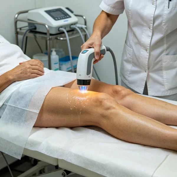
When applying a high-intensity laser for muscle pain associated with secondary myofascial contracture, the clinical priority is restoring perfusion and interrupting peripheral pain loops. Fast mechanical scanning combined with higher pulse frequencies (2,500 Hz to 10,000 Hz) downregulates overactive motor endplates, helping release protective muscle guarding around the pelvis.
Conversely, structural fiber disruptions require structural repair. Research led by Karu and colleagues on photobiological cellular signaling demonstrates that myoblast differentiation and extracellular matrix remodeling depend on specific energy densities at the target tissue layer, typically between 8 and 15 Joules per square centimeter. Insufficient power fails to trigger cellular signaling cascades, while uncontrolled thermal exposure risks additional tissue damage. Clinicians use targeted laser therapy for rotator cuff tears and severe hamstring sprains to supply sustained, controlled photon doses directly into injured tissues while preserving normal cellular function.
Clinical Case Documentation: Sports Medicine Rehabilitation
The documented clinical data below reflects an outpatient rehabilitation course for a competitive athlete presenting with a chronic, non-healing hamstring strain, treated using a multi-wavelength Class IV rehabilitation laser system equipped with a quartz massage roller.
Patient Profile and Clinical Baseline
- Case Identifier: SPORTS-MED-2026-1102
- Patient Age / Sex: 28-Year-Old Female
- Primary Diagnosis: Chronic Grade II Biceps Femoris Long Head Tear at the Proximal Myotendinous Junction with Secondary Ischiogluteal Bursitis and Perimuscular Adhesions.
- History: 10 weeks post-injury from track sprinting. Recurrent pain during terminal leg swing and prolonged sitting; recalcitrant to standard eccentric protocols, dry needling, and therapeutic ultrasound.
- Baseline Metrics: Numerical Pain Rating Scale (NPRS) at 7/10 during rapid hip flexion; Lower Extremity Functional Scale (LEFS) at 44/80; Active Straight Leg Raise (ASLR) limited to 52° due to posterior thigh pulling.
Multi-Stage Treatment Regimen
The rehabilitation schedule consisted of ten targeted sessions structured across four weeks, moving from early neuromodulation to high-energy tissue remodeling.
| Session No. | Clinical Phase & Objective | Wavelength Setup | Operating Power (W) | Pulse Frequency (Hz) | Duty Cycle (%) | Energy Delivered (J) | Cumulative Dose (J) |
| Session 01 | Neuromodulation & Perineural Calming | 980nm (65%) + 1064nm (35%) | 14.0 W | 8,000 Hz | 45% | 4,200 J | 4,200 J |
| Session 02 | Hematoma Dispersion & Drainage | 980nm (60%) + 1064nm (40%) | 16.0 W | 4,000 Hz | 50% | 4,800 J | 9,000 J |
| Session 03 | Pelvic Fascial Release & Capillary Flush | 980nm (50%) + 1064nm (50%) | 18.0 W | 2,000 Hz | 60% | 5,400 J | 14,400 J |
| Session 04 | Proximal Myotendinous Saturation | 980nm (30%) + 1064nm (70%) | 20.0 W | 500 Hz | 70% | 6,000 J | 20,400 J |
| Session 05 | Deep Interstitial Fibrotic Breakdown | 980nm (35%) + 1064nm (65%) | 22.0 W | 150 Hz | 75% | 6,600 J | 27,000 J |
| Session 06 | Satellite Cell Activation Induction | 980nm (25%) + 1064nm (75%) | 24.0 W | 50 Hz | 80% | 7,200 J | 34,200 J |
| Session 07 | High-Flux Matrix Reorganization | 980nm (20%) + 1064nm (80%) | 26.0 W | Continuous (CW) | 100% | 7,800 J | 42,000 J |
| Session 08 | Deep Linear Fascicle Realignment | 980nm (20%) + 1064nm (80%) | 26.0 W | Continuous (CW) | 100% | 7,800 J | 49,800 J |
| Session 09 | Dynamic Eccentric Tension Integration | 980nm (30%) + 1064nm (70%) | 22.0 W | 100 Hz | 75% | 6,600 J | 56,400 J |
| Session 10 | Final Tissue Consolidation & Discharge | 980nm (40%) + 1064nm (60%) | 18.0 W | 400 Hz | 65% | 5,400 J | 61,800 J |
Clinical Progression and Outcome Metrics
WEEK 1 (Sessions 1-3):
- Acute sitting discomfort over the ischial tuberosity reduced; NPRS dropped from 7/10 to 3/10.
- Palpation revealed softening of protective hypertonicity throughout the medial and lateral hamstrings.
- Ischiogluteal tenderness subsided following Session 3 lymphatic flushing.

WEEK 2 (Sessions 4-6):
- ASLR increased from baseline 52° to 74° without triggering posterior pain.
- Ultrasound evaluation showed resolution of local hypoechoic fluid cuffs around the proximal tear site.
- LEFS score advanced from 44 to 59.

WEEK 3 (Sessions 7-8):
- Continuous wave delivery applied alongside passive knee extension mobilization.
- Patient initiated light submaximal eccentric hamstring loading without discomfort or post-exercise stiffness.
- Palpable scar tissue at the biceps femoris proximal tendon felt noticeably smoother and more pliable.

WEEK 4 (Sessions 9-10):
- Full active hip flexion with extended knee restored (88° ASLR).
- Final NPRS recorded at 0/10 during rest and 1/10 following high-speed eccentric treadmill testing.
- LEFS score reached 77/80, clearing the athlete for full sprinting drills and competition return.
Comparative Analysis: High-Intensity Laser vs. Conventional Modalities
Evaluating high-intensity class IV laser alongside common physical therapies highlights its distinct functional profile.
Therapeutic Ultrasound (1 MHz / 3 MHz)
Therapeutic ultrasound relies on mechanical sound waves to create deep friction and micro-cavitation. While it delivers thermal effects to dense collagen, sound waves reflect significantly at bony interfaces, risking periosteal irritation. Ultrasound also cannot selectively stimulate mitochondrial respiration. High-intensity laser passes through deep tissue without acoustic rebound, driving targeted photobiomodulation directly inside cellular organelles rather than depending purely on bulk heating.
Instrument-Assisted Soft Tissue Mobilization (IASTM)
IASTM uses stainless steel tools to physically strip and shear fibrotic tissue planes. While helpful for breaking up superficial adhesions, forceful tool scraping can induce micro-bruising, increase local inflammation, and trigger involuntary muscle guarding in hyperalgesic patients. Class IV laser combined with a smooth quartz roller delivers deep mechanical compression alongside coherent light, relaxing muscular hypertonicity and encouraging lymphatic flow without mechanical bruising.
Platelet-Rich Plasma (PRP) Injections
PRP introduces concentrated autologous growth factors to accelerate local repair. Although chemically effective, the procedure is invasive, painful, and typically requires several weeks of relative unloading and activity restriction. Multi-wavelength laser therapy serves as an ideal non-invasive complement or standalone intervention, stimulating native microvascular perfusion and cellular ATP turnover to accelerate tissue remodeling without downtime or injection-related soreness.
Practical Clinical Application and Operational Takeaways
Maximizing the effectiveness of high-power laser therapy for muscle strain and deep soft-tissue pathology requires moving beyond static application methods:
- Dynamic manual compression: Firmly pressing a quartz treatment handpiece against the muscle belly compresses superficial skin and adipose tissue. This temporary displacement of fluid shortens the physical distance to the muscle fibers and cuts photonic loss from superficial water absorption.
- Directional fascia tracing: Clinicians should follow the anatomical course of the muscle fibers, moving from distal insertion sites toward proximal tendon attachments to promote directional lymphatic clearance and aid orderly collagen deposition.
- Frequency progression throughout recovery: Begin with high-frequency pulsing (4,000 Hz to 8,000 Hz) to calm irritated neural pathways, transition to lower frequencies (50 Hz to 500 Hz) to support cellular proliferation, and conclude with continuous wave delivery during the final remodeling phase to improve tissue elasticity.
Applying high-intensity laser protocols based on clear optical and anatomical principles gives sports medicine clinics a predictable path to resolving complex, stubborn muscular injuries.